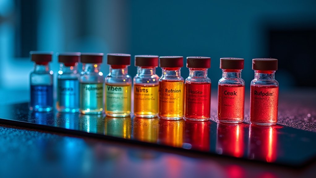
The 7 best crucial dyes for living cell observation include mitochondria-targeting BioTracker 488 Green and 633 Red, pH-responsive fluorophores for acidic organelles, lysosomal acid-sensitive BioTracker 560 Orange, enzyme-activated dyes, nuclear-permeable Hoechst 33342, DRAQ5 for long-term nuclear tracking, and plasma membrane labels like BioTracker 400 Blue. You’ll find these options maintain cell viability while providing excellent contrast and specificity. Discover how each dye uniquely reveals cellular processes without compromising your experimental integrity.
Mitochondria-Targeting Fluorescent Dyes for Metabolic Analysis
Anyone studying cellular metabolism needs reliable tools to visualize mitochondrial activity. Dyes like BioTracker 488 Green and BioTracker 633 Red offer you precise targeting of mitochondria based on membrane potential, enabling real-time assessment of these critical organelles.
These fluorescent dyes accumulate within active mitochondria, providing immediate feedback on metabolic states during live cell imaging. You’ll find they’re invaluable when identifying functional changes that indicate cellular stress or disease conditions.
Visualize metabolism in real time as dyes reveal mitochondrial stress signals during critical cellular transitions.
For thorough analysis, try co-staining techniques that combine mitochondria-specific dyes with other fluorescent markers. This approach reveals relationships between mitochondrial function and other cellular components.
You’ll gain deeper insights into processes like apoptosis, differentiation, and metabolic disorders by monitoring how mitochondrial membrane potential fluctuates under various experimental conditions.
Lysosomal Acid-Sensitive Vital Stains for Endocytosis Research
To effectively track endocytosis and lysosomal function, you’ll need specialized dyes that respond to the unique acidic environment within these organelles. BioTracker 560 Orange and BioTracker 540 Red are excellent choices as they become fluorescent upon protonation in acidic lysosomes.
These lysosomal acid-sensitive crucial stains are invaluable for live cell staining protocols, allowing you to visualize endocytic pathways and monitor degradation processes in real-time.
You can observe dynamic events like lysosomal fusion during fluorescence imaging sessions without disrupting cellular activity.
For thorough endocytosis research, combine these dyes with other fluorescent markers to simultaneously track multiple cellular processes. This approach not only enhances your understanding of cellular homeostasis but also provides insights into potential therapeutic targets for diseases characterized by lysosomal dysfunction.
Nuclear Membrane-Permeable Dyes With Low Phototoxicity
You’ll find Hoechst stains effective for short-term nuclear visualization, though they carry higher phototoxicity than newer alternatives.
SYTO dyes offer greater membrane permeability and spectral variety, making them versatile for multiplexed imaging across different cell types.
DRAQ5 outperforms many nuclear dyes with its far-red emission profile, allowing deeper tissue penetration and exceptional compatibility with other fluorophores in live-cell applications.
Hoechst Stains Comparison
Two prominent nuclear stains, Hoechst 33258 and Hoechst 33342, stand out for their ability to penetrate nuclear membranes and bind to A-T rich regions in DNA with minimal disruption to living cells.
When choosing between these Hoechst stains for your fluorescence microscopy experiments, consider these key differences:
- Hoechst 33342 provides brighter fluorescence and effectively stains both live and fixed cells
- Hoechst 33258 performs efficiently in fixed samples but can still be used in live cell applications
- Both dyes exhibit low phototoxicity, making them ideal for long-term imaging of cellular processes
- With excitation at ~350nm and emission at ~461nm, you can easily combine these dyes with other fluorescent markers for multi-color imaging
You’ll find these nuclear membrane-permeable dyes invaluable for visualizing nuclei while maintaining cell viability.
SYTO Dye Applications
SYTO dyes represent versatile tools for researchers seeking effective nuclear staining with minimal cellular disruption. These membrane-permeable fluorophores easily penetrate live cells, allowing you to observe nuclei and other cellular structures without damaging cell membranes or compromising viability.
You’ll appreciate SYTO dyes’ remarkably low phototoxicity when conducting extended imaging sessions, as they won’t greatly stress your samples during observation periods. The fluorescence intensity increases dramatically when these dyes bind to nucleic acids, providing strong signals for tracking cellular processes in real time.
For multi-color experiments, you can choose from various spectral variants that complement your existing fluorescent markers.
Consider pairing SYTO dyes with other essential stains to create thorough visualization systems that reveal complex cellular dynamics while maintaining cell health throughout your imaging sessions.
DRAQ5 Performance Analysis
DRAQ5 stands out among membrane-permeable nuclear stains for its exceptional penetration capabilities and long-term stability during live-cell imaging. When you’re monitoring cellular processes in real time, this fluorescent dye delivers superior nuclear DNA visualization without compromising cell viability.
- Exhibits minimal phototoxicity, allowing extended observation periods of live cells without damaging them.
- Emits strong near-infrared fluorescence signals that reduce spectral overlap in multiplexing experiments.
- Effectively penetrates nuclear membranes to reveal nucleolar structures and chromatin dynamics.
- Maintains consistent fluorescence intensity over time, supporting long-term cell proliferation studies.
DRAQ5’s remarkable stability and low interference with cellular functions make it ideal for researchers requiring detailed nuclear visualization in living specimens. Its compatibility with other fluorophores further enhances its utility in complex experimental designs requiring multiple markers.
Plasma Membrane Labeling Dyes for Cell Boundary Visualization
When visualizing cellular boundaries in live specimens, plasma membrane labeling dyes offer researchers a powerful tool for delineating cell morphology with exceptional clarity. Products like BioTracker 400 Blue, 490 Green, and NIR750 utilize lipophilic carbocyanines that effectively integrate into the plasma membrane without compromising cell viability.
You’ll find these dyes particularly valuable for long-term cell tracking experiments, as their low cytotoxicity preserves cellular function during extended imaging sessions. They excel in fluorescence microscopy applications, providing crisp definition of cell boundaries while allowing visualization of dynamic processes like migration and division in real time.
For more thorough analysis, consider co-staining with other fluorescent markers to simultaneously observe membrane interactions with organelles and other cellular components, enhancing your understanding of complex cellular behaviors.
Selective Cytoskeletal Stains for Live Cell Dynamics
Visualizing the intricate architecture of the cytoskeleton in living cells requires specialized fluorescent probes that minimize disruption to cellular functions. BioTracker microtubule dyes offer cell-permeable options in green and red fluorescence that don’t require washing steps, making them ideal for real-time observation.
When you’re studying dynamic cellular processes, these selective cytoskeletal stains provide essential advantages:
- Allow visualization of microtubule reorganization during cell migration and division
- Can be combined with other crucial dyes to correlate cytoskeletal changes with organelle movement
- Enable monitoring of structural responses to various experimental stimuli
- Preserve normal cell membrane interactions while providing clear imaging of internal cytoskeletal networks
You’ll gain deeper insights into endocytosis, motility, and other fundamental cellular behaviors through these non-disruptive visualization techniques.
Ph-Responsive Organelle-Specific Fluorophores
Beyond cytoskeletal visualization, understanding organelle function requires tools that respond to the cell’s internal chemistry. pH-responsive organelle-specific fluorophores offer remarkable specificity by exploiting the distinct pH environments within cellular compartments.
When you’re tracking lysosomal activity in live cells, these dyes intensify their fluorescence in acidic conditions, providing real-time insights into organelle dynamics. You’ll find these fluorophores particularly valuable for monitoring autophagy and apoptosis without compromising cell viability.
As you observe cellular processes over time, pH-responsive dyes reveal essential information about metabolic activity and cellular health.
They’re compatible with extended imaging sessions, allowing you to track organelle behavior throughout experimental timeframes. By integrating these fluorophores into your imaging workflow, you’ll gain deeper understanding of organelle-specific functions and their role in cellular physiology.
Enzyme-Activated Dyes for Real-Time Cellular Activity Monitoring
Unlike static staining methods, enzyme-activated dyes transform cellular imaging by revealing metabolic processes as they occur. You’ll find these compounds invaluable when you need to stain live cells without compromising their viability.
What makes these dyes particularly useful:
- Initially non-fluorescent until specific intracellular enzymes activate them, producing bright fluorescent signals
- Freely permeate cell membranes, allowing you to monitor cellular activity in real-time
- Remain inside living cells after activation, enabling tracking through multiple cell divisions
- Provide clear identification of viable cell populations, as seen with Calcein AM’s fluorescence when cleaved by esterases
These properties make enzyme-activated dyes exceptional tools when you’re studying dynamic cellular processes like migration, metabolism, and proliferation without disturbing the natural cellular environment.
Frequently Asked Questions
What Dyes Are Used for Cell Viability?
You can use DNA-binding dyes like 7-AAD or PI, amine-reactive dyes like Zombie Violet or LIVE/DEAD Fixable Far Red, and enzyme-activated dyes like Calcein AM to assess cell viability.
Does DAPI Only Stain Live Cells?
No, DAPI doesn’t stain live cells effectively. It’s membrane-impermeable and primarily stains dead cells with compromised membranes. You’ll need alternative dyes like Calcein AM for visualizing live cells.
What Is the Viability Dye for Cell Sorting?
For cell sorting, you’ll want to use LIVE/DEAD Fixable Viability Dyes. They’ll bind to surface proteins in live cells while penetrating dead ones, creating clear separation that remains stable even after fixation.
What Dye Is Used in Vital Nerve Staining?
For essential nerve staining, you’ll typically use Neutral Red, Methylene Blue, or fluorescent dyes like Calcein AM. These dyes penetrate living neurons without toxicity, allowing you to observe cellular structures and assess neuronal viability.
In Summary
You’ve seen how these seven crucial dyes transform live cell research, allowing you to observe cellular processes without disruption. Whether you’re tracking mitochondrial function, visualizing lysosomes, or monitoring enzyme activity, these dyes offer powerful windows into cellular life. They’ll enhance your imaging capabilities while maintaining cell viability. Choose the right dye for your specific needs, and you’ll reveal new insights into dynamic cellular behaviors under physiological conditions.


Leave a Reply