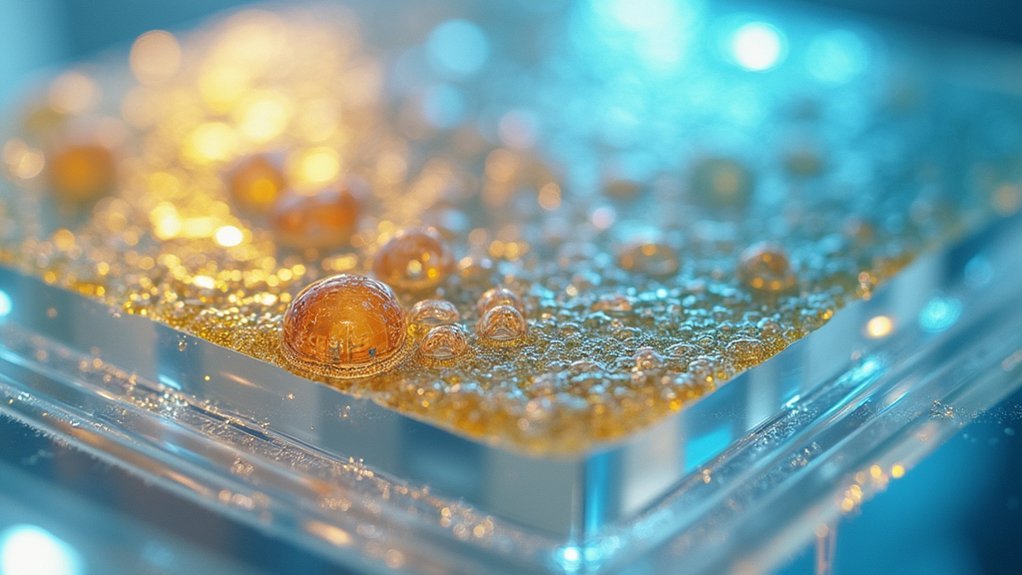
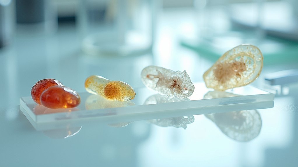
Specimen transparency for scientific imaging is achieved by removing light-scattering lipids while preserving structural integrity. You’ll need to manipulate molecular structures using organic solvents, water-based detergents, or hydrogels like in the HYBRiD technique. These methods maintain fluorescent signals and eliminate opacity, allowing visualization of complex biological structures without specialized equipment. Modern clearing approaches have revolutionized fields like neuroscience by revealing previously hidden features. The journey from opaque tissue to crystal-clear specimen unfolds through fascinating chemical interactions.
The Science Behind Tissue Transparency
While biological samples naturally appear opaque to the naked eye, achieving transparency involves a sophisticated manipulation of their molecular structure. The key challenge is removing lipids that scatter light while preserving the sample’s molecular integrity.
Traditional tissue clearing methods use organic solvents like xylene that quickly create transparency but often diminish fluorescence signals essential for detailed imaging. Water-based alternatives better preserve fluorescence but struggle with non-brain tissues and require longer processing times.
The innovative HYBRiD technique offers a breakthrough by combining organic solvents with water-based detergents and hydrogels. This approach maintains both transparency and fluorescence, making it ideal for larger biological samples.
Breakthrough HYBRiD method unites opposing approaches, preserving both transparency and fluorescence signals for unprecedented biological visualization.
These advancements have revolutionized fields like neuroscience, allowing you to visualize complex structures that were previously hidden within opaque tissues.
Traditional Clearing Methods and Their Limitations
Although widely adopted in laboratories worldwide, traditional tissue-clearing techniques present significant challenges for researchers seeking ideal imaging results.
When you’re working to achieve transparency in biological specimens, you’ll encounter several fundamental limitations:
- Solvent trade-offs – Organic solvents like xylene effectively remove lipids causing tissue opacity but often diminish fluorescence signals and introduce artifacts.
- Incomplete clearing – Water-based solvents preserve fluorescence better but struggle with complex non-brain tissues, limiting whole-organ imaging.
- Practicality concerns – Many traditional methods require hazardous chemicals and specialized equipment, making routine laboratory implementation difficult.
- Limited scalability – Conventional approaches often fail when visualizing proteins and structures in larger specimens, restricting applications for whole-animal imaging.
These limitations highlight why researchers continue seeking improved clearing methods for diverse biological specimens.
Modern Approaches to Specimen Clearing
Recent advances in specimen clearing have dramatically transformed how researchers visualize complex biological structures. The innovative HYBRiD method, developed by Scripps Research, stands at the forefront of these advancements.
You’ll find this technique uniquely combines organic solvents with water-based detergents and hydrogels in a sequential process that preserves your biological samples while enhancing tissue clearing.
Unlike traditional methods that often sacrifice fluorescence for transparency, HYBRiD maintains fluorescent signals while effectively clearing non-brain tissues. You can now process large tissue samples without perfusion, simplifying the imaging of whole animals.
When paired with immunocytochemistry, this approach offers unprecedented visualization of specific proteins within complex structures. The technique’s accessibility makes it practical for routine scientific imaging across various laboratory settings, allowing you to observe intricate cellular interactions with remarkable clarity.
The Revolutionary HYBRiD Technique
The HYBRiD technique revolutionizes specimen clearing by ingeniously combining organic solvents, water-based detergents, and hydrogels to achieve remarkable transparency while preserving protein structures.
You’ll appreciate how this approach simplifies tissue clearing to basic laboratory equipment—often just a jar and shaker—making advanced imaging accessible for more researchers.
This breakthrough method enables you to visualize complex biological structures throughout entire organisms, as demonstrated by its success imaging SARS-CoV-2 infected cells in whole mouse bodies.
HYBRiD Clearing Mechanics
While many clearing techniques require specialized equipment and complex protocols, HYBRiD stands out with its elegant simplicity and powerful results. This innovative approach achieves remarkable transparency in biological samples through a sequential combination of organic solvents, water-based detergents, and hydrogels—all while preserving protein integrity.
You’ll appreciate HYBRiD’s practical advantages:
- Simplified clearing process requiring just a jar and benchtop shaker
- Perfusion-free clearing enabling visualization of complex structures like SARS-CoV-2 in whole mouse chests
- Enhanced visualization of genetically encoded fluorescent reporters without antibody enhancement
- Effective imaging of large specimens opening new avenues for neuroscience research
This technique’s ability to maintain structural integrity while achieving exceptional transparency makes it invaluable for studying both healthy and disease-related biological processes.
Tissue-Wide Imaging Possibilities
Revolutionary in its approach, HYBRiD clearing transforms how you’ll visualize complex biological structures across entire tissues and organisms. This technique achieves exceptional transparency in biological samples while maintaining protein integrity, allowing you to examine whole animals without perfusion.
You’ll appreciate how HYBRiD simplifies tissue-clearing into a process manageable with basic equipment—just jars and shakers—making imaging large body parts practical for routine laboratory use. The method supports direct fluorescent imaging of genetically encoded reporters without antibody enhancement, dramatically improving workflow efficiency.
Current applications extend beyond basic research, with scientists tracing nerve pathways and studying pathogen spread in complex tissues. Whether you’re investigating SARS-CoV-2 infection patterns or neural connections, HYBRiD offers unprecedented access to structural information previously hidden within opaque biological specimens.
Chemical Agents That Enhance Specimen Visibility
You’ll find that organic solvents like xylene effectively remove opacity-causing lipids but can diminish fluorescent signals in your specimens.
Water-based detergents offer an alternative approach that better preserves fluorescence, though they’re typically less effective at clearing non-brain tissues than their solvent counterparts.
The incorporation of hydrogels in modern techniques like HYBRiD maintains protein structure integrity while enhancing transparency, giving you the best of both chemical approaches.
Solvents Versus Detergent Methods
When scientists need to visualize cellular structures clearly, they face a critical choice between solvent-based and detergent-based clearing methods. Traditional organic solvents like xylene effectively remove lipids for tissue-clearing but often diminish fluorescent signals.
Water-based techniques preserve these signals better but struggle with non-brain tissues.
The innovative HYBRiD method offers a practical solution by combining both approaches:
- Merges organic solvents with water-based detergents and hydrogels
- Maintains molecular integrity while achieving superior transparency
- Preserves protein retention for accurate visualization of biological structures
- Enables simpler, scalable protocols suitable for routine laboratory use
You’ll find this hybrid approach particularly valuable when working with larger specimens where traditional methods fall short, providing clearer images without introducing artifacts that could compromise your research findings.
Hydrogel Preserves Structure
At the core of effective tissue clearing lies the hydrogel matrix, which serves as a protective scaffold that maintains structural integrity while tissues become transparent.
When you’re working with the HYBRiD tissue-clearing method, these water-based hydrogels protect molecular integrity throughout the transparency process, ensuring proteins and anatomical features remain intact for imaging.
You’ll notice hydrogels prevent the loss of fluorescent signals that typically occurs with traditional organic solvents. By preserving structural relationships within tissues, they enable clearer visualization of complex biological processes.
What makes hydrogels particularly valuable is their compatibility with sequential solvents and detergents without requiring perfusion—making the technique accessible for routine laboratory applications.
You’ll appreciate the simplicity of the HYBRiD approach, as achieving high transparency requires minimal equipment—just a basic shaker and jars.
Preserving Structural Integrity During Clearing
While achieving optical transparency in biological specimens is essential for detailed imaging, maintaining structural integrity throughout the clearing process remains an equally important challenge.
The HYBRid technique addresses this concern by leveraging hydrogels as protective scaffolds during tissue-clearing.
Hydrogels serve as molecular guardians in the HYBRid approach, maintaining structural fidelity while tissues transition to transparency.
When you’re preparing biological samples for imaging, consider these key advantages:
- Hydrogels protect molecular structures while solvents and detergents remove opacity-causing lipids
- Fluorescent signals remain intact, enabling detailed visualization of complex structures
- The method eliminates the need for perfusion, further preserving tissue integrity
- Reduced hazardous chemical usage makes the protocol safer and more accessible
This approach guarantees your specimens achieve transparency without sacrificing structural details, allowing for thorough imaging of even large samples while preserving their natural composition.
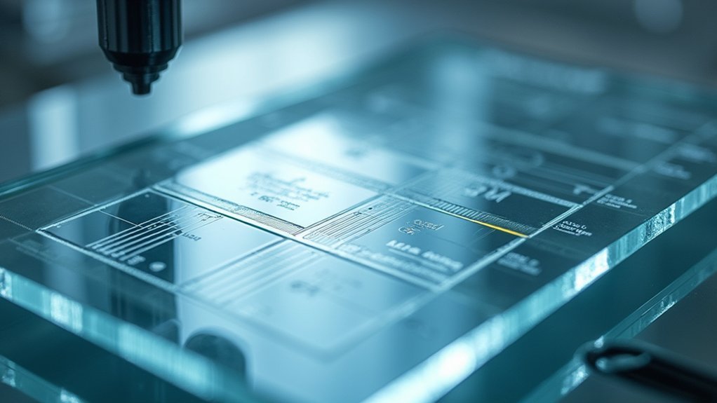
From Bench to Image: The Complete Workflow
Transforming biological specimens from opaque tissues to crystal-clear imaging subjects requires a systematic approach. Your workflow begins with specimen fixation, preserving the sample in its life-like state before processing.
Next, apply tissue-clearing techniques that remove opaque elements while protecting delicate molecular structures—the innovative HYBRiD method incorporates hydrogels for enhanced preservation during clearing.
Once transparency is achieved, advanced imaging techniques reveal previously hidden biological structures. Fluorescent signals can be captured with remarkable clarity, and 3-D reconstruction allows you to visualize complex anatomical relationships from multiple angles.
This complete bench-to-image workflow—reception, fixation, processing, embedding, sectioning, and staining—ensures each step builds toward producing scientific images with exceptional detail and accuracy, revolutionizing how we observe and understand microscopic worlds.
Applications in Advanced Biological Imaging
The implementation of HYBRiD tissue-clearing technology has opened unprecedented windows into complex biological systems. This revolutionary method preserves protein integrity while making biological samples transparent, allowing you to visualize structures that were previously impossible to see intact.
Key applications include:
- Neuroscience mapping – Trace complete neuron pathways through entire organs without sectioning
- Infectious disease research – Visualize SARS-CoV-2 infection throughout whole mouse chests
- Gene expression studies – Direct fluorescent imaging without antibody enhancement
- Developmental biology – Observe intricate structural relationships in intact organisms
You’ll find HYBRiD particularly valuable when you need thorough visualization of fluorescent markers across large specimens.
The method’s versatility continues to expand through collaborations exploring advanced imaging applications in multiple scientific disciplines.
Frequently Asked Questions
What Is a Substance That Is Added to a Specimen to Make It More Clearly Visible?
You’ll use clearing agents like xylene to enhance specimen visibility. These remove lipids causing opacity. You can also add hydrogels or fluorescent markers for better transparency during imaging procedures.
What Are the Various Stages Involved in the Preparation of a Cell Material for Microscopy?
You’ll prepare cells through fixation to preserve structure, dehydration and clearing to remove opacity, embedding for stability, sectioning with a microtome, and finally staining to enhance visibility of cellular components.
In Summary
You’ve now seen how transparency techniques transform specimen imaging by removing opaque elements while preserving structure. Whether you’re using traditional clearing methods or cutting-edge approaches like HYBRiD, you’ll achieve clearer visualization of biological samples. As you apply these techniques in your lab, you’ll notice dramatic improvements in image quality and data collection. The science of transparency continues to evolve, offering you increasingly powerful tools for scientific discovery.

Leave a Reply