
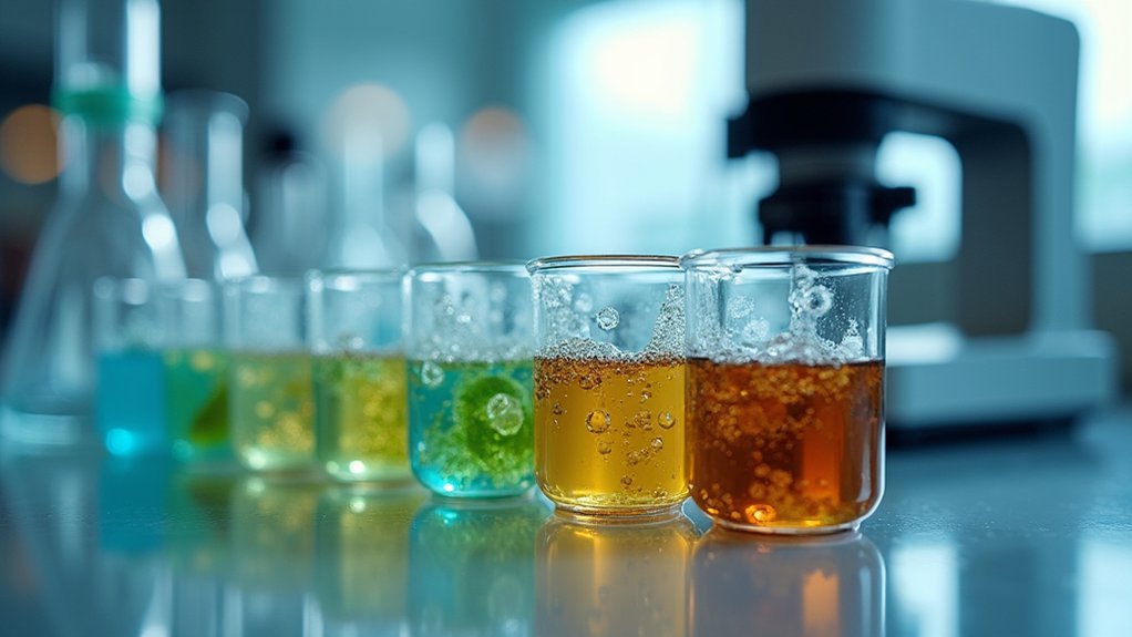
For perfect specimen sections, start with proper fixation (6-24 hours based on size), followed by thorough dehydration through graded ethanol solutions. Guarantee complete clearing with xylene before embedding in 60°C paraffin. Use a well-maintained microtome with sharp blades at 20-30° angles, and float sections in a 38-42°C water bath. You’ll need specialized techniques for challenging tissues like decalcification for hard specimens. The following techniques will transform your histological preparations from adequate to exceptional.
Proper Tissue Fixation and Preservation

Because the foundation of successful sectioning begins well before a specimen reaches the microtome, proper tissue fixation deserves your careful attention.
You’ll need fresh fixatives like formaldehyde, with incubation times ranging from 6-24 hours depending on specimen size.
The quality of your histological analysis directly depends on how well you’ve preserved morphological integrity.
Watch for signs of over-fixation (mushy tissue) or under-fixation (brittleness), as both compromise sectioning results.
If you discover under-fixed tissues, you can salvage them by de-paraffinizing, re-fixing, and re-embedding.
For specimens containing mineral deposits, apply decalcifying agents to the block surface during fixation to enhance microtomy outcomes.
Remember that fixation protocols should be tailored to specific tissue types to guarantee ideal preservation and staining quality in your final sections.
Mastering the Dehydration Process
After tissues are properly fixed, you’ll need to tackle the critical dehydration phase, which removes water from your specimens to prepare them for embedding.
Begin by immersing samples in a gradual ethanol solution series: 70%, 90%, and finally three changes of 100% ethanol. Allow 1-2 hours per step, depending on tissue size, ensuring thorough penetration.
Proper tissue dehydration requires progressive ethanol exposure, with sufficient time for complete penetration at each concentration level.
Pay close attention to this process, as incomplete dehydration can cause tissue shrinkage and distortion, compromising your histological sections.
For improved efficiency, consider using vacuum processing to enhance ethanol penetration uniformly throughout your specimens.
Don’t overlook the importance of monitoring the concentration and quality of your ethanol solutions—contamination can severely impact water content removal.
Regular quality checks will help maintain the integrity of your specimens and ultimately lead to superior sectioning results.
Effective Clearing and Infiltration Techniques
Two essential steps bridge dehydration and successful sectioning: clearing and infiltration. After completing your ethanol dehydration series, you’ll need to replace the ethanol with a clearing agent like xylene, which facilitates embedding medium penetration without distorting tissue morphology.
| Process | Key Considerations |
|---|---|
| Clearing | Replace ethanol with xylene to enhance paraffin infiltration |
| Temperature | Maintain around 60°C for molten wax; cool to 20°C during embedding |
| Vacuum Processing | Reduces air bubbles; guarantees even distribution |
| Monitoring | Prevents overheating that impacts section quality |
For best results, verify complete dehydration through the 70%, 90%, and 100% ethanol series before clearing. Using vacuum processing during infiltration improves penetration efficiency. Remember that proper temperature control of your embedding medium is critical—overheating can damage tissue structure and compromise your final section quality.
Optimizing Paraffin Embedding Procedures
Temperature control at approximately 60°C during paraffin embedding is essential for proper wax infiltration, which you’ll need to maintain consistently to avoid tissue damage or inadequate penetration.
You’ll achieve better sectioning results by selecting molds that match your specimen size and orientation needs, ensuring proper tissue placement and block formation.
Implementing vacuum infiltration can dramatically improve your paraffin penetration, especially for dense tissues, by removing trapped air and allowing complete wax infiltration throughout the entire specimen.
Temperature Control Matters
Since reliable sectioning depends on precise thermal management, controlling temperatures throughout the paraffin embedding process becomes essential for quality histological results.
You’ll achieve ideal section spreading by maintaining your water bath between 38-42°C—cold water prevents proper spreading while excessive heat melts the paraffin and compromises quality.
Chill your paraffin blocks on ice rather than freezing them. This simple temperature adjustment enhances tissue consistency and promotes better ribbon formation during sectioning.
Freezers cause rapid cooling that leads to cracking and artifacts in your specimens.
Remember to monitor your water bath temperature regularly, making adjustments based on your specific conditions.
For high-quality sections, verify both tissue and wax maintain similar temperatures during cutting—this synchronization facilitates effective sectioning while preserving structural integrity.
Proper Mold Selection
Choosing the right mold directly impacts your sectioning success and ultimately affects the quality of your histological results.
Select molds that properly fit your specimens to guarantee ideal support during the embedding process. Avoid oversized molds that require excessive paraffin, as they can compromise stability during microtomy.
For precise tissue orientation, use molds with flat surfaces that allow you to position the area of interest downward for accurate sectioning. Molds with labeled or raised areas help you maintain proper orientation throughout processing.
Remember that thicker-walled molds provide better stability but require longer time for paraffin to solidify.
Always use clean molds free from debris before embedding. Contaminants interfere with proper wax infiltration and compromise the quality of your sections.
This attention to detail guarantees successful specimen preparation.
Vacuum Infiltration Benefits
While standard embedding techniques can produce acceptable results, vacuum infiltration elevates the quality of your paraffin sections to superior levels.
By creating pressure differentials, you’ll greatly reduce tissue processing times while ensuring thorough saturation of the embedding medium throughout the specimen.
You’ll eliminate troublesome air bubbles that often cause sectioning artifacts and compromise diagnostic evaluation.
This technique allows for lower temperature processing, preserving delicate tissue morphology by minimizing heat-induced damage to cellular structures.
For consistent, high-quality paraffin blocks, monitor vacuum conditions regularly to maintain ideal infiltration rates.
The improved penetration of paraffin into tissue yields more reliable sections with fewer artifacts, ultimately enhancing your ability to evaluate specimens accurately.
This attention to embedding quality pays dividends when you’re facing challenging diagnostic cases requiring pristine tissue presentation.
Temperature Control for Perfect Sectioning
Every successful histology section begins with proper temperature management. To achieve ideal spreading of your sections, maintain water bath temperature between 38-42°C—cold water prevents proper dispersion while excessive heat melts paraffin, compromising quality.
Before sectioning, cool your paraffin blocks on aluminum foil over ice for at least five minutes. This cooling step guarantees solid consistency, preventing compression during cutting and preserving tissue integrity.
Don’t freeze blocks, however, as this causes cracking and artifacts.
During embedding and sectioning, regularly check temperatures to prevent overheating that damages tissue morphology. For best results, keep the paraffin wax and embedded tissue at similar temperatures to guarantee even cuts.
These temperature control practices will greatly enhance your sectioning outcomes and produce high-quality slides for examination.
Microtome Selection and Blade Maintenance
When choosing between rotary and sliding microtomes, you’ll find rotary models offer better efficiency for routine work while sliding types excel at harder specimens.
You can optimize your blade angle by adjusting it between 3-8 degrees for paraffin sections, testing incremental changes until achieving clean, wrinkle-free cuts.
Remember that proper blade maintenance, including regular cleaning and correct alignment, isn’t just good practice—it’s essential for consistent, high-quality sections that preserve specimen morphology.
Rotary vs. Sliding Microtomes
Choosing the right microtome for your sectioning needs can greatly impact the quality of your histological specimens. Rotary microtomes excel at paraffin-embedded tissue sectioning, delivering precise cuts between 4-10 micrometers thick. When working with larger or harder specimens, you’ll find sliding microtomes offer superior control through their unique block-sliding mechanism.
| Feature | Rotary Microtomes | Sliding Microtomes |
|---|---|---|
| Ideal for | Routine paraffin sections | Larger, harder specimens |
| Movement | Rotates blade | Slides specimen block |
| Section thickness | 4-10 μm typically | Variable, more control |
| Stability | Good for thin sections | Superior for tough tissues |
| Learning curve | Moderate | Steeper, more technical |
For both types, proper setup and alignment are vital to prevent chatter or distortion. Maintain your cutting area and replace blades when “train lines” appear to guarantee ideal section quality.
Blade Angle Optimization
Three critical factors determine the quality of your microtome sections: blade angle, sharpness, and material selection. For ideal microtomy results, maintain your blade angle between 20-30 degrees to achieve clean cuts and minimize tissue section distortion.
You’ll notice immediate improvements in section quality when using the correct angle.
Don’t overlook regular blade inspection—even minor “train lines” signal it’s time for replacement. Choose your blade material wisely; tungsten carbide offers superior longevity compared to stainless steel, though at a higher cost.
Consistency in your cutting speed and pressure settings is equally important. Establish standardized parameters and avoid frequent adjustments that can produce uneven sections.
Remember that proper blade angle idealization isn’t just about the initial setup—it requires ongoing attention to maintain precision throughout your sectioning work.
Water Bath Techniques for Wrinkle-Free Sections
A properly maintained water bath serves as the cornerstone of producing wrinkle-free histological sections. You’ll need to keep the water bath temperature between 38-42ºC to guarantee ideal spreading of paraffin sections without compromising adhesion.
Monitor the temperature regularly during sectioning to prevent cold water from interfering with proper section flattening.
- Replace flotation bath water frequently to eliminate contaminants that affect section quality.
- Use clean deionized water (5-9°C) for floating sectioned ribbons to remove wrinkles effectively.
- Gently roll sections on slides 3-5 times to promote adhesion while eliminating wrinkles.
Remember that proper water bath management directly impacts your results.
Both temperature and cleanliness contribute considerably to producing high-quality, wrinkle-free sections that will provide accurate histological data in your downstream applications.
Specialized Methods for Challenging Specimens

While water bath techniques solve many sectioning challenges, certain specimens require specialized approaches. When facing friable tissues, you’ll want to use tape transfer systems that stabilize the specimen surface before sectioning begins. For hard tissues, apply decalcifying agents and guarantee thorough rinsing before mounting to your microtome.
| Specimen Type | Challenge | Solution |
|---|---|---|
| Friable Tissue | Fragmentation | Tape transfer systems |
| Hard Tissue | Resistance | Decalcifying agents, slower cutting speeds |
| Thick Specimens | Compression artifacts | Moisture techniques, extended cooling |
You can prevent sectioning issues in challenging specimens by keeping exposed tissue surfaces moist with deionized water. For tougher specimens, adjust your microtome to slower speeds. Consider extending ethanol immersion during dehydration to improve flatness and eliminate wrinkles in your final sections.
Mounting and Drying Sections on Slides
Once you’ve successfully sectioned your specimen, proper mounting technique becomes essential for preserving tissue integrity. Float your section ribbons in deionized water at 5-9°C to eliminate wrinkles before transferring to charged microscope slides, which enhance adhesion. Briefly drain excess water to prevent movement during the drying process.
- Always monitor drying temperature carefully to avoid overheating that leads to uneven staining or nuclear meltdown.
- Limit floating time in the water bath to maintain ideal tissue morphology.
- Allow sections to dry at 37°C overnight for best results.
The mounting and drying stages are just as important as the initial sectioning. Your attention to detail during these steps directly impacts staining quality and overall specimen analysis outcomes.
Frequently Asked Questions
What Is the Process of Tissue Sectioning?
You’ll section tissue by cooling paraffin blocks on ice, cutting them with a sharp microtome blade, floating sections in a water bath (5-9°C), and placing them on slides to dry.
What Is the Best Clearing Agent?
While xylene remains standard, you’ll find less toxic alternatives like limonene-based solvents or aliphatic hydrocarbons increasingly preferable. Choose based on your specimen type, safety requirements, and whether you’re prioritizing processing speed or tissue preservation.
What Are the Different Types of Specimen Processing?
You’ll encounter several specimen processing types: fixation to preserve tissue, dehydration to remove water, clearing with agents like xylene, infiltration with embedding media, and embedding to prepare for sectioning.
What Are the Techniques of Histology?
Histology techniques you’ll employ include fixation with formaldehyde, dehydration through graded alcohols, clearing with xylene, paraffin embedding, sectioning with a microtome, and staining to visualize cellular structures under microscopic examination.
In Summary
You’ve now mastered the essential techniques for successful specimen sectioning. Remember to prioritize proper fixation, dehydration, and embedding before attempting to cut. Keep your microtome well-maintained, control your temperature variables, and use appropriate water bath techniques. Don’t forget that challenging specimens may require specialized approaches. With practice and attention to these details, you’ll consistently produce high-quality, wrinkle-free sections for your microscopic examinations.


Leave a Reply