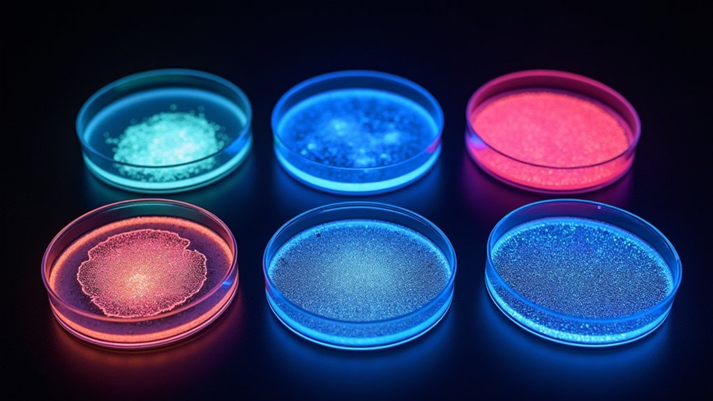
Fluorescence imaging utilizes five primary lighting methods: Widefield Epi-illumination provides accessible high-contrast imaging but with background interference; Confocal Laser Scanning uses pinhole apertures for improved signal clarity; Two-Photon Excitation achieves deeper tissue penetration with reduced photobleaching; Light Sheet Microscopy minimizes phototoxicity for live-cell studies; and TIRF enables surface-selective visualization with exceptional z-axis resolution. Each technique balances specific advantages for different biological applications, offering specialized solutions to common fluorescence imaging challenges.
Widefield Epi-Illumination: The Foundation of Fluorescence Microscopy
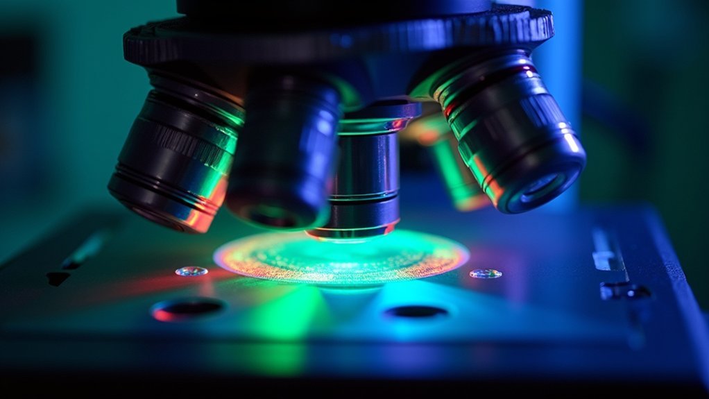
Microscopy’s cornerstone technique, widefield epi-illumination stands as the most accessible entry point into the world of fluorescence imaging.
You’ll find excitation light directed onto your sample from above the objective lens, creating even illumination across your entire field of view while capturing emitted light through the same lens.
This approach delivers high-contrast images with bright signals against dark backgrounds, making it ideal for visualizing various biological samples.
You can leverage multiple light sources, including LEDs that enable precise wavelength control and rapid switching between fluorophores.
While widefield fluorescence microscopy offers excellent rapid imaging capabilities for cell surveys and pathology applications, you’ll need to manage limitations like out-of-focus light interference and photobleaching by optimizing your imaging settings for the best results.
Confocal Laser Scanning: Optimizing Signal-to-Noise Ratio
When you need to elevate your imaging precision beyond what widefield methods can provide, confocal laser scanning microscopy (CLSM) offers remarkable advantages in signal quality. By employing a pinhole aperture that excludes out-of-focus light, CLSM dramatically enhances signal-to-noise ratio, delivering clear images with high spatial resolution.
| Feature | Benefit | Consideration |
|---|---|---|
| Pinhole aperture | Eliminates background noise | Reduced light throughput |
| Point-by-point scanning | High spatial resolution | Slower acquisition |
| Adjustable laser intensity | Precise fluorophore excitation | Potential photobleaching |
You’ll find CLSM particularly valuable for multi-color imaging applications, where simultaneous detection of multiple fluorophores is required. Remember to carefully calibrate your detector gain and excitation parameters to optimize signal quality while preventing photobleaching of your valuable samples.
Two-Photon Excitation for Deep Tissue Imaging
While conventional fluorescence techniques struggle with dense tissue penetration, two-photon excitation microscopy excels by leveraging near-infrared light to reach depths up to 1 mm in biological specimens.
Two-photon microscopy penetrates where standard techniques fail, using near-infrared light to illuminate biological structures deep within tissue.
You’ll find this approach reduces photobleaching considerably as it requires simultaneous absorption of two photons, minimizing overall light exposure to your samples.
When you focus excitation light to a precise point, you’ll achieve enhanced spatial resolution and optical sectioning capabilities, allowing you to visualize distinct layers within thick specimens.
The confined focal volume dramatically improves signal-to-noise ratio, creating clearer images in complex environments.
Two-photon microscopy is particularly valuable for neuroscience research, where you need to observe cellular interactions and neural dynamics in live tissue imaging without disrupting the native environment.
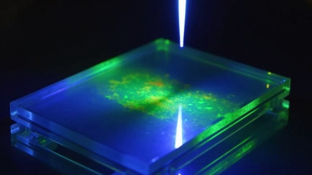
Light Sheet Illumination: Minimizing Phototoxicity
Unlike traditional illumination methods that flood specimens with light, light sheet microscopy directs a thin plane of excitation light across your sample from the side. This approach selectively excites fluorophores only in the focal plane, dramatically reducing phototoxicity and photobleaching in your specimens.
You’ll find light sheet microscopy particularly valuable for live-cell imaging, where minimizing exposure to intense light is vital. The technique captures entire fields of view in a single exposure, enabling rapid imaging while preserving cellular viability.
An evanescent wave generated during illumination guarantees that only fluorophores near the focal plane become excited. This method achieves impressive imaging depths of several hundred micrometers in thick biological tissues, allowing you to observe dynamic processes with minimal disruption to your sample’s natural state.
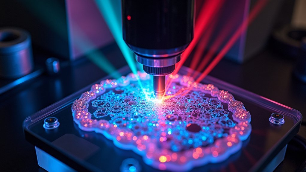
Total Internal Reflection Fluorescence (TIRF): Surface-Selective Excitation
Three primary features make TIRF microscopy exceptionally powerful for imaging cellular surfaces. First, it creates an evanescent wave that excites only fluorophores within 100-200 nm of the glass-water interface. Second, it dramatically reduces background fluorescence from deeper sample regions. Third, it provides remarkable high spatial resolution in the z-axis, perfect for visualizing surface-associated molecules.
| Feature | Benefit | Application |
|---|---|---|
| Limited penetration depth | Selective illumination | Cell membrane dynamics |
| Surface-selective excitation | Enhanced signal-to-noise ratio | Protein-protein interactions |
| Minimal illumination volume | Reduced phototoxicity | Extended live-cell imaging |
You’ll find TIRF particularly valuable when studying dynamic processes at the cell membrane. The technique’s ability to isolate thin optical sections enables real-time observation of low-abundance targets that would otherwise be obscured by cellular autofluorescence.
Frequently Asked Questions
What Kind of Light Is Used in Fluorescence Microscopy?
In fluorescence microscopy, you’ll use specific light sources like LEDs, mercury arc-lamps, or tungsten-halogen lamps. They emit wavelengths that excite fluorophores, with LEDs offering better control and longer lifespans than traditional sources.
What Are the Methods of Fluorescence Imaging?
You’ll encounter four main fluorescence imaging methods: Widefield (fast, affordable), Confocal (improved clarity, point-by-point scanning), Multiphoton (deeper penetration using near-infrared light), and TIRF (surface visualization with excellent z-resolution).
What Are the Light Sources Used in Fluorescence Spectroscopy?
You’ll commonly use LEDs, mercury arc-lamps, and tungsten-halogen lamps in fluorescence spectroscopy. LEDs offer control and longevity, mercury lamps emit strong UV light, while tungsten-halogen provides even illumination across wavelengths.
What Is the Best Camera for Fluorescence Microscopy?
You’ll want an sCMOS or EMCCD camera with high quantum efficiency (>70%), low noise, and sufficient cooling. Choose larger pixels for sensitivity or smaller pixels for resolution depending on your specific fluorescence application.
In Summary
You’ve now explored five essential lighting techniques that power today’s fluorescence imaging. Whether you’re examining cell membranes with TIRF, reducing photodamage with light sheet methods, or penetrating deep tissues using two-photon excitation, your choice of illumination critically impacts your results. By understanding each method’s strengths, you’ll select the best approach for your specific biological questions and specimen characteristics.


Leave a Reply