Positive phase contrast makes cells appear bright against a dark background, enhancing visibility of delicate structures without staining. You’ll see clearer cellular boundaries, organelles, and membranes compared to brightfield or negative phase contrast. It’s ideal for live cell observation as it preserves natural conditions while revealing fine details through refractive index differences. The technique minimizes halo artifacts that can distort cellular edges, offering superior detail for dynamic cellular processes. Discover why researchers consistently choose this method for critical cell studies.
Principles of Positive Phase Contrast in Cellular Imaging
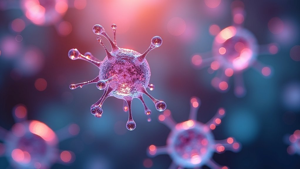
While traditional microscopy often struggles to reveal unstained cellular structures, positive phase contrast transforms invisible details into bright, clearly defined features against a dark background.
This technique works by manipulating the phase of light waves that pass through your specimens. When light travels through cellular components with different refractive index values, positive phase contrast enhances these differences through constructive interference.
You’ll notice the advantage immediately when observing live cells—organelles and cellular boundaries appear luminous against a darker field without any chemical staining.
This preservation of natural cellular states is vital for studying dynamic processes like cell division or motility. The technique effectively converts phase differences, which your eyes can’t normally detect, into amplitude differences that you can easily see and document.
Enhanced Visibility of Cellular Structures and Boundaries
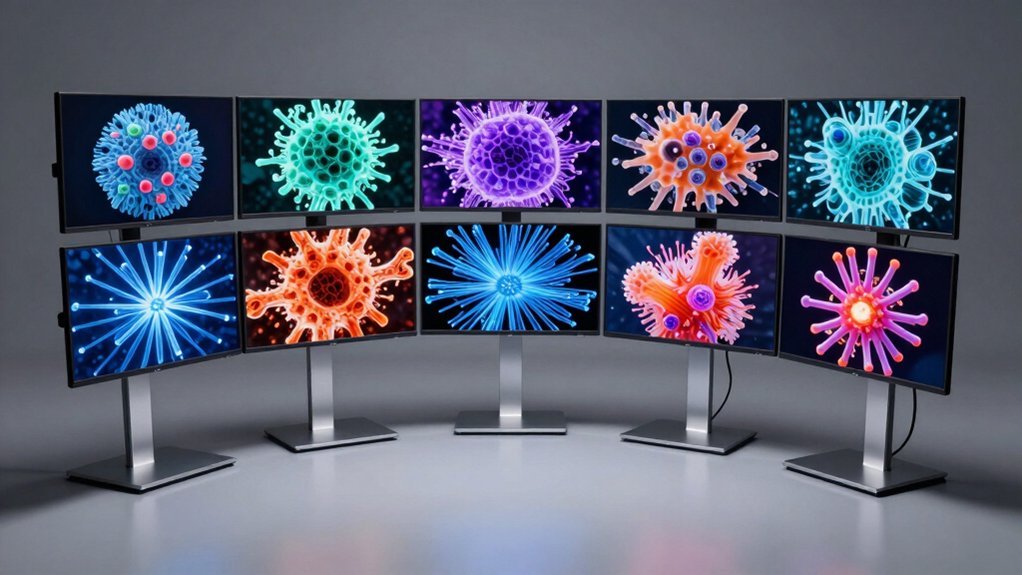
Because positive phase contrast illuminates cellular details with remarkable clarity, you’ll immediately notice how organelles and membranes stand out as bright, well-defined structures against the darker background.
This technique transforms subtle differences in refractive index within cells into visible brightness variations, allowing you to easily distinguish between nuclei, mitochondria, and vacuoles without staining.
You’ll find that positive phase contrast can greatly enhance contrast between adjacent cellular structures, making spatial relationships more apparent.
The technique reveals cellular boundaries with exceptional definition, enabling you to observe morphological changes in living specimens.
When you’re studying microbiology or cytology samples, you’ll appreciate how previously obscured features become readily identifiable, providing valuable insights into cell structure and function without compromising cellular integrity.
Advantages Over Traditional Brightfield Microscopy
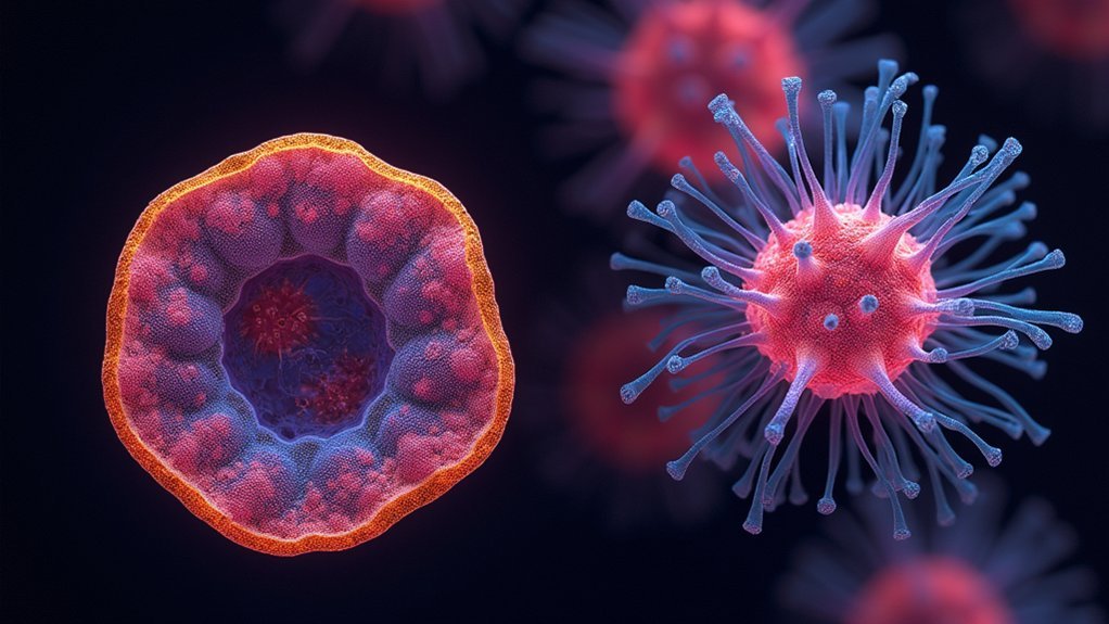
When you use positive phase contrast microscopy, you’ll observe superior cellular detail that traditional brightfield methods simply can’t match.
You won’t need to stain your specimens, allowing for non-destructive observation of living cells in their natural state without altering their functionality.
The technique provides considerably enhanced contrast based on refractive index differences, letting you clearly distinguish organelles and fine structures that would remain virtually invisible under conventional brightfield conditions.
Superior Cellular Detail
Although traditional brightfield microscopy has served scientists for centuries, positive phase contrast microscopy represents a significant leap forward in cellular visualization technology.
You’ll see cellular structures with unprecedented clarity as the technique transforms subtle refractive index differences into visible contrast.
The advanced method reveals organelles and cytoplasmic details that remain invisible in brightfield observation.
When examining living cells, you’ll notice nuclei and internal structures appearing bright against a dark background—creating sharp definition without staining chemicals.
This preservation of natural cellular conditions allows you to observe authentic behaviors and morphological features.
Unlike traditional methods that might miss subtle variations in cell architecture, positive phase contrast reveals these nuances clearly.
You’re able to document cellular processes in their native state, avoiding the artifacts and distortions that staining procedures often introduce.
Non-destructive Cell Observation
Perhaps the most significant advantage of positive phase contrast microscopy lies in its non-destructive approach to cellular observation.
Unlike traditional brightfield microscopy, which struggles to reveal details in transparent specimens, positive phase contrast manipulates non-diffracted light to enhance visibility without compromising cellular integrity.
You’ll find this technique particularly valuable when studying live cells, as it eliminates the need for stains that could alter natural cellular processes or introduce artifacts.
When examining unstained specimens, positive phase contrast creates striking contrast between cellular structures and their surrounding medium, allowing you to observe organelles and internal details in their native state.
This non-destructive imaging capability enables you to monitor cellular dynamics over extended periods, making it ideal for studying developmental processes, motility, and real-time cellular responses without disrupting the very behaviors you’re investigating.
Enhanced Contrast Without Staining
The remarkable advantage of positive phase contrast microscopy becomes immediately apparent when comparing it to traditional brightfield techniques. You’ll observe transparent specimens with striking clarity without needing to stain them, preserving cellular integrity while enhancing natural visibility.
| Feature | Brightfield Microscopy | Positive Phase Contrast |
|---|---|---|
| Visibility | Poor for transparent cells | High for transparent cells |
| Staining | Required for detail | Not required |
| Background | Bright background | Dark background |
| Cell appearance | Low contrast, difficult to see | Bright cells, enhanced contrast |
| Live cell imaging | Limited by staining toxicity | Excellent for real-time processes |
Positive phase contrast converts subtle phase shifts into amplitude differences, allowing you to distinguish organelles and structures that remain invisible in brightfield conditions. This technique delivers superior visualization of living cells in their natural state, enabling you to study dynamic cellular processes without introducing artifacts.
Comparing Positive vs. Negative Phase Contrast for Cell Observation
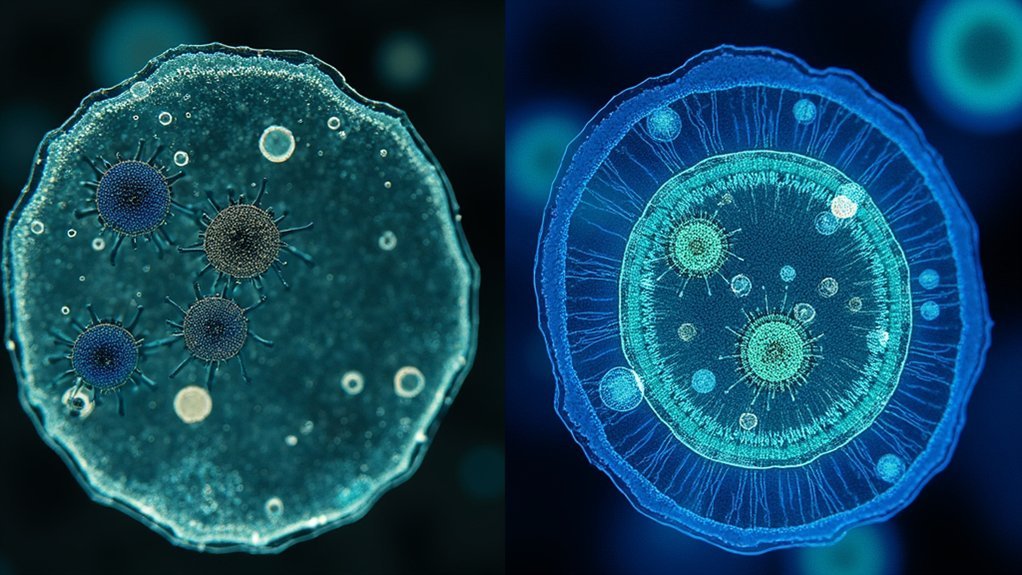
When you’re examining cells, positive phase contrast creates bright cells against a dark background, providing sharper edge definition than negative phase contrast.
This enhanced contrast occurs because positive phase contrast advances light waves passing through specimens, while negative phase contrast retards these waves, resulting in fundamentally different image characteristics.
You’ll notice the bright halos in positive phase contrast greatly improve your ability to distinguish cellular outlines and internal structures, making it particularly valuable for observing thicker specimens and live cells.
Image Clarity Differences
How does positive phase contrast transform our ability to observe cellular structures? It radically enhances image clarity by advancing light wave phases, creating bright structures against dark backgrounds.
You’ll notice cellular structures appear remarkably more defined and distinct, making it easier to differentiate between closely positioned organelles that might blur together under negative phase contrast.
When you’re examining microorganisms, positive phase contrast reveals fine details that would otherwise remain obscured. It’s particularly valuable because it minimizes halo effects that can distort cellular boundaries in negative phase contrast imaging.
For cytologists studying live cells, this technique provides clearer delineation without requiring stains, allowing you to observe cellular processes in real-time.
The enhanced contrast between cellular components gives you unprecedented precision when monitoring division and motility in living specimens.
Cell Edge Detection
Cell edge detection represents one of the most striking differences between positive and negative phase contrast techniques. When you’re examining live cells, positive phase contrast creates bright halos around cell boundaries, greatly enhancing your ability to distinguish cellular edges from their surroundings.
| Feature | Positive Phase Contrast | Negative Phase Contrast |
|---|---|---|
| Edge Appearance | Bright halos | Dark outlines |
| Background | Darker | Lighter |
| Detail Visibility | Higher | Lower |
| Dynamic Process Tracking | Excellent | Good |
| Structural Differentiation | More pronounced | Less defined |
The bright halos produced in positive phase contrast aren’t just aesthetically pleasing—they’re functionally superior for cell edge detection. You’ll notice that cellular boundaries appear more pronounced, allowing you to track morphological changes in real-time without staining. This clear delineation enables you to observe subtler structural differences that might otherwise go undetected with negative phase contrast techniques.
Contrast Mechanism Explained
Although both techniques utilize phase shifts, the fundamental mechanism behind positive phase contrast transforms invisible cellular details into brilliant, easily observable structures.
When light passes through your specimens, the differences in refractive indices between cellular components create minute phase changes that become amplified and visible.
In positive phase contrast images, the light waves advancing through cellular structures appear brighter against a darker background, enhancing the visibility of organelles and membrane boundaries.
This contrast technique makes cellular details more intuitive to interpret compared to negative phase contrast, where structures appear dark against a bright field.
The changes in refractive properties are translated into amplitude variations that your eye can detect, revealing dynamic cellular processes without requiring stains that might disrupt natural cell behavior.
Optimal Applications in Live Cell Research and Monitoring
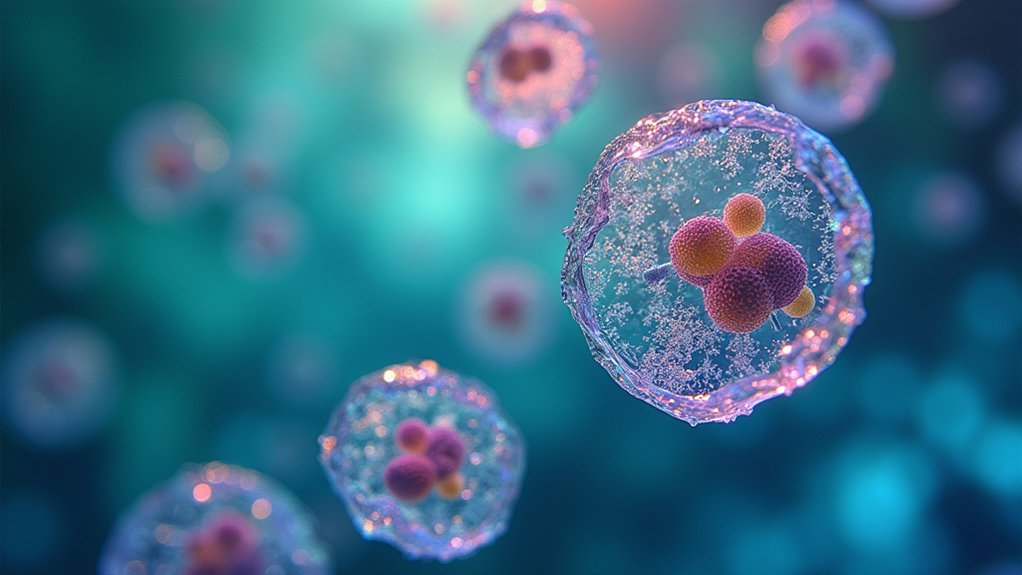
While many imaging techniques require specimen preparation that can disrupt cellular processes, positive phase contrast microscopy stands out as an indispensable tool for live cell research applications.
Positive phase contrast microscopy enables real-time cellular observation without disruptive preparation techniques.
You’ll find this technique particularly valuable when studying dynamic cellular structures in real time, as it produces bright images against dark backgrounds without requiring potentially harmful stains.
When monitoring bacterial growth or tracking mitosis, positive phase contrast allows you to observe microorganisms in their natural state. This preservation of physiological conditions is vital for longitudinal studies in tissue engineering and cell biology.
You can accurately analyze cell motility and morphological changes while avoiding artifacts like halos that might interfere with image interpretation.
For research requiring intact, functioning live cells with clearly visible internal components, positive phase contrast offers unmatched advantages in both clarity and specimen preservation.
Technical Considerations for Quality Positive Phase Contrast Imaging
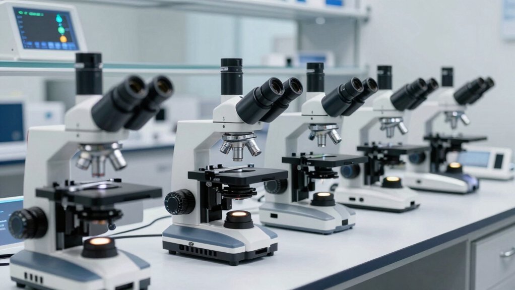
To achieve the exceptional cellular visualization that positive phase contrast microscopy offers, specific technical considerations must be addressed. You’ll need to carefully align the condenser annulus with the phase ring to guarantee ideal contrast of transparent cellular structures.
| Component | Key Consideration |
|---|---|
| Objectives | Select proper Ph-marked objectives (Ph1, Ph2, Ph3) |
| Alignment | Center condenser annulus precisely with phase ring |
| Light Source | Use appropriate intensity to balance contrast |
| Specimen | Prepare thin samples to minimize phase artifacts |
When working with positive phase contrast, proper alignment is critical—misalignment reduces image clarity and contrast. Your microscope’s phase ring transforms phase differences into brightness variations, making transparent cellular details visible without staining. This preservation of cells in their natural state makes this technique invaluable for observing dynamic cellular processes in real time.
Frequently Asked Questions
What Is the Difference Between Positive and Negative Phase Contrast?
Positive phase contrast shows cells as bright objects on a dark background, while negative phase contrast displays them as dark objects on a bright background. You’ll see clearer cellular details with positive contrast.
Why Is Phase Contrast Better Than Bright-Field?
Phase contrast is better than bright-field because you can see living cells without staining them. It converts phase shifts into visible contrast, revealing cellular structures that you’d miss with bright-field’s absorption-based imaging.
What Are the Advantages of Phase Contrast?
Phase contrast advantages include letting you observe living cells without staining, providing better visibility of transparent structures, allowing real-time monitoring of cellular processes, and minimizing artifacts compared to other microscopy techniques.
How Is Negative Phase Different From Positive Phase?
In negative phase contrast, you’ll see cells appear dark against a bright background, while in positive phase, they’re bright against a dark background. Negative phase retards light waves, whereas positive phase advances them.
In Summary
You’ll find positive phase contrast superior for cell imaging because it creates bright cells against dark backgrounds, enhancing visibility of cellular boundaries and internal structures. It’s particularly valuable when you’re studying living, unstained specimens where detail is essential. For most biological applications, you’ll achieve better contrast and clearer visualization of cellular morphology than with negative phase contrast or traditional brightfield techniques.


Leave a Reply