
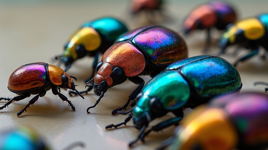
To capture stunning polarized specimens, position your light at a 90-degree angle to maximize polarization effects. Use low ISO (100-400) with apertures between f/8-f/16, and mount your camera on a tripod for stability. Select circular polarizers with multi-coating for ideal results. Rotate specimens gradually to reveal changing birefringence patterns. Document multiple polarizer angles by rotating in 15-30 degree increments. Maintain clean optics and proper focus for crystal-clear details. These techniques will transform your ordinary specimens into extraordinary scientific art.
Master Your Light Source Placement for Maximum Polarization

Why do some polarized specimen images appear vibrant and detailed while others fall flat? The secret often lies in your light source positioning. For best results, place your light at a precise 90-degree angle to the specimen—this critical alignment maximizes the polarized light effect and creates stunning contrast.
Enhance this setup by attaching a polarizing filter to your camera lens, which selectively blocks reflected light and reveals birefringent details that would otherwise remain hidden.
A polarizing filter transforms your lens into a discovery tool, revealing hidden birefringent structures invisible to the naked eye.
Experiment with your light’s distance; moving it closer intensifies polarization effects, while positioning it farther creates softer contrasts.
The position of your camera relative to both the light and specimen greatly influences the polarizer’s effect. Try rotating your polarizer while viewing through the lens to find the perfect orientation that transforms ordinary specimens into extraordinary images.
Select the Optimal Camera Settings for Microscope Photography
Perfect light positioning forms only half of the polarized imaging equation—your camera settings must complement this precise arrangement.
Start with a low ISO (100-400) to minimize noise when working with polarizing filters that naturally reduce light transmission.
Adjust your exposure by setting the aperture between f/8 and f/16 to achieve greater depth of field, ensuring more of your specimen remains crisp throughout.
Select a shutter speed between 1/30 and 1/200 seconds depending on your light intensity to capture sharp details without blur.
To reduce glare and capture the most accurate polarization effects, stabilize your camera on a tripod, especially significant at slower shutter speeds.
Utilize live view mode for precise focusing adjustments before capturing your final image—this real-time preview helps perfect composition and reveals subtle polarization details.
Choose the Right Polarizing Filters for Your Specimen Type
You’ll get the best results by matching your polarizing filter to both your equipment and specimen type.
For crystalline or birefringent materials, circular polarizers with multi-coating will reveal intricate structural details while minimizing unwanted flare.
Remember that different specimens respond uniquely to polarized light, so you might need to experiment with filter orientation to highlight specific features in minerals, crystals, or biological tissues.
Filter Selection Essentials
When capturing polarized specimens, selecting the right polarizing filter becomes as essential as choosing the proper lens for your camera.
For ideal results with modern digital cameras, prioritize circular polarizer filters that won’t interfere with your auto-focusing and metering functions.
Always check your lens cap for the correct diameter size before making your filter selection.
If you’re working with a wide-angle lens, invest in slim-profile polarizing filters to prevent vignetting and guarantee even polarization across your entire frame.
While premium brands like B+W offer superior durability, you’ll find suitable alternatives from Tiffen and Hoya if you’re budget-conscious.
Remember to keep your polarizing filters handy whenever shooting in bright conditions—they’re invaluable tools for enhancing contrast and color saturation when documenting specimens in the field.
Match Material Properties
When capturing polarized specimens, selecting the right polarizing filter becomes as essential as choosing the proper lens for your camera. The material properties of your specimen directly influence your filter choice—specimens with birefringence require polarizers that effectively block specific light waves.
Consider investing in high-quality polarizers from brands like B+W to minimize optical distortion and enhance clarity. Though more expensive, they’ll deliver superior results that justify the cost.
Circular polarizers are ideal as they’re compatible with modern digital cameras and easier to adjust in the field.
Remember that the angle of light incidence greatly affects your results. You’ll need to experiment with rotating and repositioning your polarizers to achieve ideal contrast.
For varied specimen sizes, larger polarizers (3-4 inches) typically provide better coverage and more consistent results.
Position Specimens to Reveal Birefringent Properties
The proper positioning of specimens between polarizers forms the foundation of successful polarized photography. When capturing birefringent properties, you’ll need to place your specimen between two polarizers oriented at 90° to each other—the polarizer and analyzer. This crossed-polarizer setup creates the necessary conditions for observing stunning optical effects.
- Rotate your specimen gradually while maintaining its position between the polarizers to reveal changing colors and stress patterns.
- Ensure consistent, bright lighting to maximize contrast in the birefringent patterns.
- Experiment with different angles between the specimen and polarizers to uncover unique phase shifts.
- Try various materials like plastics, minerals, and stressed glass—each will display distinctive birefringent characteristics.
Calibrate Your Focus for Crystal-Clear Microscopic Detail

To achieve crystal-clear polarized images, you’ll need to master precise focusing adjustments that compensate for the unique optical properties of birefringent materials.
Start with lower magnification objectives to locate your specimen, then switch to higher power once you’ve confirmed proper polarizer-analyzer alignment and specimen orientation.
You’ll get the sharpest results by making micro-adjustments to your focus while rotating the stage, allowing you to capture the exact moment when birefringence reveals the specimen’s internal structure.
Precise Focusing Techniques
Achieving exceptional clarity with polarized specimens depends largely on your focusing precision. When working with birefringent materials, proper calibration isn’t just helpful—it’s crucial for revealing their true structural details.
You’ll need to master both technical setup and focusing techniques to capture stunning polarized images.
- Use the fine focus knob for micro-adjustments while viewing specimens, allowing you to find the perfect focal plane for maximum clarity.
- Implement focus stacking to enhance depth of field by combining multiple images taken at different focal planes.
- Verify your calibration with a stage micrometer to verify accuracy across different magnifications.
- Maintain clean optics by regularly removing dust and debris from all components to prevent interference with light paths.
Objective Selection Strategy
Beyond precise focusing techniques, your objective selection forms the backbone of exceptional polarized microscopy. Choose objectives specifically designed for polarized light microscopy to maximize the contrast and clarity of birefringent specimens. Higher numerical aperture lenses capture finer crystalline details and dramatically improve resolution under polarized conditions.
Don’t overlook calibration—properly aligned objectives minimize aberrations that can distort your crystalline structures. As you work, rotate both the stage and analyzer to find the ideal viewing angle that enhances birefringence effects in your specimen. This rotation technique often reveals structural details invisible from a static position.
Maintain your equipment meticulously; even minor smudges can create artifacts that compromise your polarized images. Regular cleaning and inspection of objectives guarantees the optical clarity needed for truly stunning polarized microscopy results.
Capture the Full Color Spectrum With Proper White Balance
While polarized specimens showcase a stunning array of vivid colors, you’ll only capture their true visual splendor with proper white balance settings. Proper white balance eliminates color casts that can distort how accurately your images represent the specimen’s actual appearance under polarized light.
- Use a gray card or white reference surface to manually set your camera’s white balance, ensuring colors remain true to life.
- Adjust for your specific lighting conditions – whether using natural sunlight or artificial sources, each requires different white balance settings.
- Experiment with camera presets like daylight, cloudy, or fluorescent to find the most flattering representation.
- Fine-tune in post-processing if needed, making subtle adjustments to achieve the most accurate color rendition of your polarized specimens.
Document Multiple Polarizer Angles for Complete Analysis

To fully understand the intricate structures and stress patterns within your specimens, you’ll need to capture images at multiple polarizer angles. Documenting multiple polarizer angles reveals details that might otherwise remain hidden with a single orientation.
Rotate the filter in 15-30 degree increments to capture a wide range of effects. Use a rotational sample stage to easily adjust specimen positions while methodically recording your observations.
Pay special attention to angles producing the most pronounced birefringence effects, as these highlight critical structural characteristics.
Maintain a detailed log of specific angles and their corresponding visual results. This systematic approach allows you to analyze and compare how different orientations affect various specimens, creating a thorough dataset that enhances your understanding of the material’s properties and internal stresses.
Frequently Asked Questions
What Is the Most Common Technique for Polarizing Light?
You’ll usually polarize light using two polarizing filters: a polarizer for filtering incoming light and an analyzer for examining the effects. Position them at 90 degrees to each other for best results.
How Do You Take Polarized Photos?
You’ll need two polarizing filters: one on your lens and another as an analyzer. Position them at 90° to each other, adjust for ideal light conditions, and compensate for the 1-2 stop light reduction.
What Angle Should You Shoot From the Sun With a Polarizing Filter to Get the Best Effect of the Filter?
For the best polarizing effect, you’ll want to position yourself at a 90-degree angle from the sun. This maximizes the filter’s ability to reduce glare and enhance colors in your photos.
What Is the Polarized Light Microscope Technique?
You’ll use polarized light microscope technique by placing your specimen between two polarizers (polarizer and analyzer) to reveal birefringence in anisotropic materials, allowing you to observe unique optical properties and crystalline structures.
In Summary
You’re now equipped to transform ordinary specimens into vibrant scientific masterpieces. By controlling your light, optimizing settings, and selecting appropriate filters, you’ll reveal hidden details invisible to the naked eye. Remember, it’s the careful positioning and documenting at multiple angles that’ll give you thorough data. With these techniques, you’ve got everything needed to capture polarized specimens that are both scientifically valuable and visually stunning.


Leave a Reply