
For ideal polarized microscope photos, use light sources with 4500K-5500K color temperature to enhance birefringent contrast. Implement Köhler illumination for even light distribution and carefully adjust your aperture diaphragm to balance resolution with depth of field. Keep optical surfaces clean and manage glare with proper filter positioning. LED systems offer adjustable settings and longer lifespans than tungsten lights. Regular maintenance guarantees consistent results that will reveal crystal structures in stunning detail.
Understanding Polarized Light Sources and Their Impact on Microscope Photography
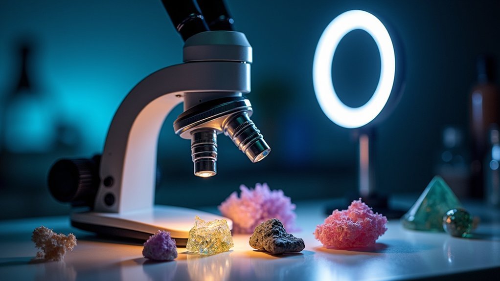
When working with polarized light microscopy, your choice of light source dramatically affects the quality of your images. Selecting the ideal color temperature for your specific specimen enhances contrast and clarity, revealing subtle details in birefringent materials that might otherwise remain hidden.
To achieve exceptional quality images, you’ll need to properly implement Köhler illumination, ensuring even lighting distribution and maintaining perfect optical alignment.
Proper Köhler illumination is non-negotiable for capturing fine details in polarized microscopy—it’s the foundation of uniform specimen illumination.
Don’t overlook the aperture diaphragm—adjusting it correctly balances resolving power with depth of field while boosting contrast.
Regular maintenance of your light sources is essential. Clean lenses and replace bulbs when needed to maintain consistent intensity.
Remember to calibrate your camera’s white balance and exposure settings to match your polarized light conditions for accurate color reproduction.
Selecting the Ideal Color Temperature for Birefringent Specimen Imaging
When you’re examining birefringent specimens, selecting the proper color temperature between 4000K-5000K will greatly enhance your image clarity and color accuracy.
Tungsten lights offer warmer tones that can soften certain crystalline structures, while LEDs provide more consistent output with less heat generation and better energy efficiency.
Different wavelengths directly affect how birefringent materials split light, so you’ll need to adjust your light source accordingly for ideal visualization of your specific specimen type.
Subheading Discussion Points
Although many factors influence successful polarized light microscopy, choosing the appropriate color temperature stands as a critical decision for ideal birefringent specimen imaging.
You’ll want to aim for the 4000K-6500K range, with 5500K being particularly effective as it mimics natural daylight and guarantees true color representation of your specimens.
When working with birefringent specimens, a consistent light source prevents imaging fluctuations that could compromise your results.
Be certain to calibrate your color temperature settings alongside proper aperture diaphragm adjustments to achieve peak resolution and clarity.
This combination greatly reduces color casting in your images, allowing for more precise interpretation of birefringence patterns.
Remember that polarized light microscopy demands this careful balance of optical settings—your specimen’s subtle structural details will be much more visible with properly refined lighting.
Tungsten Vs LED Performance
Choosing between tungsten and LED illumination presents a critical decision point for polarized light microscopy applications.
Tungsten’s 3200K color temperature produces a warm spectrum that can enhance contrast in birefringent specimens, potentially improving image quality for certain biological and mineral samples.
LED systems offer greater flexibility with adjustable color temperatures (2700K-6500K), allowing you to tailor illumination to specific specimens. For detailed structural imaging, you’ll benefit from cooler LED temperatures, while maintaining control over light intensity to prevent saturation.
Consider practical factors too: tungsten sources require warm-up time and frequent replacement, while LEDs provide longer lifespan and generate less heat—particularly valuable for temperature-sensitive birefringent materials.
Your choice ultimately depends on balancing natural color rendering (tungsten’s strength) against the brightness and maintenance advantages LEDs deliver.
Wavelength Effects on Birefringence
The spectral characteristics of your light source directly impact the quality and interpretability of birefringent specimens under polarized light microscopy.
When imaging birefringent materials, choose light sources with color temperatures between 4500K and 5500K to enhance contrast and detail visibility.
Shorter wavelengths (blue light) produce more pronounced birefringence effects than longer wavelengths (red light), making blue filters particularly effective for enhancing crystalline structures while reducing background interference.
This occurs because the refractive index of specimens varies with wavelength, creating distinct interaction patterns across the visible spectrum.
You’ll achieve ideal results by adjusting both the intensity and wavelength of your light source to match your specimen’s specific characteristics.
This calibration guarantees well-defined interference colors and clearer visualization of structural details, greatly improving your polarized microscopy outcomes.
Köhler Illumination Setup for Enhanced Polarized Light Distribution
When properly configured, Köhler illumination transforms polarized light microscopy from an ordinary observation technique into a powerful analytical tool. You’ll achieve superior contrast and resolution by guaranteeing uniform light distribution across your specimen.
To optimize your polarized light microscope, first focus on your sample, then adjust the field diaphragm to match your field of view before centering the light source.
Focus first, adjust diaphragm second, center light source last—the essential sequence for optimal polarized microscopy.
- Adjust the aperture diaphragm carefully to balance resolution and contrast
- Clean optical components regularly to prevent artifacts and light scattering
- Center the field diaphragm precisely to guarantee even illumination
- Monitor light intensity to prevent overexposure of birefringent structures
- Check alignment of polarizer and analyzer to maximize polarization effects
Remember that proper Köhler setup minimizes artifacts while enhancing the visibility of birefringent structures, allowing you to capture the subtle details that make polarized microscopy invaluable.
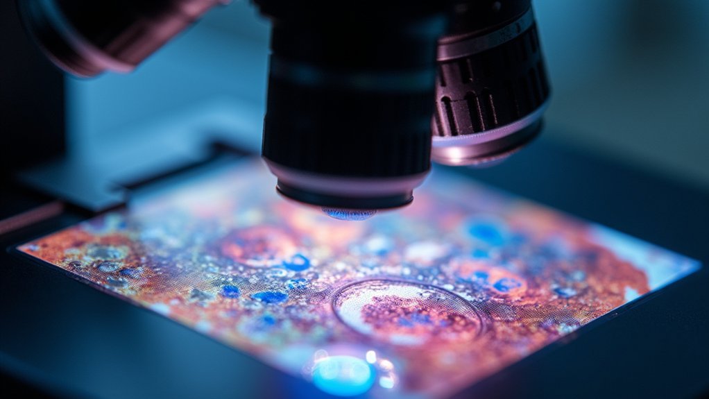
Balancing Light Intensity to Reveal Crystal Structures

When examining crystal structures, you’ll need to select a light source with the appropriate intensity and color temperature for your specimen’s characteristics.
Your microscope’s color temperature settings will greatly impact how birefringent features appear, with cooler temperatures often enhancing subtle structural details in transparent crystals.
Adjusting your light source’s intensity in conjunction with ideal color temperature lets you balance contrast and clarity, preventing both the washout effect of excessive illumination and the murky appearance of insufficient light.
Light Source Considerations
Properly balanced light intensity serves as the foundation for revealing intricate crystal structures in microscopy. When working with polarized light, your illumination source must complement the optical properties of your specimen to achieve ideal results.
- Choose a color temperature around 5500K for accurate birefringent material representation.
- Adjust intensity based on sample thickness—too bright saturates images, too dim obscures details.
- Implement Köhler illumination through proper condenser lens positioning to guarantee even light distribution.
- Align your polarizing filters precisely to maximize contrast in birefringent features.
- Maintain your light sources regularly, as variations in output can dramatically affect image quality.
Optimizing Color Temperature
Why does color temperature matter so critically in polarized light microscopy? At 5500K-6500K, your light source mimics natural daylight, revealing the true colors and details of birefringent structures in your specimens.
This ideal range guarantees accurate visualization of crystal structures and their optical properties.
You’ll need to carefully balance light intensity—too bright and you’ll wash out delicate details; too dim and you’ll lose essential contrast.
Implement Köhler illumination to distribute light evenly across your sample, creating consistent illumination and enhancing image quality.
Regular calibration prevents color temperature drift that can compromise your observations.
When fine-tuning brightness, consider adding neutral density filters to reduce intensity without altering the color temperature, allowing you to capture intricate crystal details with precision.
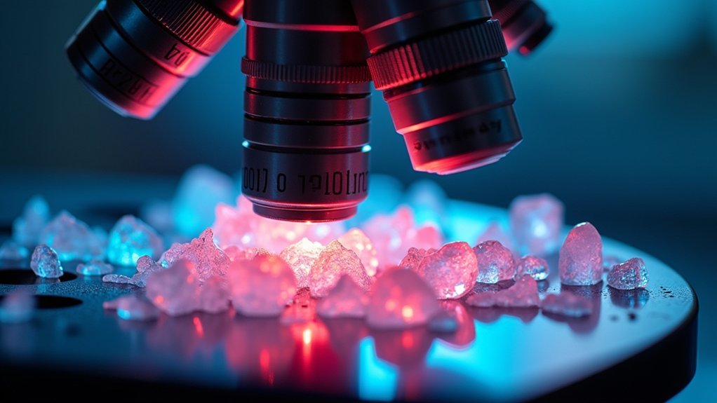
Aperture Diaphragm Adjustments for Optimal Contrast in Polarized Imaging
The aperture diaphragm serves as the critical control point for achieving exceptional contrast in polarized light microscopy.
You’ll need to carefully balance your settings to reveal birefringent structures with maximum clarity while maintaining adequate illumination. A smaller aperture increases contrast but reduces brightness—find your ideal middle ground for peak resolution.
- Adjust aperture to control light entering the system and depth of field
- Reduce aperture size to enhance definition of birefringent structures
- Minimize light scattering by fine-tuning aperture settings
- Set diaphragm to prevent overexposure while preserving detail visibility
- Recalibrate regularly as sample thickness changes affect peak settings
Managing Reflection and Glare in Polarized Microscope Setups
Effective glare management represents a considerable challenge in polarized microscopy, even with perfectly adjusted aperture settings. To overcome this obstacle, position your light sources at ideal angles that minimize direct reflection off specimen surfaces.
Managing glare in polarized microscopy requires strategic light source positioning to minimize specimen surface reflections—even with optimal aperture adjustments.
You’ll find that installing polarizing filters on these light sources considerably reduces unwanted reflections while enhancing the quality of polarized light entering your microscope.
Don’t underestimate the importance of a clean workspace—regularly wipe down all optical surfaces, especially the polarizer and analyzer components. Dust and fingerprints create additional glare points that compromise image quality.
Fine-tune your aperture diaphragm to control incoming light volume, and experiment with different intensity settings until you achieve the perfect balance between adequate illumination and glare reduction.
These adjustments will dramatically improve contrast and detail in your polarized microscopy images.
Light Source Maintenance Procedures for Consistent Polarized Imaging
Because microscope illumination systems deteriorate gradually over time, you’ll need a systematic maintenance routine to guarantee consistent polarized imaging results.
Your light source directly affects image clarity and contrast in polarized microscopy, making regular maintenance essential.
- Clean optical components weekly to remove dust that scatters light and degrades image quality
- Adjust your light source’s color temperature to match specific sample requirements
- Implement Köhler illumination techniques for even lighting distribution across your field of view
- Regulate light intensity to prevent overexposure while still revealing fine details in birefringent materials
- Schedule quarterly maintenance checks including bulb replacement and alignment verification
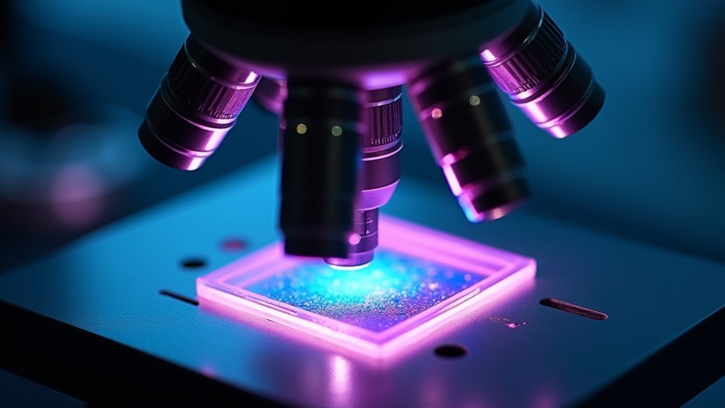
Specialized Filters and Their Effects on Light Source Quality
Selecting the right filter materials, whether glass, polymer, or crystal-based, will greatly impact your polarized microscopy results through varying degrees of optical clarity and durability.
You’ll notice distinct RGB polarization effects when using different filter combinations, which can either enhance or diminish specific spectral components in your specimen visualization.
The thickness of your chosen filters isn’t merely a physical characteristic—it directly affects light transmission efficiency, wavelength selectivity, and ultimately the resolution of your microscopic images.
Filter Material Selection Matters
While light sources provide the foundation for microscopy illumination, the materials used in your filters ultimately determine image quality and experimental success.
When selecting filters for polarized light microscopy, consider materials that reduce background noise while enhancing specific wavelengths critical to your specimen’s visualization.
- Choose high-quality PVA polarizers to guarantee effective light polarization with minimal distortion.
- Incorporate bandpass filters to isolate precise wavelengths needed for your specimens.
- Select materials with high transmittance ratings to maximize usable light reaching your sample.
- Use neutral density filters to control light intensity and prevent overexposure.
- Consider specialized filter materials that can eliminate unwanted wavelengths while enhancing desired spectra.
The right filter materials transform ordinary microscope illumination into precisely controlled light that reveals subtle structural details invisible under standard conditions.
RGB Polarization Effects
Beyond filter material selection, specialized RGB polarization filters open up remarkable visualization possibilities by manipulating the wavelength-dependent nature of birefringence. You’ll achieve superior image clarity when you match your light source’s color temperature to the peak transmission of your chosen filters.
| Filter Type | Effect on Sample | Application |
|---|---|---|
| Red filters | Enhances contrast in dense structures | Crystallography |
| Green filters | Improves detail in mid-range features | Geological samples |
| Blue filters | Highlights fine structural elements | Biological specimens |
These specialized filters introduce phase shifts that dramatically improve visualization of anisotropic and birefringent materials. By combining RGB filters with high-quality light sources, you’ll optimize illumination quality and capture sharper polarized light microscopy images with enhanced color differentiation. This approach allows you to isolate specific wavelengths, revealing compositional characteristics that would otherwise remain hidden.
Filter Thickness Considerations
As light traverses through specialized filters, the thickness of your filter materials profoundly impacts image quality and contrast in polarized light microscopy.
When you’re fine-tuning your setup, remember that filter thickness directly affects the optical path and phase relationships of transmitted light.
- Select filters with ideal thickness to minimize absorption while maximizing contrast
- Consider how thicker filters may introduce unwanted interference patterns
- Calculate appropriate thickness when using compensators to enhance birefringent structures
- Verify your filter thickness complements your light source’s color temperature
- Balance thickness requirements against the need for full spectrum transmission
Adapting Light Sources for Different Polarized Microscopy Techniques
Since polarized microscopy techniques demand specific illumination conditions, selecting and configuring the appropriate light source becomes essential for ideal results.
You’ll need different setups depending on your technique—LEDs work well for fluorescence while halogen lamps excel in brightfield applications to maximize contrast when examining birefringent specimens.
Optimize your optical path using Köhler illumination to guarantee even light distribution across your sample.
When working with biological specimens, aim for a color temperature around 5500K, but consider lower temperatures for mineralogical samples.
Always adjust your light intensity carefully—overexposure washes out detail while underexposure obscures critical features.
Neutral density filters can help fine-tune illumination levels.
Remember to clean lenses regularly and verify proper alignment to maintain consistent illumination quality throughout your microscopy sessions.
Troubleshooting Common Light Source Issues in Polarized Photomicrography
When your polarized photomicrographs lack clarity or show unexpected artifacts, the light source is often the culprit. Effective troubleshooting starts with understanding common illumination problems that affect image quality.
Light source issues frequently cause poor polarized photomicrographs. Proper diagnosis begins with recognizing typical illumination challenges.
- Check for flickering by inspecting electrical connections and replace faulty bulbs to maintain consistent illumination.
- Clean optical surfaces regularly to prevent dust from scattering light and reducing contrast.
- Verify color temperature is approximately 5500K for natural representation of birefringent materials.
- Adjust the aperture diaphragm to balance resolving power and depth of field for your specific specimen.
- Monitor light intensity to avoid oversaturation or underexposure that can mask critical details.
Frequently Asked Questions
How Do I Adjust the Light Source on My Microscope?
You’ll adjust your microscope’s light source by using Köhler illumination, modifying the aperture diaphragm, and experimenting with brightness levels. Keep components clean and choose the right color temperature for your specific sample.
How Does a Polarized Light Microscope Produce an Image?
A polarized light microscope produces images when light passes through a polarizer, interacts with your birefringent specimen, and recombines at the analyzer, creating interference patterns that reveal optical properties through color and brightness differences.
How Can You Improve the Quality of an Image on a Microscope?
You can improve microscope image quality by adjusting light intensity, optimizing Köhler illumination, fine-tuning the aperture diaphragm, cleaning all optical surfaces, and experimenting with camera settings to match what you see through the eyepiece.
What Are the Disadvantages of Polarizing Microscopes?
You’ll face limitations with polarizing microscopes: they’re ineffective for isotropic materials, require specialized training, have complex calibration, offer limited field of view, and depend on specific optical components that can easily misalign.
In Summary
You’ll achieve stunning polarized microscope photos by carefully optimizing your light sources. Focus on selecting appropriate color temperatures, implementing proper Köhler illumination, and fine-tuning your aperture diaphragm settings. Don’t overlook regular maintenance of your equipment and the strategic use of specialized filters. With these adjustments, you’ll reveal intricate crystal structures and enhance birefringent specimen details that would otherwise remain invisible under standard illumination conditions.


Leave a Reply