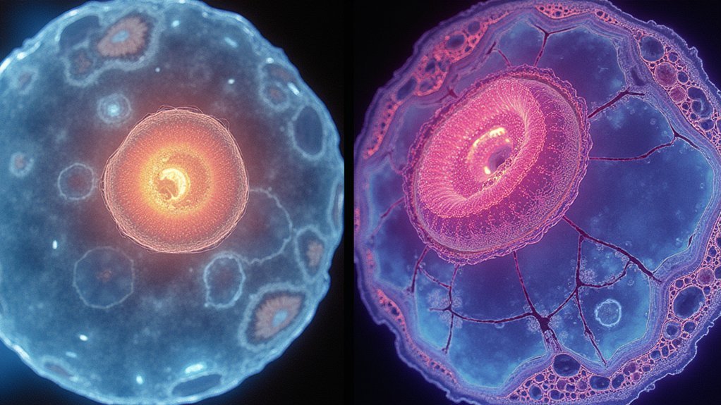
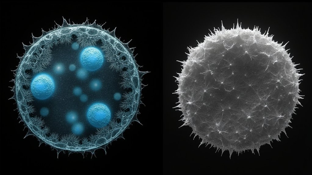
Phase contrast microscopy excels at visualizing unstained transparent specimens by transforming phase differences into visible brightness variations, while brightfield requires staining to achieve contrast. You’ll see clearer cell membranes, organelles, and nuclear boundaries with phase contrast, making it superior for live cell imaging and time-lapse studies. Brightfield offers simplicity and accessibility but struggles with transparent samples. The right choice depends on your specimen type and whether preserving natural cellular states matters for your research.
The Fundamental Principles of Phase Contrast Microscopy

Invisibility becomes visibility under phase contrast microscopy, a revolutionary technique that transforms transparent specimens into clearly observable structures. Developed by Frits Zernike in 1934 (earning him the Nobel Prize for Physics in 1953), this contrast technique cleverly converts phase differences in light into brightness variations.
When you examine biological specimens through light microscopy, the system uses an annular aperture and quarter-wave phase plate to create phase shifts. These shifts produce destructive interference, resulting in high contrast images without staining.
You’ll find it particularly effective for thin specimens (5-10 micrometers) where subtle refractive index differences between cells and their surroundings become visible. Look for specialized objectives marked with green Ph1, Ph2, or Ph3 inscriptions to guarantee peak performance when using this invaluable life science tool.
Understanding Brightfield Microscopy Mechanics
While many advanced microscopy techniques exist today, brightfield microscopy remains the cornerstone of laboratory visualization. When you use a brightfield microscope, you’re viewing specimens against a bright background where they appear darker due to their optical properties. The illuminator beneath the stage directs light upward through your specimen, while the condenser lens focuses this light for ideal visibility.
You’ll calculate total magnification by multiplying the ocular lens power (typically 10×) by the objective lens power (commonly 40×), giving you 400× magnification.
Though effective for stained samples, brightfield microscopy struggles with transparent biological specimens that phase contrast microscopy handles well.
Brightfield reveals stained structures clearly but falters with transparent specimens where phase contrast excels.
The technique’s effectiveness relies on light absorption by chromophores or added stains that enhance contrast. Most brightfield systems can achieve magnifications up to 1000×, though resolution limitations become apparent at higher powers.
Visualizing Unstained Specimens: Why Phase Contrast Excels

While brightfield microscopy struggles with transparent specimens, phase contrast transforms invisible phase differences into visible brightness variations you can easily detect.
You’ll notice living cells and their internal structures appear with remarkable clarity without requiring toxic stains that might alter cellular behavior or kill specimens.
This preservation of natural cellular states makes phase contrast microscopy your ideal tool for studying dynamic processes like cell division, migration, and morphological changes in real time.
Contrast Enhancement Principles
Because most biological specimens appear nearly transparent under conventional brightfield microscopy, phase contrast microscopy emerged as a revolutionary technique for visualizing unstained samples.
While brightfield microscopy relies on light absorption to create contrast, phase contrast imaging utilizes optical density variations and converts phase shifts into brightness differences.
The key to this contrast enhancement lies in the specialized optical system that includes an annular aperture and quarter-wave phase plate.
When light passes through specimens with refractive index differences, the diffracted and non-diffracted light waves undergo opposite phase shifts, creating high-contrast images.
This makes phase contrast particularly valuable for observing living cells and fine cellular structures that would otherwise remain invisible.
You’ll see remarkable detail in transparent specimens without the need for stains that might damage or alter delicate biological samples.
Living Cell Visualization
Three key advantages make phase contrast microscopy the gold standard for visualizing living cells. You’ll observe unstained biological specimens with remarkable clarity as the system converts phase shifts into brightness differences, revealing intricate cellular morphology without harmful stains.
Unlike brightfield techniques, phase contrast excels at detecting subtle variations in optical density, making previously invisible structures visible without compromising cell viability.
| Feature | Phase Contrast | Brightfield |
|---|---|---|
| Contrast | Enhanced without stains | Requires staining |
| Cell Viability | Maintains live cells | Often compromises |
| Detail Level | Reveals fine structures | Limited for unstained samples |
This non-invasive contrast enhancement enables real-time observation of cellular processes, allowing you to study dynamic interactions in their natural state—critical for advancing cell biology research.
Contrast Enhancement Techniques in Both Imaging Methods
Contrasting approaches to visibility enhancement define the fundamental difference between brightfield and phase contrast microscopy. In brightfield microscopy, you’ll rely on staining techniques to absorb light and create contrast, whereas phase contrast microscopy converts optical path differences into brightness variations without requiring stains for biological samples.
- Phase contrast utilizes specialized components—annular apertures and quarter-wave phase plates—to transform invisible phase shifts into visible contrast.
- Brightfield depends on standard optics and light absorption, limiting its effectiveness with transparent specimens at high magnifications.
- When observing live cells, phase contrast preserves cellular integrity by eliminating toxic staining procedures.
- For routine educational or diagnostic work, brightfield offers simplicity while phase contrast delivers superior detail in unstained specimens.
Cellular Structure Visibility Comparison

When you examine cellular structures, phase contrast microscopy offers superior membrane definition with crisp boundaries that reveal delicate cell peripheries without staining.
You’ll notice intracellular components like organelles and cytoplasmic granules appear with enhanced clarity in phase contrast images, while these same structures often remain invisible or indistinct in brightfield unless heavily stained.
The nucleus and its boundaries stand out dramatically in phase contrast due to differences in optical density, providing you with essential morphological information that brightfield imaging frequently obscures in unstained preparations.
Membrane Definition Capabilities
Since membrane visualization remains a critical aspect of cellular research, the methods used to observe these structures greatly impact data quality and interpretation.
Phase contrast microscopy excels at defining cellular membranes by transforming phase shifts into visible contrast, allowing you to see unstained specimens with remarkable clarity. You’ll notice membranes appear darker against lighter backgrounds due to differences in optical density, creating high-contrast visuals essential for detailed analysis.
- Phase contrast reveals membrane dynamics in live cell imaging without disrupting natural processes
- Brightfield requires stains that may introduce artifacts or kill cells, limiting physiological relevance
- Optical density differences in phase contrast highlight even subtle membrane structures invisible in brightfield
- You’ll avoid toxic stains with phase contrast, preserving cellular integrity while achieving superior membrane definition
Intracellular Component Visualization
Moving from membrane examination to the interior of the cell reveals another significant difference between these microscopy techniques.
When you’re studying intracellular components, phase contrast microscopy excels by converting light phase differences into visible brightness variations. This creates high-contrast images of cellular structures without requiring stains that might interfere with natural processes.
You’ll notice that brightfield microscopy struggles with unstained samples, as it depends on absorption differences that often fail to highlight fine intracellular components. These limitations become apparent when observing transparent organelles that remain virtually invisible under brightfield conditions.
Phase contrast allows you to visualize the dynamic activity within living cells in real-time, revealing organelles and structural details approximately 5-10 micrometers thick with remarkable clarity—something brightfield simply can’t match without potentially harmful staining procedures.
Nucleus Boundary Clarity
Examining nucleus boundaries reveals one of the most striking differences between these microscopy techniques.
While brightfield illumination struggles with distinguishing the nucleus from surrounding cytoplasm in unstained specimens, phase contrast microscopy transforms subtle phase differences into visible intensity variations.
You’ll notice phase contrast images offer superior clarity of the nucleus boundary without requiring stains. This technique excels at revealing:
- Fine details of the nuclear envelope invisible in brightfield
- Chromatin structure variations within the nucleus
- Sharp delineation between nucleus and cytoplasm based on refractive index differences
- Dynamic changes in nuclear morphology during cellular processes
Even at similar magnifications (up to 1000×), phase contrast microscopy delivers markedly better resolution of these essential cellular structures.
This makes it particularly valuable for studying live cells where nucleus boundary details can provide vital insights.
Applications in Live Cell Imaging and Time-Lapse Photography

When researchers need to observe living cells in their natural state, phase contrast microscopy emerges as the superior choice over brightfield imaging.
You’ll find that phase contrast converts light phase shifts into brightness variations, allowing you to visualize transparent specimens without staining—preserving cell viability and natural behavior.
Unlike brightfield microscopy, which requires stains that may alter dynamic biological processes, phase contrast delivers high contrast imagery of living cells without interference.
This advantage becomes vital in time-lapse photography, where you’re tracking cell morphology changes over extended periods. The phototoxicity and staining requirements of brightfield can compromise long-term observations.
Phase contrast microscopy excels at revealing real-time cell interactions and movements, making it invaluable for developmental biology and physiology research where unaltered cellular behavior is essential.
Equipment Requirements and Setup Considerations
The technical requirements for each microscopy method vary considerably in complexity and specialized components.
When you’re setting up a brightfield microscope, you’ll need only basic optical components—a light source, condenser, and objectives—making it accessible for routine lab work.
In contrast, phase contrast microscopes demand specialized equipment including annular apertures and quarter-wave phase plates specifically designed to visualize transparent specimens without staining.
- You’ll need precise alignment of optical components in phase contrast to detect phase differences effectively.
- Brightfield setups work with straightforward configurations but struggle with unstained samples.
- Phase contrast excels at imaging living cells without dyes, essential for time-lapse studies.
- Proper calibration and matching phase contrast objectives with corresponding phase stops guarantees ideal imaging results.
Image Quality and Resolution Limitations

Although both microscopy techniques reveal cellular structures, they differ dramatically in how they present image details and their inherent limitations.
When examining unstained specimens, brightfield microscopy often produces poor image quality due to limited contrast, with resolution typically capping at 1000× magnification and struggling to reveal features in thicker samples.
Phase contrast excels by delivering high-contrast imaging without requiring dyes, transforming invisible phase shifts into visible brightness variations. You’ll appreciate how it reveals fine structural details in specimens 5-10 micrometers thick, making it superior for live cell observation.
However, be aware of characteristic artifacts like halos that can obscure boundaries in phase contrast images. Your choice between these techniques should ultimately depend on your specimen type and whether resolution or contrast is your priority.
Choosing the Right Technique for Your Specimen Type
Selecting between phase contrast and brightfield microscopy hinges primarily on your specimen’s inherent properties and your research objectives.
For unstained biological specimens, especially living cells, phase contrast offers superior visualization by converting optical density differences into visible contrast without toxic staining.
Brightfield microscopy, though widely accessible, requires staining for adequate contrast with transparent samples.
- Use phase contrast when you’re observing dynamic cellular processes in live specimens 5-10 micrometers thick.
- Choose brightfield when working with stained preparations or specimens with natural absorption properties.
- Consider phase contrast for teaching applications where high-contrast images of cellular structures are needed without preparation.
- Opt for brightfield microscopy when specimen thickness exceeds the ideal range for phase contrast or when color information is essential.
Frequently Asked Questions
Why Is Phase Contrast Better Than Bright Field?
Phase contrast is better than bright field because it enhances transparent specimens’ visibility without staining. You’ll see living cells’ fine structures clearly as phase shifts convert to brightness variations, unlike brightfield’s limited absorption-based contrast.
What Is the Advantage of Using Phase Contrast Microscopy Over Brightfield Quizlet?
You’ll find phase contrast microscopy gives you better visibility of unstained living cells, as it converts phase differences to brightness variations without toxic stains, letting you observe dynamic cellular processes in real-time with higher contrast.
Which Microscope Is Best for Viewing an Unstained Specimen?
For viewing an unstained specimen, you’ll want to use a phase contrast microscope. It’s specifically designed to convert phase differences into visible brightness differences, allowing you to see transparent structures without staining them first.
Is Dic Better Than Phase Contrast?
Yes, DIC is better than phase contrast for most applications. You’ll get enhanced 3D visualization, finer details, and fewer artifacts like halos. It’s superior for thick specimens and provides better optical sectioning capabilities.
In Summary
You’ll find both techniques valuable in your microscopy work, but they serve different purposes. Choose brightfield for stained specimens and routine observations where simplicity matters. Opt for phase contrast when you’re examining unstained, transparent specimens or need enhanced cellular detail. Consider your specific research needs, specimen characteristics, and budget constraints when deciding which imaging method will deliver your best results.


Leave a Reply