The best specimens for phase contrast microscopy include live cultured cells (5-10μm thick), protozoa, unstained tissue sections, bacteria, biofilms, thin cytological smears, and transparent aquatic organisms. You’ll get excellent results with transparent, unstained samples that maintain their natural state. For clearest images, guarantee even specimen distribution and proper thickness to enhance light transmission. Green filters can improve contrast when differentiating live from dead cells. Discover how these preparation techniques reveal intricate cellular details invisible to conventional microscopy.
7 Best Specimens For Phase Contrast Imaging Success

While traditional microscopy often requires staining that can damage or alter specimens, phase contrast imaging excels with transparent, unstained samples that maintain their natural state.
You’ll achieve high contrast with living cells, particularly cheek lining cells, which allow observation of dynamic cellular processes without artificial modifications.
Thin tissue slices (5-10 micrometers) are ideal candidates, providing superior light transmission while highlighting structural details through refractive index differences.
Transparent specimens like bacteria and protozoa reveal their morphology and movement patterns clearly under phase contrast microscopy.
For forensic applications, unstained tissue and materials like paint chips or textile fibers show subtle compositional differences invisible under brightfield techniques.
Live Cultured Cells: Ideal Thickness and Preparation Methods
Because live cultured cells provide dynamic insights into cellular processes, they’re among the most valuable specimens for phase contrast microscopy.
For ideal imaging, maintain cells at an ideal thickness of 5-10 micrometers, which reveals intracellular organelles without distortion.
You’ll achieve better results by distributing cells evenly in culture dishes and avoiding overcrowding that obscures cellular details.
Always maintain a physiological environment with temperature control to preserve natural cell morphology during observation.
Phase contrast excels with specimens exhibiting subtle refractive index differences, making it perfect for examining unstained live cells.
To enhance your imaging quality, use specialized growth media that supports cell viability while minimizing background interference.
This combination of proper preparation methods and environmental control guarantees you’ll capture clear, high-contrast images of dynamic cellular activities.
Protozoa and Microorganisms: Capturing Dynamic Cellular Movement
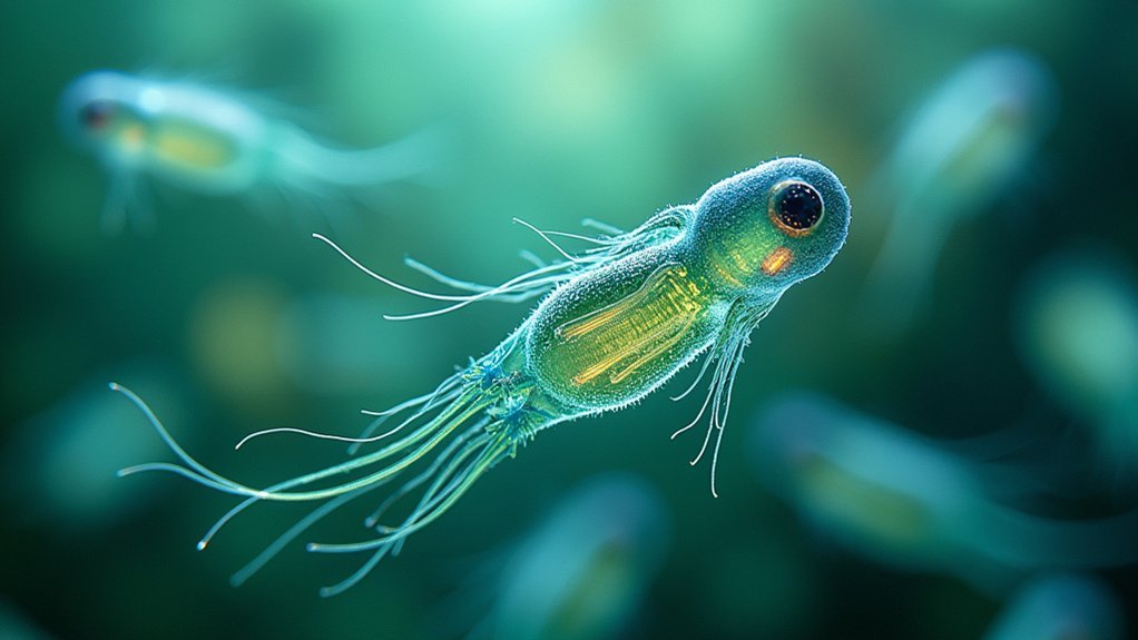
Since they display remarkable morphological details without requiring stains, protozoa and microorganisms represent exceptional specimens for phase contrast microscopy. You’ll observe dynamic movement of living microorganisms in their natural state, with thickness ranging from 5-10 micrometers—ideal for detecting subtle phase shifts.
| Organism | Key Features | Ideal Observation |
|---|---|---|
| Amoeba | Pseudopod formation | Cellular movement |
| Paramecium | Cilia motion | Feeding behaviors |
| Bacteria | Cell division | Colony formation |
| Yeast | Budding process | Reproduction cycles |
Phase contrast microscopy enhances visualization of these specimens through their varying refractive indices. You can witness cellular processes like division and motility in real-time, while studying how these organisms interact with their environment without the interference of artificial staining.
Unstained Tissue Sections: Achieving Optimal Contrast Without Fixatives
Unstained tissue sections maintain native cellular architecture that would otherwise be altered by chemical fixatives, giving you a more accurate view of biological structures in their natural state.
You’ll need to carefully consider section thickness, as sections between 5-10 micrometers provide the ideal balance between structural integrity and light transmission for phase contrast imaging.
These thin, unstained preparations reveal subtle details of cell morphology and organization that remain invisible in conventional brightfield microscopy, especially when proper mounting techniques are employed.
Native Cell Architecture Preservation
While fixed and stained specimens dominate conventional microscopy, unstained tissue sections represent the gold standard for phase contrast imaging when preserving native cellular architecture is crucial.
You’ll capture biological specimens in their natural state without introducing artifacts from fixatives or dyes. Phase contrast microscopy excels with transparent biological specimens approximately 5-10 micrometers thick, converting phase shifts into visible intensity differences. This contrast enhancement reveals delicate structures invisible to conventional bright-field methods.
For peak results, verify the specimen’s refractive index closely matches the surrounding medium to minimize distracting halos.
The true advantage lies in observing dynamic processes like cell division and migration in real time. By maintaining cellular integrity, you’ll witness living cells functioning naturally—something impossible with fixed preparations that fundamentally capture only a static moment in cellular history.
Thickness Considerations Matter
The success of phase contrast imaging hinges dramatically on specimen thickness, with the ideal range falling between 5-10 micrometers for unstained tissue sections.
When you’re preparing specimens, remember this critical dimension guarantees maximum contrast without requiring fixatives.
If your specimens are too thick, they’ll produce excessive phase shifts that distort your images, compromising clarity and detail.
By maintaining appropriate thickness, you’ll effectively visualize subtle refractive index differences between cellular components and their surrounding medium.
Thin sections enable proper light manipulation, allowing you to distinguish various structures clearly within your specimen.
You’ll also minimize frustrating artifacts like halos that can plague phase contrast microscopy.
Your careful attention to thickness parameters guarantees that unstained tissue sections reveal their natural architecture with maximum detail and minimal distortion.
Bacteria and Biofilms: Techniques for Enhanced Visualization
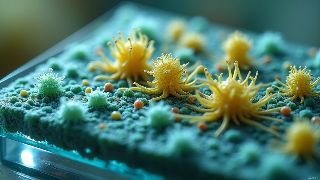
Microscopic explorers of the bacterial world find phase contrast microscopy invaluable for visualizing these transparent microorganisms without disruptive staining procedures. You’ll observe cellular structures, motility patterns, and biofilm formation in real-time as bacteria interact and respond to environmental changes.
For ideal results, configure your optical system with low numerical aperture objectives specifically designed for bacteria observation. Adding a green filter will enhance contrast, helping you differentiate between live and dead cells within complex biofilm communities.
| Technique | Application | Benefit |
|---|---|---|
| Low NA objectives | Bacterial imaging | Minimizes halo effects |
| Green filter | Biofilm analysis | Improves cell differentiation |
| Real-time observation | Motility studies | Captures dynamic behaviors |
When examining biofilms, adjust your focus gradually to assess their three-dimensional structure and density—critical for understanding these fascinating microbial communities.
Thin Cytological Smears: Maximizing Phase Difference Detection
Thin cytological smears require meticulous preparation to achieve the ideal 5-10 micrometer thickness that maximizes phase differences detectable under your microscope.
You’ll need to master cell dispersion methods that create even, single-layer distributions while avoiding clumping that can obscure fine cellular details.
When preparing your specimens, remember that consistent thickness across the slide greatly enhances contrast and allows you to visualize subtle refractive index variations in cellular components without staining artifacts.
Proper Preparation Techniques
When preparing specimens for phase contrast imaging, achieving the ideal thickness of cytological smears becomes a critical factor in successful visualization. You’ll want to aim for 5 to 10 micrometers thickness to enhance phase difference detection in your samples.
Proper fixation preserves cellular structure and prevents distortions that would otherwise compromise image quality. Verify you’ve created an even distribution of cells across your slide to facilitate consistent imaging results.
After application, use gentle drying techniques to maintain cellular morphology integrity without introducing artifacts.
For maximum contrast, match your phase plate and annular diaphragm to the specific objective magnification you’re using. This combination maximizes visibility of cellular details by enhancing the phase shifts that occur as light passes through your specimen’s varying densities.
Cell Dispersion Methods
Achieving effective cell dispersion represents the cornerstone of creating thin cytological smears that reveal intricate cellular details under phase contrast.
You’ll need to spread your cell suspension evenly to create specimen layers of 5-10 micrometers thickness, optimizing light transmission and phase shift detection.
For successful phase contrast microscopy of thin smears:
- Apply a small droplet of cell suspension to your slide and use a second slide at a 30° angle to create an even, thin layer with well-separated cellular structures.
- Confirm proper alignment of the phase annulus with your objective to accurately visualize subtle phase shifts.
- Allow smears to dry completely before viewing to prevent movement artifacts that can obscure phase contrast details.
Optimal Thickness Considerations
When preparing specimens for phase contrast microscopy, you’ll achieve superior results with thin specimens between 5-10 micrometers thick. This specific range maximizes phase shifts while preventing distortion.
Thin cytological smears greatly enhance the visibility of cellular structures by creating distinct phase differences between organelles and their surroundings. If your specimens are too thick, overlapping phase shifts will reduce image clarity and obscure fine details.
For best results, work with thin layers of unstained biological materials like cheek cells or blood smears. The ideal thickness guarantees that refractive index differences between cellular components and the surrounding medium translate effectively into visible intensity variations.
This precise thickness balance creates the perfect conditions for phase contrast microscopy to reveal structural details that would otherwise remain invisible.
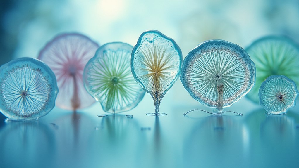
Transparent Aquatic Organisms: From Algae to Small Invertebrates
Transparent aquatic organisms represent some of the most ideal specimens for phase contrast microscopy due to their naturally low optical density. When you’re examining green algae, diatoms, or small invertebrates like water fleas (Daphnia), you’ll see internal structures with remarkable clarity without needing stains that might disrupt cellular processes or kill the specimen.
These planktonic organisms offer distinct advantages for your microscopy work:
- Living specimens remain viable during observation, allowing you to study motility and division in real-time.
- Small invertebrates like copepods and rotifers display clear morphological features that become instantly distinguishable.
- Diatoms reveal intricate silica cell walls and unique patterns that would remain invisible under traditional brightfield conditions.
This approach considerably contributes to marine biology by enabling direct observation of ecological interactions.
Frequently Asked Questions
What Type of Specimen Is Best for Phase Contrast Microscopy?
You’ll get the best results from phase contrast microscopy when using thin, transparent, living specimens like bacteria, protozoa, or cultured cells where subtle refractive index differences create natural contrast without staining.
How Do You Get a Good Image Under Phase Contrast Microscope?
To get a good image under phase contrast microscope, you’ll need thin specimens (5-10μm), proper phase objectives (Ph1-Ph3), aligned condenser annulus, green filter for enhanced contrast, and well-maintained optical components for maximum clarity.
What Organism Can Be Seen in a Phase Contrast Microscope?
You’ll see living bacteria, protozoa, yeast, cultured human cells (fibroblasts, epithelial), and subcellular structures clearly with a phase contrast microscope. It’s excellent for observing transparent microorganisms without staining them.
What Are the Best Practical Applications of a Phase Contrast Microscope?
You’ll find phase contrast microscopes excel for observing live cells without staining, studying bacterial motility, examining forensic fibers, analyzing transparent materials, and monitoring subcellular structures in real-time. They’re invaluable for dynamic biological research.
In Summary
You’ll find your phase contrast microscopy truly shines with these seven specimen types. By focusing on transparent samples that create natural phase differences, you’re opening up detailed cellular structures without stains or fixatives. Whether you’re examining live cells, protozoa, or unstained tissues, remember that proper preparation and appropriate thickness are your keys to achieving stunning contrast and revealing nature’s hidden microscopic details.


Leave a Reply