
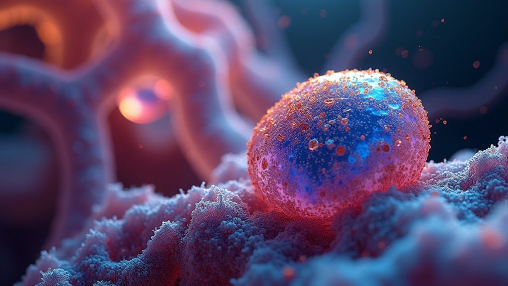
Phase contrast time-lapse imaging transforms your cell culture observations from static snapshots into dynamic cellular narratives that reveal hidden behavioral patterns and biological responses over hours or days. You’ll capture up to 50,000 images with proper interval timing, light intensity control, vibration reduction, computer-aided annotation, automated mitosis detection, growth factor visualization, and artifact mitigation techniques. These seven approaches will dramatically enhance your ability to document cellular behavior in unprecedented detail. The techniques below will elevate your imaging from ordinary to extraordinary.
Advanced Time-Lapse Capture Methods for Cell Lineage Tracking
While traditional microscopy provides static snapshots of cellular processes, advanced time-lapse capture methods have revolutionized cell lineage tracking by documenting cell behavior over extended periods.
You’ll find these techniques can capture nearly 50,000 phase contrast images over 3.5 days at 5-minute intervals, revealing how C2C12 myoblast cells respond to different growth factors.
This approach enables you to observe dynamic cellular processes that would otherwise remain hidden. The resulting datasets—comprising 48 annotated image sequences—facilitate development of sophisticated cell tracking algorithms.
However, you’ll face challenges requiring customized solutions, as conventional methods often yield false positives. The three-module tracking system (segmentation, mitosis detection, and association) demands significant effort, with a single fully annotated sequence requiring 60-90 hours to complete.
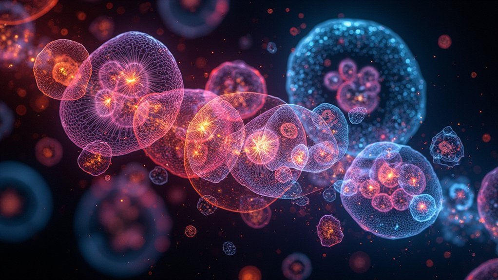
Multi-Day Phase Contrast Imaging of Myoblast Dynamics
Because cellular differentiation unfolds gradually over multiple days, the extensive dataset of 49,919 phase-contrast images captures C2C12 myoblast dynamics with unprecedented temporal resolution.
You’ll find these high-quality 16-bit images (1392 × 1040 pixels) document approximately 3.5 days of continuous cell behavior under four distinct growth factor conditions.
The time-lapse imaging protocol enables you to track complex cellular responses to Control, FGF2, BMP2, and combined FGF2+BMP2 treatments across multiple replicates.
This innovative approach combines rigorous manual annotation with automated tracking-by-detection methods, enhancing segmentation accuracy by removing imaging artifacts.
The resulting XML annotations and CSV worksheets provide you with powerful resources for developing your own cell tracking algorithms and deepening your understanding of myoblast dynamics in response to biological signals.
Optimizing Acquisition Parameters for Brilliant Cellular Detail
You’ll need to carefully select frame intervals that capture the relevant cellular events while preventing excessive photobleaching during time-lapse sequences.
Balancing exposure time is critical—too short won’t reveal subtle details, while too long causes blurring and phototoxicity during your 15-20 z-stack sections.
When controlling light intensity, aim for the minimum illumination required to maintain an acceptable signal-to-noise ratio across your 200-cycle acquisition, adjusting laser power and pinhole settings for each unique experimental context.
Frame Interval Selection
When selecting frame intervals for phase contrast time-lapse imaging, researchers must balance capturing sufficient cellular detail against generating manageable data volumes. The conventional 5-minute interval provides an excellent starting point for most experiments, allowing you to document dynamic cellular behaviors while keeping data sets reasonable.
Consider your specific research objectives when determining intervals. For studying rapid events like mitosis, you’ll need shorter intervals, while cell migration studies might permit longer ones.
Remember that extended observations—potentially spanning 3.5 days with 48 image sequences—require consistent interval settings for coherent analysis.
Phase-contrast imaging’s advantage is capturing natural cell dynamics without fluorescent labeling, but proper calibration of shutter synchronization and frame rates remains crucial to minimize motion artifacts and guarantee high-quality results.
Exposure Time Balance
The delicate balance of exposure time represents the cornerstone of successful phase contrast time-lapse imaging. You’ll need to carefully calibrate your illumination strength to maximize signal quality without inducing photobleaching or cellular damage.
For ideal z-stacks, adjust exposure parameters to enhance your signal-to-noise ratio while considering that typical imaging sequences may require 200 cycles to generate one hour of high-quality footage.
When working with quantitative phase contrast (qDPC), minimize motion artifacts by optimizing your exposure settings during multiple intensity measurements.
The dual-color linear-gradient pupil designs offer significant advantages, reducing required frames and accelerating imaging speeds up to 12 times faster than conventional methods.
Remember to maintain appropriate z-slice thickness (50-60 μM) while balancing exposure to capture complete cellular structures with crystal-clear phase contrast definition.
Light Intensity Control
Light intensity control operates as the central pillar for generating brilliant phase contrast images across extended time periods.
You’ll need to carefully adjust illumination levels to achieve ideal signal-to-noise ratios without triggering photobleaching or cell death. When capturing cell morphology, remember that too much light destroys the very details you’re trying to document.
Fine-tune adjustable parameters like pinhole size and laser power for each specific experiment.
Your z-stacks should typically comprise 15-20 sections over 50-60 μM depth ranges, with illumination strength precisely calibrated to minimize background noise.
Consider employing partially coherent light in quantitative differential phase contrast microscopy to reduce speckle noise and enhance lateral resolution.
For faster acquisition and higher quality, implement a dual-color linear-gradient pupil system that reduces necessary intensity measurements while maintaining isotropic phase contrast response.
Computer-Aided Annotation Techniques for Time-Lapse Sequences
Developing effective annotation methods represents a critical challenge in phase contrast time-lapse imaging due to the substantial time investment required.
When you’re tracking cell division across hundreds of frames during image acquisition, manual annotation alone becomes prohibitively time-consuming—annotating a single cell lineage takes 2-3 hours, with complete sequence annotation requiring 60-90 hours.
Today’s automated and manual cell tracking solutions employ standardized annotation schemes that combine efficiency with accuracy:
- Three-module tracking systems (segmentation, mitosis detection, and association) enhance cell identification precision
- Consistent labeling systems using 18 different cell states provide clarity in tracking complex cellular dynamics
- XML formatting facilitates easier analysis and sharing of annotation data between researchers
This hybrid computer-aided approach markedly reduces workload while maintaining the high-quality annotations essential for meaningful analysis.
Mitigating Artifacts in Extended Phase Contrast Sessions
Extended phase contrast imaging requires you to address several common artifacts that can compromise your data quality.
You’ll need to minimize halo effects by implementing dual-color linear-gradient pupil designs, while controlling environmental vibrations through proper isolation systems and calibration of your imaging setup.
To prevent photobleaching during long time-lapse sequences, you should optimize your illumination settings and utilize partially coherent light sources that reduce both light exposure and speckle artifacts.
Halo Effect Reduction
Although phase contrast imaging revolutionized cellular visualization, the persistent halo effect remains a significant challenge during extended imaging sessions.
You’ll notice significant improvements by implementing partially coherent light sources, which effectively reduce speckle noise while enhancing overall image quality during time-lapse experiments.
For ideal results in your extended phase contrast sessions:
- Adjust illumination settings strategically—modify pinhole size and laser power to balance signal-to-noise ratio
- Implement dual-color linear-gradient pupil designs to improve isotropic phase contrast response
- Regularly calibrate your optical setup to minimize color leakage between image sensor channels
Consider utilizing the weak object transfer function (WOTF) approach for faster phase retrieval with fewer intensity measurements.
This technique reduces motion artifacts that typically contribute to halo effects in long-duration imaging, providing cleaner results for your time-lapse studies.
Environmental Vibration Control
While cellular dynamics become increasingly apparent through phase contrast techniques, environmental vibrations can severely compromise image quality during time-lapse experiments.
You’ll notice these vibrations manifest as motion artifacts that obscure subtle cellular behaviors you’re trying to capture.
Implement a vibration isolation table to greatly enhance stability of your imaging setup.
Combine soft materials with rigid structures in your microscope’s base to effectively dampen unwanted movements.
You’ll achieve more consistent results by monitoring and minimizing airflow around your imaging area, as even gentle air currents can introduce disruptive vibrations.
Regular calibration in a controlled environment guarantees any residual artifacts are minimized.
These measures collectively preserve image clarity throughout extended imaging sessions, allowing you to collect reliable phase contrast data for thorough analysis.
Photobleaching Prevention
Phase contrast imaging sessions face another major challenge beyond vibrations: photobleaching. When capturing time-lapse sequences, particularly with fluorescent images, signal degradation can greatly compromise your data quality.
To minimize photobleaching effects, you’ll need to optimize your imaging strategy.
Balance these critical parameters in your setup:
- Reduce exposure time while maintaining adequate signal-to-noise ratio
- Increase intervals between time-lapse captures to decrease cumulative light exposure
- Employ spinning-disk confocal microscopy for faster acquisition speeds
Growth Factor Visualization Through Differential Contrast Enhancement
Because cellular responses to growth factors occur at microscopic scales with minimal optical contrast, differential phase contrast (DPC) microscopy has emerged as a critical tool for visualizing these subtle interactions. When you’re studying growth factors like FGF2 and BMP2, you’ll find that quantitative DPC (qDPC) delivers superior results with just two intensity measurements—dramatically reducing your imaging time while maintaining resolution.
| Growth Factor | Phase Contrast Quality | Cell Behavior Visibility |
|---|---|---|
| FGF2 | High definition | Proliferation evident |
| BMP2 | Sharp boundaries | Differentiation clear |
| None (control) | Baseline clarity | Natural dynamics visible |
The dual-color linear-gradient pupil design enhances isotropic phase contrast response, allowing you to capture the 49,919 images needed for long-term imaging without differential interference complications. This approach reveals mitotic events with unprecedented clarity across 3.5 days of continuous observation.
Automated Mitosis Detection in Long-Duration Cell Cultures
Since manually annotating cell division events can consume up to 90 hours for a single sequence, automated mitosis detection has become indispensable for long-term phase contrast imaging.
Automated mitosis detection transforms time-consuming manual annotation into efficient analysis for extended cell monitoring studies.
The in-house tracking system employs a three-step approach that identifies cytokinesis, considerably enhancing accuracy when monitoring cellular dynamics over extended periods.
You’ll find this technique especially valuable because it:
- Constructs candidate patch sequences based on pixel intensity to identify potential mitotic regions in time-lapse images
- Employs probabilistic modeling with machine learning to evaluate visual features of candidate patches
- Integrates parent-daughter relationship tracking during cell division events
Frequently Asked Questions
What Is the Time-Lapse Imaging Technique?
Time-lapse imaging is your method for capturing image sequences at regular intervals that you’ll compile into a movie, allowing you to observe dynamic biological processes that unfold too slowly for real-time viewing.
How Do You Get a Good Image Under Phase Contrast Microscope?
For good phase contrast images, you’ll need to adjust your illumination strength carefully, calibrate your optical setup precisely, and use the right pinhole size. Don’t forget to verify image quality with software like ImageJ.
What Is Phase Contrast Technique?
Phase contrast technique converts light phase shifts through transparent specimens into visible contrast. You’ll see cellular structures without staining, making it ideal for observing living cells during your experiments with minimal disruption.
What Is Time-Lapse Confocal Imaging?
Time-lapse confocal imaging lets you capture sequential 3D images over time, using laser scanning and pinholes to eliminate background light. You’ll see cellular processes unfold with high contrast and spatial resolution.
In Summary
You’ll find these seven phase contrast time-lapse techniques transform your cell imaging capabilities. Whether you’re tracking lineages, observing myoblast dynamics, or automating mitosis detection, these methods deliver exceptional clarity for extended periods. They’ll help you overcome common artifacts while enhancing visualization of subtle cellular interactions. By optimizing your acquisition parameters and applying these advanced techniques, you’ll capture stunning details that reveal cellular secrets previously hidden from view.
Leave a Reply