
The main components of contrast optics include the light source system, condenser assembly, phase plates and rings, objective lens configurations, and differential interference contrast prisms. These elements work together to manipulate optical path differences, creating visible contrast from transparent specimens. You’ll need properly adjusted annular diaphragms and phase plates to achieve ideal results. The condenser alignment and aperture settings greatly impact your image quality and resolution. Discover how these components transform invisible details into striking visual information.
Numeric List of 8 Second-Level Headings
Contrast optics relies on eight fundamental components that form its structural backbone. When you’re studying microscopy techniques, you’ll need to understand these essential elements:
- Light Source Systems
- Condenser Assembly
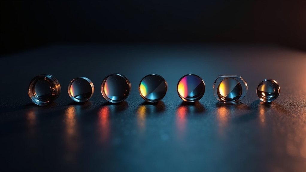
- Phase Plates and Rings
- Specimen Mounting Platforms
- Objective Lens Configurations
- Differential Interference Contrast Prisms
- Image Formation Mechanisms
- Refractive Index Compensation Tools
Each component plays a vital role in enhancing visibility of transparent specimens by exploiting differences in refractive index.
Phase contrast optics transforms these subtle variations into amplitude differences your eyes can detect. The differential interference contrast components create a three-dimensional effect by separating light paths.
The magic of microscopy lies in making the invisible visible through clever manipulation of light’s fundamental properties.
You’ll find these elements working together in sophisticated arrangements to reveal structural details that would otherwise remain invisible in conventional optical microscopy.
The Science Behind Optical Path Differences in Phase Objects
When you observe transparent specimens through phase contrast microscopy, you’re witnessing phase shift mechanics in action as light waves travel through materials with different refractive indices.
The optical path difference (OPD), calculated as t(n(s) – n(m)), quantifies how much the light’s phase changes when passing through your specimen compared to the surrounding medium.
These phase shifts, measured as δ = (2π/λ)(OPD), create the critical contrast that lets you see cellular structures and other transparent objects that would otherwise remain invisible in conventional brightfield microscopy.
Phase Shift Mechanics
Understanding the invisible becomes possible through phase shift mechanics, the fundamental principle that allows us to visualize transparent specimens under a microscope.
When light passes through a phase object, the optical path difference (OPD) occurs based on the formula OPD = t(n(s) – n(m)), where specimen thickness and refractive index variations become essential. This creates a phase difference δ = (2π/λ)(OPD), which conventional microscopes can’t detect directly.
Phase contrast microscopy exploits diffraction effects at cell edges, converting these phase shifts into visible intensity differences. The optical gradient generated at boundaries between different refractive indices transforms the otherwise invisible phase information into detectable contrasts.
Using coherent light sources enhances this effect, making subtle structural details apparent even in unstained specimens that don’t absorb light but only alter its phase.
OPD Quantification Methods
Accurately measuring optical path differences forms the backbone of modern phase contrast imaging.
When you’re quantifying OPD, you’ll use the formula OPD = t(n(s) – n(m)), where t represents specimen thickness and the difference between refractive indices of the specimen and surrounding medium creates the path difference.
This OPD translates to a phase difference (δ) calculated as δ = (2π/λ)(OPD), where λ is the light wavelength.
Even transparent specimens with minimal thickness can generate significant OPD due to refractive index variations. When a zero OPD occurs (identical refractive indices), phase differences become undetectable to your eye.
The brilliance of phase contrast microscopy lies in its ability to convert these otherwise invisible phase shifts into measurable intensity differences, enabling visualization of unstained specimens that would remain hidden in conventional microscopy.
Essential Components of Phase Contrast Microscope Systems
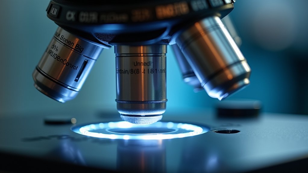
The intricate world of cellular structures becomes visible through specialized optical components in a phase contrast microscope. This system relies on an annulus diaphragm that creates a ring of light, transforming phase shifts into image contrast.
You’ll find the phase plate positioned strategically in the optical path, selectively altering light phases from transparent specimens. Your condenser lens focuses light onto the sample, with its aperture settings critical for ideal contrast without sacrificing resolution.
The objective lens, which should have a high numerical aperture, captures diffracted light to resolve fine details. This intelligent optical design allows you to visualize cellular structures without staining, preserving their natural morphology.
The harmonious interaction between these components enables the observation of living samples in their native state.
Annular Rings and Phase Plates: How They Transform Contrast
When light passes through transparent specimens, annular rings and phase plates work together to transform invisible phase differences into visible contrast. These optical elements are essential in phase contrast microscopy for visualizing unstained specimens that you couldn’t see with standard brightfield techniques.
- Annular rings create a specialized ring of light that interacts with your transparent specimens, modifying the phase relationships of passing light waves.
- Phase plates introduce controlled phase shifts, converting these phase changes into brightness variations.
- Contrast enhancement occurs when these components work in tandem, revealing cellular structures like membranes and organelles.
- Live cell imaging benefits tremendously from this technique, allowing you to observe dynamic biological processes without the artifacts introduced by staining.
Light Manipulation Techniques in Modern Contrast Methods

Beyond the foundational elements of annular rings and phase plates, modern microscopy offers a rich array of sophisticated light manipulation techniques that transform how you’ll see transparent specimens.
Phase contrast converts subtle light shifts into visible amplitude changes, dramatically improving the specimen image.
Phase contrast microscopy transforms invisible optical phase variations into visible intensity differences, revealing cellular details otherwise hidden from view.
You’ll find differential interference contrast (DIC) particularly valuable when examining structures with varying refractive index, as it creates striking three-dimensional effects using polarized light.
Darkfield microscopy enhances contrast by capturing only scattered light against a black background, revealing details invisible in brightfield.
For ideal optical transfer function, you can adjust the condenser aperture to sharpen light-specimen interaction, though this affects resolution.
Modern contrast methods also incorporate fluorescent markers and color filters, offering contrast enhancing capabilities that let you visualize specific cellular structures with remarkable clarity.
Refractive Index Variations and Their Impact on Image Formation
Understanding refractive index variations forms the cornerstone of effective contrast microscopy, as these subtle differences determine how light interacts with your specimen.
When light passes through biological materials with refractive indices greater than 1.0, phase shifts occur that you can leverage for contrast enhancing.
- Small refractive index differences between specimen components create optical path differences (OPD) that make transparent specimens visible.
- Phase shifts resulting from these variations form the basis for techniques like phase contrast microscopy.
- The optical transfer function (OTF) and modulation transfer function (MTF) directly respond to refractive index changes, affecting image contrast at increasing spatial frequencies.
- Capturing diffracted light at wide angles improves resolution of fine details, especially when working with specimens having varied refractive properties.
Comparing Brightfield, Darkfield, and Differential Interference Methods
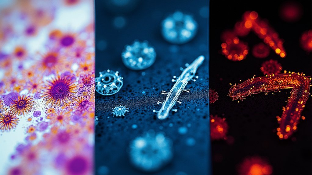
Although all microscopy techniques aim to enhance specimen visibility, brightfield, darkfield, and differential interference contrast methods employ fundamentally different optical principles to achieve this goal. When imaging specimens, each technique offers unique advantages for visualizing structural details in biological specimens.
| Technique | Optical Principles | Best Application |
|---|---|---|
| Brightfield microscopy | Uses transmitted light; captures light intensity variations | Stained specimens with good absorption |
| Darkfield microscopy | Employs oblique illumination; highlights scattered light | Transparent or colorless samples requiring contrast enhancing |
| Differential Interference Contrast | Utilizes polarized light; reveals phase differences | Unstained specimens needing pseudo-3D appearance |
You’ll find that brightfield works well with absorptive samples, while darkfield excels with transparent specimens. DIC bridges the gap by creating detailed relief-like images without staining requirements.
Optimizing Condenser Settings for Maximum Phase Visibility
When examining transparent or unstained specimens, proper condenser adjustment becomes the cornerstone of achieving outstanding phase visibility in microscopic imaging.
You’ll need to fine-tune your contrast optics to convert invisible phase differences into visible intensity variations.
For ideal results:
- Adjust the condenser aperture carefully—smaller apertures enhance contrast but may sacrifice resolution, so find the balance that works for your specimen.
- Position your condenser for optimal light capture, ensuring perfect alignment with the phase ring to convert phase shifts into visible differences.
- Utilize a high numerical aperture condenser to collect more diffracted light, essential for resolving fine structural details.
- Regularly calibrate your settings to account for refractive index differences between mounting media and specimens.
Frequently Asked Questions
What Are the Components of Contrast?
Contrast components include light absorption, specimen brightness, reflectance, refractive index differences, illumination techniques, and optical system parameters. You’ll need these elements to achieve the minimum 2% visibility threshold in microscopy images.
What Are the Main Components of a Fiber Optics System?
A fiber optics system’s main components include a light-carrying core, reflective cladding with lower refractive index, protective outer jacket, light sources, connectors, and receivers. You’ll find these elements in all optical fiber installations.
What Is Contrast Optics?
Contrast optics are microscopy techniques you’ll use to enhance visibility of transparent specimens without staining. They convert phase shifts into brightness differences, making subtle features visible that you’d otherwise miss in standard brightfield microscopy.
What Are the Two Major Components of the Phase Contrast Microscope?
The two major components of the phase contrast microscope are the phase plate and the annular diaphragm. They work together to convert phase shifts in light into amplitude changes you can see.
In Summary
You’ve now explored the foundational elements of contrast optics, from phase plates to annular rings. By understanding how these components manipulate light paths and exploit refractive index differences, you’ll achieve dramatically improved visibility of transparent specimens. Remember to fine-tune your condenser settings and select the appropriate contrast method for your specific samples. With practice, you’ll transform invisible phase objects into clearly defined, high-contrast images.


Leave a Reply