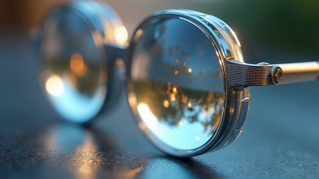
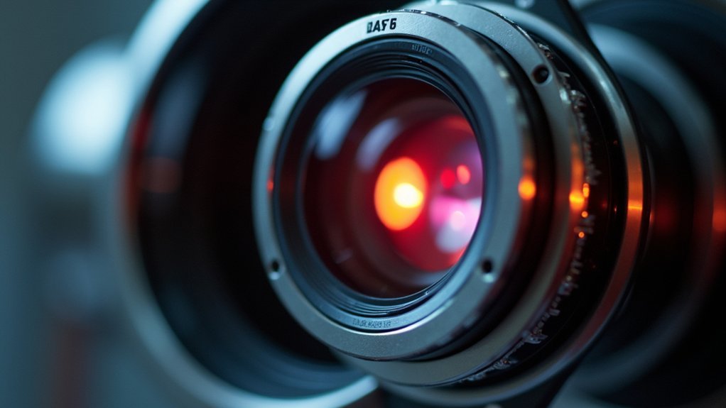
The main components of contrast optics include phase rings in objective lenses, condenser annular diaphragms, phase plates, and aperture controls. These elements work together to transform invisible phase shifts into visible amplitude differences, making transparent specimens observable without staining. You’ll find specialized objectives designed specifically for phase contrast, along with Köhler illumination systems that guarantee uniform light distribution. Understanding these components will reveal your ability to visualize previously hidden cellular structures.
Numeric List of 12 Second-Level Headings
Twelve distinct sections comprise our exploration of contrast optics fundamentals.
You’ll find our content organized into these critical second-level headings:
- Historical Development of Optical Contrast
- Basic Principles of Light Manipulation
- Brightfield Microscopy Fundamentals
- Darkfield Techniques and Applications
- Phase Contrast Optics Mechanisms
- Differential Interference Contrast Systems
- Polarized Light Configurations
- Hoffman Modulation Contrast
- Rheinberg Illumination Methods
- Oblique Illumination Approaches
- Digital Contrast Enhancement
- Future Trends in Contrast Microscopy
This structured approach guarantees you’ll grasp both traditional and cutting-edge contrast techniques.
Each section builds upon previous concepts, creating a thorough understanding of how various optical contrast methodologies reveal specimen details that would otherwise remain invisible under standard illumination conditions.
Fundamentals of Phase Contrast Technology
When observing transparent specimens through conventional microscopes, you’ll often encounter a frustrating limitation—cells and other unstained biological structures remain nearly invisible despite being physically present. Phase contrast microscopy solves this problem by transforming phase shifts into amplitude changes that your eyes can detect as brightness variations.
| Phase Object Property | How It Affects Imaging |
|---|---|
| Refractive index differences | Create phase shifts in light waves |
| Transparent appearance | Invisible in brightfield microscopy |
| No natural amplitude change | Requires phase ring intervention |
| Live cell compatibility | Enables observation without staining |
The phase contrast microscope’s essential component is the phase ring within the objective lens. This ingenious element converts otherwise invisible phase differences into visible intensity variations. You’ll achieve excellent results when using objectives with higher numerical apertures, allowing you to observe living cells’ dynamic processes without disrupting their natural morphology.
The Role of Phase Rings in Contrast Enhancement
Phase rings form the heart of contrast enhancement in modern microscopy systems. Located in the objective’s back focal plane, these specialized components transform invisible phase shifts into visible amplitude differences when light passes through transparent specimens.
You’ll notice the effectiveness of phase rings through the distinctive bright halos they create around cells and other phase objects. This visibility enhancement occurs without staining, making them invaluable for observing living specimens.
For ideal contrast enhancement, you must properly align the phase rings with your specimen and the optical path. Any misalignment will immediately reduce image quality.
Different phase ring configurations are available depending on your specimen characteristics and desired contrast levels. This flexibility allows you to tailor your microscopy approach to specific research needs while maintaining exceptional clarity.
Condenser Annular Diaphragms: Function and Optimization
Integral to achieving ideal contrast in microscopy, condenser annular diaphragms work by precisely controlling the illumination pathway to your specimen. They create annular light beams that greatly enhance visibility of transparent structures, especially in phase contrast microscopy.
You’ll achieve best results by matching the condenser aperture diaphragm opening to your objective’s numerical aperture. This careful alignment guarantees that light reaches your specimen at the proper angle, maximizing contrast while maintaining resolution.
Precise condenser-to-objective alignment creates the perfect balance of contrast and resolution for revealing specimen details.
When properly adjusted, these diaphragms enable techniques like differential interference contrast to reveal subtle specimen details that would otherwise remain invisible.
Remember that imprecise diaphragm settings can dramatically reduce contrast quality. Fine-tuning this component allows you to manipulate light coherence and refine the illumination conditions for your specific microscopy application.
Phase Plates and Their Light-Shifting Properties
At the core of contrast enhancement techniques, phase plates serve as critical optical elements that transform invisible phase differences into visible intensity variations.
When you’re examining transparent specimens like living cells, these specialized components manipulate light waves to reveal structures that would otherwise remain hidden.
The effectiveness of phase plates in contrast optics depends on three key factors:
- The precise thickness variations engineered to create specific optical path differences
- The wavelength of illumination light interacting with the phase plate
- The refractive index difference between your specimen and its surrounding medium
You’ll notice distinctive bright halos forming around cellular structures when using phase plates correctly.
This characteristic effect helps you distinguish fine details and internal components that brightfield microscopy alone can’t reveal.
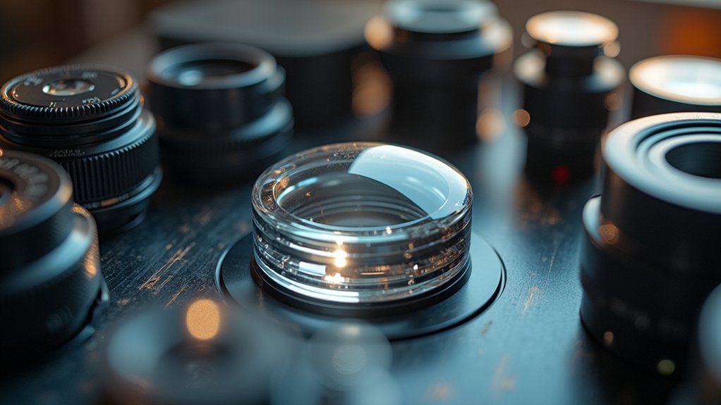
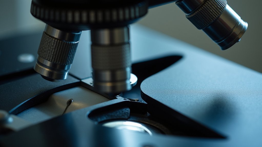
Specialized Objectives for Phase Contrast Imaging
When you’re working with transparent biological specimens, specialized objectives designed specifically for phase contrast imaging become essential tools in your microscopy arsenal.
These objectives convert phase shifts in light into visible intensity differences, revealing cellular structures that would otherwise remain invisible.
You’ll notice these specialized objectives feature high numerical aperture values to capture maximum diffracted light, critical for resolving fine details in phase contrast microscopy.
They incorporate annular apertures that create characteristic halos around phase objects, enhancing structural boundaries.
For ideal results, you’ll need to properly align these objectives with your optical system.
Many are calibrated for specific refractive indices, ensuring they’ll work effectively across various biological samples.
With proper setup, you’ll achieve the contrast necessary to visualize transparent specimens with remarkable clarity.
Kohler Illumination Systems in Contrast Formation
Beyond specialized objectives, the illumination system plays a fundamental role in creating effective contrast. Kohler illumination maximizes your microscopic imaging by ensuring uniform light distribution across the specimen plane, markedly enhancing contrast while reducing glare and hot spots.
This sophisticated system minimizes optical aberrations through:
- Precise focusing of the light source onto the condenser, which then projects an evenly distributed illumination onto your specimen.
- Adjustment capabilities for the condenser aperture diaphragm, allowing you to control numerical aperture and directly influence contrast.
- Careful alignment of all optical components to achieve peak clarity, essential for detailed specimen analysis.
You’ll find that proper Kohler illumination setup transforms your microscopy work by revealing fine structural details that might otherwise remain invisible under standard illumination conditions.
Amplitude vs. Phase Objects: Visualization Differences
Understanding the fundamental difference between amplitude and phase objects is essential for choosing the right microscopy technique for your specimens.
Amplitude objects, typically stained samples, absorb light and alter its amplitude, making them clearly visible in brightfield microscopy with high contrast.
Phase objects, however, only change light’s phase without affecting amplitude. This occurs due to differences in refractive index between the specimen and its medium. You’ll find that unstained living cells are classic phase objects, appearing nearly invisible in conventional microscopy due to minimal light absorption.
When examining phase objects, you’ll need specialized techniques like phase contrast microscopy, which converts phase shifts into amplitude differences. This creates visible halos around transparent specimens, greatly enhancing their visibility despite their transparent nature.
Microscope Aperture Controls for Optimal Contrast
The precise manipulation of aperture controls serves as the cornerstone of achieving ideal contrast in microscopy. Your condenser aperture directly affects the light illuminating your specimen—reduce it to enhance contrast, but be aware that excessive reduction may compromise resolution.
Similarly, the field aperture limits incoming light, reducing stray illumination and improving detail visibility.
When adjusting aperture controls for maximal contrast, consider:
- Specimen type – Different samples require specific lighting conditions
- Balance of resolution and contrast – Find the sweet spot where details remain sharp
- Optical aberration management – Proper aperture settings minimize distortions
You’ll need to make continual adjustments based on your specific imaging needs, as the perfect balance between brightness, resolution, and contrast varies with each specimen you observe.
Refractive Index Influence on Specimen Visibility
While aperture controls provide the mechanical foundation for contrast, refractive index differences form the optical basis of specimen visibility in microscopy. The refractive index—the ratio of light’s speed in vacuum versus in a specimen—determines how light bends as it passes through your sample.
When light encounters materials with varying refractive indices, it undergoes phase shifts that create natural contrast. You’ll notice that specimens with higher refractive indices bend light more dramatically, enhancing edges and internal structures. This principle is particularly essential in phase contrast microscopy, where these shifts are amplified.
The optical path difference (OPD) calculated from specimen thickness and refractive index directly affects image contrast. By selecting mounting media with appropriate refractive properties, you can markedly improve visibility of otherwise transparent specimens.
Contrast Enhancement Through Digital Processing
Digital image processing transforms your microscopy observations through powerful algorithms that enhance contrast where traditional optics fall short.
You’ll find histogram equalization techniques particularly valuable for redistributing pixel intensities, maximizing visibility in previously indistinct specimen regions.
Edge detection methods complement these approaches by emphasizing structural boundaries, allowing you to discern fine cellular interfaces and morphological details with unprecedented clarity.
Algorithm Types and Applications
Modern contrast enhancement algorithms transform how we visualize microscopic specimens by manipulating digital image properties at the pixel level. These sophisticated tools redistribute pixel intensities to reveal details in biological samples that would otherwise remain hidden to the human eye.
The most effective algorithms for microscopy include:
- Histogram equalization – Redistributes brightness values across the entire image, ideal for specimens with poor initial contrast.
- Adaptive histogram equalization – Applies localized contrast enhancement to different regions independently.
- Contrast-limited adaptive histogram equalization (CLAHE) – Prevents over-amplification of noise while enhancing meaningful details.
You’ll find these techniques particularly valuable in live cell imaging, where they enable real-time visualization of dynamic processes while minimizing artifacts introduced during specimen preparation.
Histogram Equalization Techniques
Among the algorithms mentioned above, histogram equalization stands as a fundamental technique that transforms low-contrast microscopy images into highly detailed visualizations. This process works by redistributing pixel intensity values across the full available range, effectively stretching the contrast to reveal features that were previously hidden in overly dark or bright regions.
When you apply histogram equalization to your microscopy images, you’ll notice significant enhancement in feature visibility as the algorithm creates a more uniform distribution of brightness levels.
You can utilize this technique on both grayscale and color images, though with color specimens, it’s often better to process individual channels to maintain natural coloration.
While the method dramatically improves contrast, be mindful that it can sometimes introduce noise—balance is key for ideal results.
Edge Detection Methods
While scanning a sample under a microscope, you’ll often encounter structures whose subtle boundaries blend into surrounding tissue. Edge detection algorithms can dramatically enhance these boundaries by identifying abrupt changes in image intensity. These digital processing techniques highlight contrast differences that your eyes might otherwise miss.
Modern microscopy software incorporates several powerful edge detection methods:
- Sobel operators calculate intensity gradients between pixels, emphasizing shifts between different structures.
- Canny algorithms provide superior noise reduction while preserving essential edge information.
- Prewitt operators detect edges by measuring intensity differences in adjacent pixel neighborhoods.
When combined with techniques like adaptive contrast stretching and histogram equalization, edge detection transforms barely visible cellular boundaries into clearly defined structures.
You’ll appreciate this enhancement when examining intricate biological specimens where subtle details carry significant diagnostic value.
Comparison of Positive and Negative Phase Contrast
Despite sharing fundamental principles, positive and negative phase contrast techniques represent distinct approaches to visualizing transparent specimens.
In positive phase contrast, you’ll observe bright specimen images against a darker background as phase shifts are converted into amplitude increases. This makes it particularly effective for examining living cells and their intricate structures.
Negative phase contrast, however, produces dark specimen images against a brighter background by converting phase shifts into amplitude decreases. This alternative configuration helps you identify specific details that might remain obscured in positive contrast.
When selecting between these techniques, consider your specimen type and research objectives. The difference lies primarily in the phase plate configuration, which determines how light is manipulated and ultimately how your specimens appear under microscopic examination.
Frequently Asked Questions
What Are the Components of Contrast?
Contrast components include light intensity differences, aperture settings, specimen absorption properties, background intensity, staining techniques, and optical system quality. You’ll notice these elements work together to create visible distinction in your images.
What Are the Main Components of a Fiber Optics System?
In a fiber optics system, you’ll find three main components: the core where light travels, the cladding that reflects light back into the core, and the buffer coating that protects the fiber.
What Is Contrast Optics?
Contrast optics is a microscopy technique you’ll use to enhance visibility of transparent specimens. It converts light phase shifts into brightness differences, allowing you to see colorless structures that’d otherwise be invisible under standard illumination.
What Are the Two Major Components of the Phase Contrast Microscope?
The two major components of the phase contrast microscope you’ll need to understand are the phase plate and the annular diaphragm. Together, they convert phase shifts into visible contrast when viewing transparent specimens.
In Summary
You’ve now explored the essential components of contrast optics, from phase rings and condenser diaphragms to phase plates and aperture controls. Understanding how refractive index differences create visibility and how both hardware and digital processing enhance contrast will improve your microscopy skills. Whether you’re working with positive or negative phase contrast, you’ll achieve clearer images by mastering these fundamental elements of contrast optics.


Leave a Reply