For ideal phase contrast microscopy images, you’ll need to carefully balance exposure time and light intensity. Start with shorter exposures (50ms-2s) and gradually increase while monitoring your histogram to avoid saturation. Position your condenser at its highest setting and match its numerical aperture to your objective. Use neutral density filters to control brightness while minimizing halo artifacts. Regular calibration of your microscope components guarantees consistent results and helps you capture the fine details of transparent specimens.
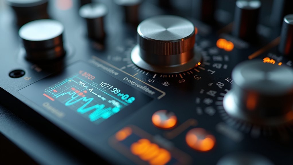
Understanding Exposure Controls in Phase Contrast Microscopy
Mastery of exposure controls forms the cornerstone of successful phase contrast microscopy.
You’ll need to carefully balance light intensity and exposure time based on your specimen’s characteristics to achieve high contrast while minimizing phototoxicity in living samples.
Monitor your signal intensity distribution using histograms—look for a good spread without saturation, indicating proper exposure.
While longer exposures can enhance visibility, they may damage living specimens, requiring you to compromise between image quality and sample preservation.
Adjust gain settings judiciously; higher gain amplifies both signal and noise, potentially degrading your images.
Don’t overlook the importance of regular calibration by checking for stray light and aligning optical components.
This maintenance guarantees your phase contrast microscopy delivers consistently superior results with ideal exposure control.
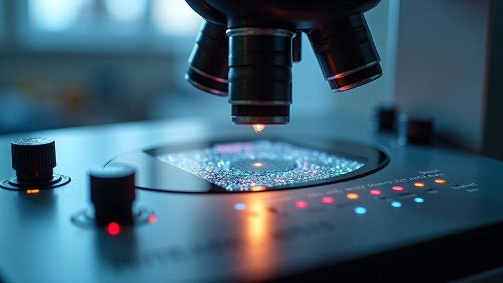
Optimizing Light Intensity and Condenser Settings
With your exposure fundamentals established, proper light intensity and condenser settings become your next focus in phase contrast microscopy. Using a halogen light source or LED with adjustable output allows you to fine-tune brightness without causing photobleaching in live specimens.
Position your condenser at its highest setting to fully illuminate the annular aperture, enhancing contrast in your images.
For best phase contrast results:
- Match your condenser’s numerical aperture to the objective you’re using, ensuring sufficient light reaches your specimen.
- Adjust light intensity gradually until details appear crisp without overexposure.
- Monitor your histogram during imaging to prevent image saturation that can obscure fine structural details.
Remember to regularly check your condenser aperture diaphragm as you switch between objectives to maintain ideal illumination conditions.
Managing Exposure Time for Transparent Specimens
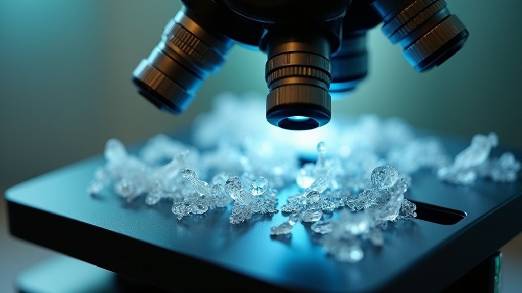
Transparent specimens present unique challenges in phase contrast microscopy, as their low natural contrast requires precise exposure timing to reveal structural details without causing damage.
You’ll need to optimize exposure time based on specimen thickness, typically ranging from 50 ms to several seconds.
Use your camera’s histogram during acquisition to verify you’re neither underexposing (crowded low signal) nor overexposing (saturation).
The histogram serves as your exposure compass, guiding you between the shoals of underexposure and the rocks of signal saturation.
Start with lower exposure times and gradually increase while evaluating image quality in real-time.
Remember that objectives with higher numerical aperture may require shorter exposure times to prevent overexposure.
Regular calibration of camera settings is essential for consistent results.
Mitigating Halo Artifacts Through Camera Adjustments
Although phase contrast microscopy reveals transparent structures effectively, it often introduces bright halos around specimen edges that can obscure essential details. Your camera settings play a vital role in minimizing these unwanted artifacts while preserving image quality.
To reduce halo artifacts in your phase contrast images:
- Optimize exposure settings – Avoid overexposure which intensifies halos around high phase shift areas. Select an exposure time that captures detail without saturating the sensor.
- Adjust gain controls – Fine-tune your camera’s gain to reduce noise, which helps decrease the visibility of halo artifacts in your final images.
- Control light intensity – Implement neutral density filters to manage the amount of light reaching your camera, preventing excessive brightness that exacerbates halo effects.
Regular calibration of your microscope’s phase contrast components further enhances these adjustments.
Advanced Techniques for High-Contrast Image Capture

While basic exposure settings form the foundation of good phase contrast imaging, mastering advanced camera techniques greatly elevates your image quality.
You’ll achieve superior results by refining exposure times to prevent saturation—check your histogram for balanced signal distribution without clipping.
Use your phase contrast microscope’s high numerical aperture (NA) objectives to maximize light collection, dramatically improving image contrast.
When implementing binning, you’ll gain sensitivity but remember this trades off with resolution.
Don’t overlook illumination settings—regularly calibrate your condenser aperture and diaphragm to maintain ideal contrast without introducing optical artifacts.
Apply gain judiciously; while it enhances signal, excessive gain amplifies background noise.
These advanced techniques, when properly executed, transform standard phase contrast images into high-quality scientific data with remarkable clarity and detail.
Frequently Asked Questions
How Do You Get a Good Image Under Phase Contrast Microscope?
You’ll get a good image by aligning the condenser annulus with the phase ring, using moderate light intensity, adjusting exposure time carefully, monitoring the histogram, and selecting higher numerical aperture objectives.
Is Phase Contrast Worth It?
Yes, phase contrast is worth it! You’ll see living cells without killing them with stains. It’s cost-effective and gives excellent detail for transparent specimens, though you’ll need to evaluate your sample thickness.
Is Kohler Illumination Phase Contrast?
No, Köhler illumination isn’t phase contrast. It’s a lighting setup technique that optimizes illumination in any microscopy. You’ll use Köhler illumination with phase contrast to achieve even lighting and enhance your transparent specimen visualization.
What Is the Resolution Limit for Phase Contrast?
You’ll find phase contrast microscopy limited to approximately 200 nanometers resolution due to light’s diffraction limit. Your objective lens’s numerical aperture (NA) directly impacts this—higher NA values provide better resolution.
In Summary
You’ll achieve the best phase contrast images when you balance your exposure controls properly. Start with ideal light intensity and correct condenser alignment, then fine-tune your camera’s exposure time and gain settings. Remember to adjust your settings for each specimen’s transparency level, and don’t hesitate to use post-processing for challenging samples. With practice, you’ll consistently capture clear, artifact-free images with outstanding detail.


Leave a Reply