For phase contrast imaging, you’ll need C-Mount adapters specifically designed to preserve phase contrast properties. Select magnifications between 0.5x and 1.0x to balance field of view with detail. Ascertain compatibility with your microscope’s trinocular port and match the adapter to your camera’s sensor size to prevent vignetting. Quality adapters like AmScope CA-NIK-SLR or specialized Olympus options maintain optical clarity and light transmission. The right adapter choice dramatically impacts your imaging results and research quality.
Understanding the Basics of Phase Contrast Imaging
Three key components form the foundation of phase contrast imaging: specialized optics, light manipulation, and precise alignment.
Phase contrast imaging excellence demands specialized optics, masterful light manipulation, and meticulous alignment—the trinity of cellular visualization without stains.
When you observe live cells through this technique, you’re witnessing light phase shifts converted into visible intensity variations, eliminating the need for potentially harmful staining processes.
The system relies on critical optical components like phase plates and annular rings that create contrast based on refractive index differences in your specimen.
For peak results, you’ll need a pre-centered phase contrast slider working in harmony with standardized magnification rings (10x, 20x, and 40x).
To document these observations, LM digital adapters serve as essential camera adapters, ensuring high resolution image capture of your microscopy work, preserving the enhanced visibility and contrast that makes phase contrast imaging so valuable.
Key Features of Camera Adapters for Phase Contrast Microscopy
When selecting a camera adapter for phase contrast microscopy, you’ll need to evaluate C-Mount adapter magnification options ranging from 0.5x to 1.0x to achieve ideal field of view and resolution for your specimens.
Your choice of trinocular port matters considerably, as it determines how light is distributed between eyepieces and camera, affecting both visual observation and image capture quality.
Modern adapters offer quick-switch trinocular port selection, allowing you to seamlessly shift between direct visual examination and digital imaging without disturbing your specimen’s position.
C-Mount Adapter Magnification
Selecting the proper magnification for your C-Mount adapter stands as a critical factor in achieving ideal phase contrast imaging results. Most adapters offer various magnifications—typically 1.2x, 1.5x, and 2.25x—that directly affect how the optical image projects onto your camera sensor.
When choosing your adapter’s magnification, you’ll need to take into account your microscope’s numerical aperture and your specific imaging needs. Higher magnifications reveal finer details but may reduce the field of view, while lower magnifications capture more of your specimen at once.
The internal optics of quality C-mount adapters are designed to minimize chromatic aberration while enhancing contrast—essential for clear phase contrast imaging. Remember that the adapter must match your camera’s sensor size to avoid vignetting or lost resolution, ensuring the optical pathway remains optimized.
Trinocular Port Selection
The trinocular port serves as the critical interface between your microscope and camera system for phase contrast imaging. When selecting a trinocular setup, you’ll need camera adapters specifically designed to maintain optical quality for visualizing transparent specimens without staining.
Look for LM digital adapters that support your preferred camera type—whether DSLR or mirrorless—while ensuring they’re compatible with standard phase contrast magnifications (10x, 20x, and 40x). These adapters should seamlessly integrate with your trinocular port, allowing you to simultaneously view specimens through eyepieces while capturing digital images.
For maximum efficiency, choose adapters that incorporate a pre-centered phase contrast slider. This feature eliminates time-consuming re-centering procedures, streamlining your workflow when examining live cells and other transparent samples under phase contrast microscopy.
Trinocular Tube Adapters vs. Direct C-Mount Options
Because microscopy workflows vary widely between researchers, choosing between trinocular tube adapters and direct C-mount options becomes a critical decision for phase contrast imaging success.
Trinocular tube adapters excel by allowing you to simultaneously view specimens through eyepieces while capturing images—particularly valuable when examining live specimens that require real-time observation.
In contrast, direct C-Mount options connect cameras directly to your microscope’s photo port, creating a more compact setup but potentially limiting simultaneous viewing.
The standardized optical design of C-Mount adapters projects phase contrast images directly to the camera sensor, ensuring high-quality image capture.
Your choice ultimately depends on workflow priorities—opt for trinocular setups when you need enhanced flexibility for specimen observation, or choose C-Mount configurations when you prefer compactness and streamlined image acquisition.
Matching Camera Sensors to Phase Contrast Optical Paths
When selecting a camera adapter for phase contrast imaging, you’ll need to take into account your camera’s sensor size to prevent vignetting and guarantee full capture of the microscope’s optical field.
Your sensor’s resolution must align with the microscope’s resolving power to accurately record the subtle contrast differences phase imaging reveals.
The adapter’s optical design should maintain the numerical aperture relationship between your objective lenses and camera sensor, preserving both resolution and contrast in your captured images.
Sensor Size Considerations
Selecting the right camera sensor size for phase contrast imaging represents a critical decision that directly impacts your final image quality. You’ll need to match your camera’s sensor to your microscope’s optical path using an appropriate C-Mount adapter.
| Sensor Size | Recommended Adapter | Optical Considerations |
|---|---|---|
| 1/4″ | 0.25x – 0.5x | Narrower field of view |
| 1/2″ | 1.0x | Standard field of view |
| 2/3″ | 1.0x | Enhanced detail capture |
| 1″ | 0.63x – 1.0x | Wide field of view |
| 36×24mm | Custom adapter | Maximum resolution |
When selecting an adapter, remember that back focal length must align with your phase contrast system. Larger sensors in high-resolution cameras can capture finer details but require specialized adapters to prevent vignetting and maintain proper phase ring alignment throughout the optical path.
Optimizing Resolution Capture
Beyond sensor size alone, the strategic matching of camera sensors to phase contrast optical paths determines your ultimate resolution capabilities.
When selecting camera adapters, verify they’re compatible with your microscope’s optical configuration to achieve maximum clarity in your phase contrast imaging.
For best results, you’ll need to align your camera’s sensor precisely with the optical path—dedicated LM digital adapters can greatly enhance your image quality.
Match the adapter’s magnification to your objectives, typically using standardized ring slits for 10x, 20x, and 40x magnifications.
Don’t compromise on quality—adapters with a plan achromatic optical system minimize aberrations while preserving image fidelity.
Pair these with cameras featuring high pixel counts and good ISO sensitivity to capture detailed images, especially when working with specimens in low-light conditions.
High-Resolution Imaging Requirements for Phase Contrast
Although phase contrast microscopy revolutionized the observation of transparent specimens, achieving ideal results demands specific high-resolution imaging components. You’ll need camera adapters specifically designed to maintain optical quality when viewing unstained samples. These adapters preserve the delicate contrast relationships that make cellular structures visible.
| Component | Requirement | Benefit |
|---|---|---|
| Camera | High pixel count | Enhanced detail resolution |
| Adapter | Pre-centered sliders | Eliminates recalibration |
| System Compatibility | DSLRs/Mirrorless | Maximized dynamic range |
When selecting equipment, prioritize adapters with standardized ring slits that support consistent magnification at 10x, 40x, and especially 20x levels. The combination of proper adapters with modern cameras dramatically improves light sensitivity, allowing you to capture subtle variations in transparent specimens that would otherwise remain invisible.
Adapter Magnification Factors and Their Effects on Image Quality
When selecting camera adapters for phase contrast imaging, you’ll need to balance magnification factors with resolution capabilities to prevent introducing unwanted artifacts.
Higher magnification options like the 2X AmScope adapter can enhance detail capture, but you should opt for low-distortion models that maintain optical integrity across the field of view.
Your microscope’s cropping factor will also impact the final image quality, requiring consideration of how the adapter’s magnification interacts with your camera sensor size to preserve the phase contrast effect.
Magnification and Resolution Balance
As you select a camera adapter for phase contrast imaging, understanding the relationship between magnification factors and image quality becomes essential for successful microscopy.
A 2X adapter doubles the image size on your sensor, but this higher magnification must be balanced against potential resolution loss.
When working with high magnification objectives (like 40x), your camera adapter’s calibration directly affects whether details remain crisp or suffer from chromatic aberration.
The resolution you’ll achieve depends on both the adapter’s magnification and your microscope’s optical system quality.
For phase contrast imaging specifically, you’ll need adapters designed to preserve the technique’s characteristic optical properties.
Remember to match your camera’s sensor size with the adapter’s design specifications—this pairing is particularly vital in high precision microscopy where subtle cellular details matter.
Low-Distortion Adapter Options
Despite their small size, low-distortion camera adapters play a critical role in preserving phase contrast image quality. When selecting an adapter, you’ll find options with magnification factors of 1.5x or 2x that enhance resolution while maintaining minimal distortion.
The quality of optical components in these adapters guarantees accurate light transmission to your camera sensor, preserving specimen details critical for phase contrast imaging. Adapters with built-in corrections minimize chromatic aberration, essential for achieving precise detail and color accuracy.
Your choice of magnification factor affects the effective numerical aperture and working distance, directly impacting overall image quality. For best results, choose adapters specifically engineered for your microscope’s optical system to guarantee seamless integration.
This compatibility ensures maximum image fidelity when capturing the subtle contrast differences that make phase contrast techniques so valuable.
Cropping Factor Considerations
Magnification factors directly connect to the cropping phenomenon you’ll encounter in phase contrast imaging. When selecting an adapter, understand that common factors (1.2x, 1.5x, 2.25x) determine how much of your specimen is captured by your camera’s sensor size.
To calculate your cropping factor, divide your camera’s sensor size by the projected image size created by your microscope and adapter magnification factor. Higher magnification factors increase detail resolution but narrow your field of view—a critical tradeoff in phase contrast imaging.
C-Mount adapters with appropriate magnification guarantee the projected image properly fits your sensor, minimizing distortion while maximizing quality.
Always verify optical system compatibility with your phase contrast setup to prevent aberrations. The ideal adapter balances magnification needs with image quality requirements for your specific application.
Nikon-Compatible Adapters for Phase Contrast Applications
Scientists and researchers working with Nikon microscopes can greatly enhance their phase contrast imaging capabilities through specialized camera adapters. The AmScope CA-NIK-SLR and similar Nikon-compatible adapters integrate seamlessly with your Nikon SLR/DSLR cameras, offering 2X magnification power.
Specialized camera adapters like AmScope CA-NIK-SLR unlock enhanced phase contrast imaging for Nikon microscope users.
These adapters include multiple mounting sizes (23.2mm, 30mm, and 30.5mm) ensuring compatibility across various Nikon microscope models. You’ll benefit from the pre-centered phase contrast slider, which eliminates re-centering needs when capturing high-quality images.
For ideal results, pair your camera adapter with Nikon’s CFI-60 optical system, supporting higher numerical apertures and greater working distances.
Don’t forget to experiment with manual focus and exposure adjustments to maximize image clarity. These adaptations will markedly improve your phase contrast imaging outcomes.
DSLR Mounting Solutions for Olympus CKX41 Microscopes
Equipping your Olympus CKX41 microscope with the right DSLR mounting solution transforms your phase contrast imaging capabilities.
LM digital adapters specifically designed for this microscope offer seamless integration with both DSLR and mirrorless cameras, delivering high-resolution results.
You’ll appreciate the CKX41’s pre-centered phase contrast slider that eliminates re-centering when attaching your camera, making the workflow efficient.
These camera adapters support standard 10x, 20x, and 40x magnifications, optimizing your image quality for detailed specimen analysis.
For maximum flexibility, choose the trinocular tube option, which allows you to simultaneously observe specimens and capture images.
This configuration enables uninterrupted phase contrast imaging while your DSLR collects digital data through the properly mounted adapter, giving you professional-grade documentation of your microscopy work.
C-Mount Adapters With Optimal Optical Corrections
When seeking superior phase contrast imaging, you’ll find that C-Mount adapters with specialized optical corrections deliver exceptional results that standard adapters simply can’t match.
These premium adapters maintain perfect image clarity with their precisely engineered 17.5mm back focal length, guaranteeing ideal alignment with your microscope’s optical system.
The internal optics minimize aberrations, maximizing the phase contrast effect while preserving fine specimen details.
For best results, you’ll want to carefully match your C-Mount adapter’s specifications with your microscope objectives. This compatibility is essential for achieving the highest quality phase contrast images.
Don’t overlook sensor compatibility—quality C-Mount adapters work with various camera sensor sizes, helping you maintain proper magnification and field of view when capturing transparent specimens.
This versatility guarantees your investment remains useful across different imaging setups.
Light Transmission Efficiency in Phase Contrast Adapter Systems
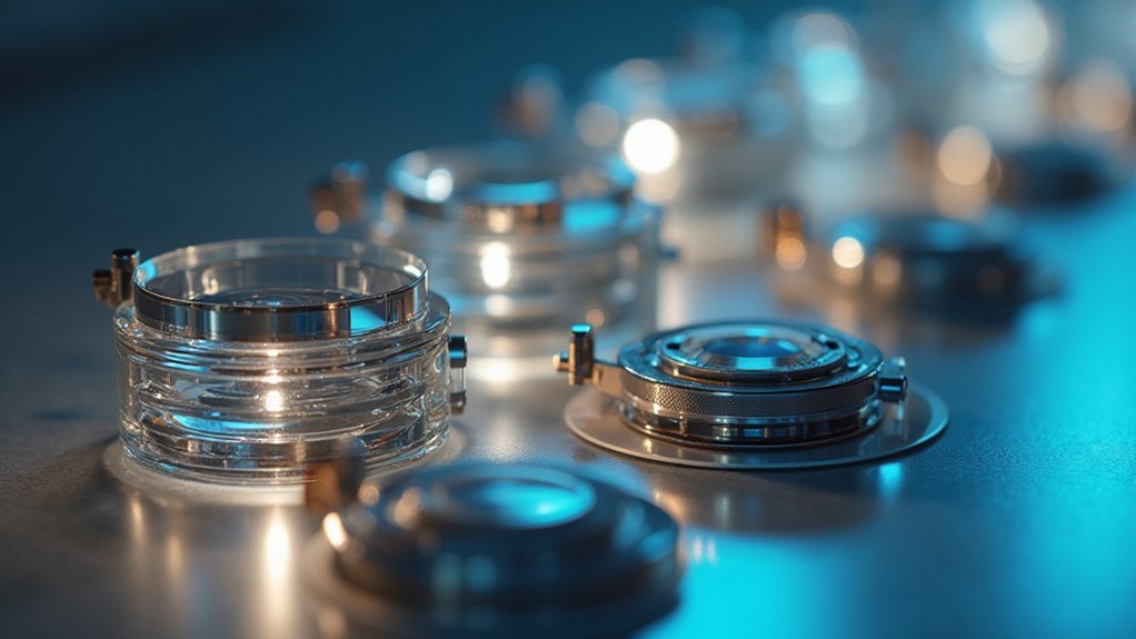
Because light utilization determines image quality in phase contrast microscopy, optimizing transmission efficiency through your adapter system becomes vital for successful imaging. High-quality adapters with pre-centered phase contrast sliders eliminate constant re-centering, maintaining consistent light pathways critical for visualizing transparent specimens.
| Magnification | Light Transmission Feature | Impact on Imaging Results |
|---|---|---|
| 10x | Standardized ring slit design | Enhanced detail visibility |
| 20x | Optimized light path geometry | Improved contrast in live cells |
| 40x | Maximum light utilization | Higher resolution of fine structures |
The optical quality of your camera adapters directly influences phase contrast imaging outcomes. When selecting adapters, prioritize those designed specifically for phase contrast imaging with superior light transmission efficiency. This guarantees you’ll capture the subtle details in transparent specimens that would otherwise remain invisible with standard brightfield techniques.
Budget-Friendly Adapter Options for Research Laboratories
While ideal light transmission remains a key consideration, research laboratories often face budget constraints when acquiring phase contrast imaging equipment.
The AmScope CA-NIK-SLR Nikon SLR/DSLR Camera Adapter offers a cost-effective solution at $400, providing 2X magnification for phase contrast imaging. This versatile adapter accommodates multiple mounting sizes (23.2mm, 30mm, 30.5mm), making it compatible with various microscope types.
You’ll find satisfactory image quality for routine phase contrast applications, though some users report chromatic aberration issues that require optimizing your camera settings.
AmScope’s online configurator helps you select the most appropriate camera adapter for your specific setup, ensuring compatibility and performance.
For even tighter budgets, consider exploring used or refurbished adapter options that can deliver adequate results while maximizing your research funding.
Advanced Adapter Systems for Fluorescence and Phase Contrast Imaging
Since research often requires both fluorescence and phase contrast techniques, advanced adapter systems now integrate capabilities for both imaging modalities.
These specialized microscope adapters feature pre-centered phase contrast sliders that eliminate re-centering, making your workflow more efficient.
Streamlined imaging without the hassle—pre-centered sliders mean less setup time and more research productivity.
You’ll appreciate the standardized ring slits for 10x, 20x, and 40x magnifications, which optimize light transmission and enhance image clarity during phase contrast imaging.
These LM microscope adapters guarantee minimal distortion while supporting both DSLR and mirrorless cameras for fluorescence observation.
The modular design of these advanced adapter systems future-proofs your investment, allowing you to upgrade components as imaging technology evolves.
This adaptability guarantees you’ll maintain cutting-edge capabilities for both fluorescence and phase contrast imaging without replacing your entire system.
Troubleshooting Common Adapter Issues in Phase Contrast Photography
Four common issues can derail your phase contrast imaging results even with high-quality camera adapters.
First, verify adapter-slider compatibility, as misalignment dramatically reduces image quality and contrast.
Second, routinely inspect optical surfaces for dust and debris that introduce unwanted artifacts in your images.
Third, adjust your camera’s exposure time and aperture settings to compensate for phase contrast’s unique lighting demands. You’ll need to experiment with these settings as each specimen requires different parameters.
Fourth, switch to manual focus instead of relying on auto-focus features that struggle with phase contrast’s subtle gradients.
If your images remain dark or lack detail, try different magnification objectives and adjust your condenser position.
Proper troubleshooting of these common adapter issues guarantees clearer, more accurate phase contrast photography results.
Frequently Asked Questions
What Is a C-Mount Adapter?
A C-mount adapter is a device you’ll use to connect your digital camera to a microscope. It has standardized 1″-32 threading and contains optics that project microscope images onto your camera’s sensor.
What Type of Adapter Is Useful for Attaching Steel Cameras to the Microscope?
For attaching steel cameras to your microscope, you’ll need a C-Mount adapter. It connects directly to the microscope’s trinocular head or photo port, ensuring proper alignment for high-quality phase contrast imaging.
How Do You Attach a Camera to a Microscope?
You’ll need to secure a compatible camera adapter between your camera and microscope. Make certain it fits your camera’s mount and the microscope’s eyepiece or trinocular port for a stable, clear imaging connection.
What Is a Microscope Adapter?
A microscope adapter is a device you’ll use to connect your camera to a microscope. It’ll guarantee proper alignment between your camera’s sensor and the microscope’s optics, allowing you to capture clear specimen images.
In Summary
You’ll need to match your adapter to both your microscope and camera specifications for ideal phase contrast results. Consider your sensor size, resolution requirements, and budget constraints when selecting between C-mount, trinocular, or specialized adapters. Don’t overlook light transmission efficiency—it’s essential for capturing the subtle contrast differences that make phase contrast imaging so valuable in cellular visualization.


Leave a Reply