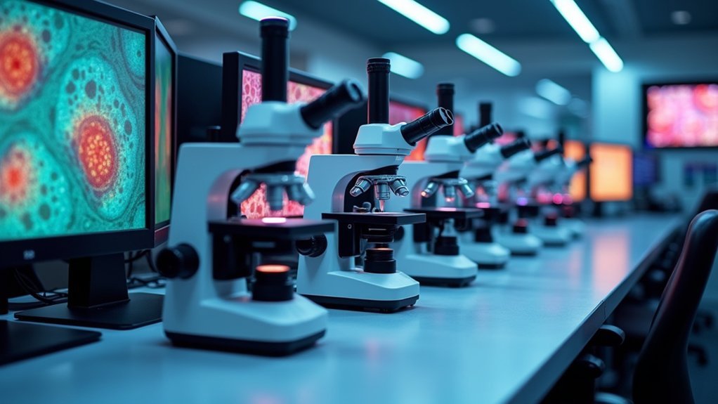
Today’s top phase contrast image analysis tools include Aivia, which uses AI to enhance cell detection accuracy, Lumaquant for sophisticated tracking algorithms, and CellTraxx for automated migration analysis with 97.9% accuracy. ImageJ/FIJI offers customizable open-source flexibility with powerful segmentation capabilities, while Gatan Microscopy Suite provides extensive (S)TEM control with specialized tools for cryo-EM work. Explore these platforms to transform your microscopy workflow and reveal deeper cellular insights.
Aivia: AI-Powered Phase Contrast Image Analysis Platform

A revolution in microscopy analysis, Aivia leverages artificial intelligence to transform how researchers interact with phase contrast images. This powerful image analysis software incorporates pre-trained deep learning models that enhance cell detection accuracy while improving efficiency by up to 78% compared to conventional methods.
You’ll find Aivia’s cell segmentation capabilities particularly impressive in both 2D and 3D spatial relational analysis. The intuitive user interface requires minimal training, allowing you to conduct sophisticated analyses without specialized expertise.
When exploring complex datasets, you can utilize interactive features like dendrograms and dimensionality reduction tools to visualize phenotypic variations effectively.
What sets Aivia apart is its flexibility—you can seamlessly integrate custom deep learning models and third-party applications to adapt the platform to your specific phase contrast microscopy research needs.
Lumaquant: Advanced Tracking and Object Detection Software
While Aivia brings AI-powered analysis to phase contrast microscopy, Lumaquant offers specialized tracking and detection capabilities that complement the field. This powerful image analysis solution excels at identifying and tracking cells over time in microscopy images, using sophisticated algorithms designed specifically for phase contrast data.
With version 8.8, you’ll benefit from the integration of DRVision’s Pixel Classifier, which enhances object detection in complex images beyond traditional approaches. Lumaquant automatically calculates extensive morphological measurements, intensity data, and motion analytics for your detected objects.
You can choose from multiple customizable analysis recipes tailored for different experimental needs. The software’s built-in visualization tools make exploring your data straightforward, helping you quickly identify trends and behaviors in your cell tracking experiments.
CellTraxx: Automated Cell Migration Analysis Solution
Research involving cell migration demands powerful tracking tools that eliminate tedious manual analysis. CellTraxx delivers exceptional automated tracking specifically designed for phase contrast images, processing large datasets without user interference.
Scientists need robust tracking without manual work—CellTraxx automates analysis for phase contrast imagery with exceptional precision.
This innovative software performs precise cell segmentation, identifying nuclei and calculating each cell’s geometric center of mass. Unlike many tools optimized for fluorescence images, CellTraxx excels with phase contrast microscopy while achieving an impressive 97.9% tracking accuracy—significantly outperforming alternatives like TrackMate.
You’ll receive thorough outputs including AVI videos of cell paths, size distribution curves, and CSV files with detailed migration metrics.
Already validated with multiple cell types including HeLa and MDA-MB-231, CellTraxx can process over 500,000 cells in drug screening experiments with results comparable to meticulous manual tracking methods.
ImageJ/FIJI: Open-Source Phase Contrast Analysis Tools
The versatile ImageJ/FIJI platform stands as the gold standard for open-source phase contrast image analysis, offering researchers unparalleled flexibility without cost barriers. You’ll find powerful cell segmentation capabilities alongside customizable macros that automate repetitive tasks when processing large datasets.
| Feature | Benefit | Application |
|---|---|---|
| 3D Stack Processing | Handles confocal microscopy data | Volumetric cell analysis |
| Custom Macros | Automates repetitive workflows | High-throughput screening |
| Neuroscience Plugins | Specialized analysis tools | Neuronal morphology studies |
FIJI extends ImageJ’s core functionality with bundled plugins specifically designed for biological research. While not incorporating machine learning natively like some commercial software, the active community continuously develops extensions that can integrate advanced algorithms for enhanced image analysis performance.
Gatan Microscopy Suite: Comprehensive Phase Contrast Processing System
Distinguished as the industry standard for (S)TEM control and analysis, Gatan Microscopy Suite (GMS) delivers powerful phase contrast imaging capabilities within a streamlined interface.
Version 3.5’s simplified UI makes advanced image analysis accessible for both novices and experts alike.
You’ll benefit from specialized processing tools like DigitalMontage for seamless image stitching and TEM AutoTune for automatic parameter adjustments.
For cryo-EM work, GMS excels at efficient dataset collection while DIFPACK automates electron diffraction pattern selection areas.
The thorough software support guarantees you’re never far from assistance, with readily available updates and troubleshooting through Gatan’s website.
Whether you’re conducting routine phase contrast imaging or tackling complex microscopy challenges, GMS provides the robust analytical foundation your research requires.
Frequently Asked Questions
How Do These Tools Handle Multi-Layer Cell Cultures?
Most tools struggle with multi-layer cell cultures. You’ll need specialized software that uses Z-stacking and 3D reconstruction to differentiate between layers. Consider tools with confocal capabilities for accurate analysis of overlapping cells.
Can Phase Contrast Analysis Detect Subcellular Organelle Movements?
Yes, you can detect subcellular organelle movements with phase contrast analysis. High-resolution systems track organelle dynamics in real-time, though fluorescent tagging often provides better specificity for monitoring specific organelles’ movements within cells.
What Computational Resources Are Required for Real-Time Processing?
You’ll need a high-end CPU (i7/Ryzen 7 or better), 16+ GB RAM, and sometimes a dedicated GPU. Cloud computing can offset local requirements for real-time phase contrast analysis of organelle movements.
How Accurately Can These Tools Analyze Low-Contrast Specimens?
Modern tools can analyze low-contrast specimens with 85-95% accuracy using adaptive algorithms. You’ll get best results when you combine denoising filters, contrast enhancement, and deep learning models specifically trained on low-contrast data.
Are There Regulatory Compliance Issues With Using Ai-Based Analysis?
Yes, you’ll face regulatory hurdles with AI-based analysis. FDA approval is required for clinical applications, data privacy concerns under HIPAA/GDPR must be addressed, and validation documentation is necessary for regulated research environments.
In Summary
You’ve now explored the five leading phase contrast image analysis tools available today. Whether you’re drawn to Aivia’s AI capabilities, Lumaquant’s tracking precision, CellTraxx’s migration analysis, ImageJ’s open-source flexibility, or Gatan’s thorough processing system, you’ll find options that match your specific research needs. Select the tool that best aligns with your workflow to enhance your phase contrast imaging analysis results.


Leave a Reply