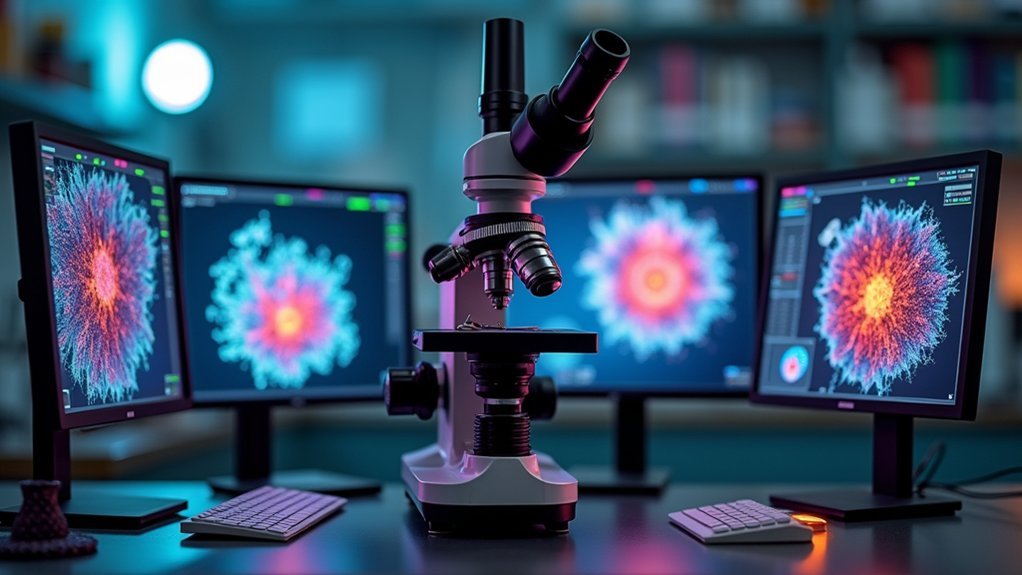
The top 5 phase contrast image analysis tools offer specialized capabilities for your microscopy needs. Aivia utilizes deep learning for enhanced accuracy, while CellTraxx excels at automated cell tracking with 98% accuracy. Lumaquant focuses on precise motility pattern tracking, ImageJ/FIJI provides cost-effective open-source solutions, and Gatan Microscopy Suite delivers professional-grade processing. Each tool combines powerful analysis capabilities with user-friendly interfaces to transform your complex microscopy data into meaningful results.
Aivia: Ai-Powered Phase Contrast Analysis With Deep Learning Models
Powerhouse of artificial intelligence, Aivia revolutionizes phase contrast image analysis through its sophisticated deep learning architecture.
You’ll benefit from greatly accelerated cell detection with enhanced accuracy when processing complex datasets that traditional methods struggle with.
This image analysis software excels in both 2D and 3D visualization, allowing you to explore cellular interactions across varied morphologies.
With 22 applications and 20 pre-trained deep learning models, you can customize analysis pipelines for your specific research requirements.
Aivia’s AI-driven cell segmentation capabilities overcome common phase contrast challenges, delivering robust object tracking and quantification.
Even without specialist expertise, you’ll quickly master the intuitive interface, enabling advanced analyses right in your laboratory.
CellTraxx: Automated Cell Tracking and Migration Analysis
While Aivia harnesses AI for broad image analysis needs, CellTraxx offers specialized automation for cell tracking in phase contrast microscopy. This powerful tool stands out among image analysis solutions by accurately segmenting and identifying nuclei without user intervention, even when fluorescence images aren’t available.
You’ll appreciate CellTraxx’s nearest neighbor approach and track-repair algorithm, which achieve impressive 98% tracking accuracy compared to gold standards. The software has proven its reliability by successfully tracking over 500,000 cells in drug screens, delivering results comparable to manual methods.
CellTraxx excels at measuring migration velocity across extensive datasets, generating thorough outputs including videos, images, and CSV files with detailed quantifications. Its nuclear-like segmentation minimizes track noise, outperforming alternatives like CP + TM, especially with challenging phase contrast imagery.
Lumaquant: Advanced Object Detection for Live Cell Imaging
Unlike broader analysis platforms, Lumaquant specializes in addressing the unique challenges of live cell imaging through phase contrast microscopy. You’ll benefit from its powerful image processing algorithms that excel in complex imaging scenarios where traditional methods fail.
| Feature | Benefit |
|---|---|
| Advanced object detection | Tracks motility patterns over time with high precision |
| Machine learning integration | Distinguishes objects accurately using Pixel Classifier |
| Automatic measurements | Calculates morphological, intensity, and lineage metrics |
| User-friendly interface | Visualizes data with built-in interactive charts |
| Thorough analysis | Reveals trends in cell behavior across time points |
Lumaquant’s specialized capabilities make it invaluable for researchers who need detailed analysis of live cell dynamics. The software’s ability to process challenging datasets while providing intuitive visualization tools streamlines your workflow from acquisition to analysis.
ImageJ/FIJI: Open-Source Solutions for Phase Contrast Quantification
ImageJ/FIJI stands as the cornerstone of open-source phase contrast image analysis, offering researchers a cost-effective alternative to proprietary solutions like Lumaquant. This versatile software provides powerful processing capabilities for quantifying cellular structures without requiring extensive coding knowledge.
When you’re analyzing phase contrast images of cells, ImageJ excels with:
- Built-in tools for brightness/contrast adjustment, cropping, and labeling that streamline your workflow
- Advanced plugins specifically designed for neuroscience applications and cell biology analysis
- Robust support for 3D confocal microscopy stacks, enabling thorough quantitative measurements
You’ll appreciate the abundance of online tutorials that make learning the analysis platform accessible regardless of your experience level. The software’s flexibility allows you to perform complex quantification tasks while maintaining an intuitive interface, making it a staple in research laboratories worldwide.
Gatan Microscopy Suite: Professional-Grade Phase Contrast Processing
Powerhouse of professional microscopy, Gatan Microscopy Suite (GMS) represents the gold standard for researchers needing sophisticated phase contrast image processing capabilities.
The newly updated version 3.5 offers you a streamlined interface that’s accessible whether you’re a novice or expert user.
Experience effortless microscopy with GMS 3.5’s intuitive interface that welcomes users at all skill levels.
You’ll find extensive support for advanced phase contrast imaging techniques, including in-situ and diffraction imaging analysis. GMS’s professional-grade tools like TEM AutoTune automatically adjust settings for ideal contrast, while DigitalMontage enables seamless stitching of complex images.
What sets GMS apart are its specialized analysis tools such as DIFPACK, which automates selection of electron diffraction patterns.
This industry-standard software delivers everything you need for thorough phase contrast image processing, from acquisition to final analysis, all within one integrated platform.
Frequently Asked Questions
How Do Phase Contrast Tools Handle Overlapping Cells During Tracking?
Phase contrast tools handle overlapping cells during tracking by using segmentation algorithms, unique cell identifiers, and predictive modeling. You’ll find they often employ machine learning to distinguish individual cells despite overlap challenges.
Can These Tools Integrate Data From Fluorescent and Phase Contrast Images?
Yes, you’ll find most modern tools can integrate phase contrast with fluorescence data. They’ll align and overlay channels, allowing you to track morphology while simultaneously measuring protein expression or other fluorescent markers in the same cells.
What Hardware Specifications Are Needed for Real-Time Phase Contrast Analysis?
For real-time phase contrast analysis, you’ll need a multi-core processor (i7/Ryzen 7+), 16GB+ RAM, dedicated GPU with 4GB+ VRAM, and SSD storage. High-speed camera interfaces like USB 3.0 or PCIe are also essential.
How Do Different Cell Types Affect Analysis Accuracy?
Cell types with distinct morphologies (neurons, fibroblasts) yield higher accuracy than those with similar appearances. You’ll need specialized algorithms for specific cell types, as thickness, density, and translucency directly impact analytical precision.
Are Cloud-Based Processing Options Available for Large Datasets?
Yes, you’ll find several cloud-based solutions for processing large phase contrast datasets. Services like Amazon AWS, Google Cloud, and specialized platforms like CellProfiler Analyst offer scalable computing power without local hardware limitations.
In Summary
You’ve discovered five powerful tools that’ll transform your phase contrast image analysis workflow. Whether you’re leveraging Aivia’s AI capabilities, tracking cell movements with CellTraxx, detecting objects via Lumaquant, utilizing ImageJ’s open-source flexibility, or processing with Gatan’s professional suite, you’re now equipped to extract meaningful data from your microscopy images. Choose the solution that best fits your specific research needs and technical requirements.


Leave a Reply