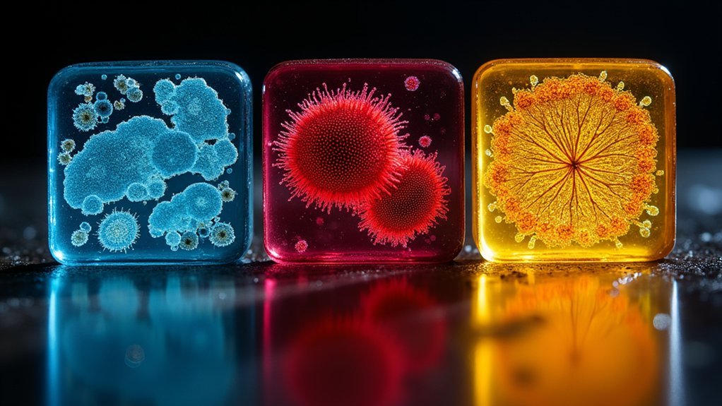
The three best negative stains for revealing cell structure are uranyl acetate, phosphotungstic acid (PTA), and ammonium molybdate. Uranyl acetate remains the gold standard for electron microscopy, providing exceptional contrast despite safety concerns. PTA offers versatility across bacteria, viruses, and animal cells without requiring heat fixation. Ammonium molybdate serves as a gentle alternative for fragile structures, preserving their native state. Each stain reveals unique cellular details that can transform your understanding of microscopic architecture.
Uranyl Acetate – The Gold Standard for Electron Microscopy
Since its introduction in the late 1950s, uranyl acetate has established itself as the gold standard for negative staining in electron microscopy. When you’re seeking high contrast images of cellular structures, this heavy metal salt delivers exceptional results by binding to macromolecular complexes while creating a dark background that highlights unstained components.
For ideal results in transmission electron microscopy (TEM), you’ll want to use concentrations between 1% and 2%, adjusting based on your specific sample. Uranyl acetate’s excellent electron density makes it invaluable for visualizing viruses, proteins, and organelles with remarkable clarity.
Remember that despite its imaging advantages, you must implement proper safety protocols when handling this reagent due to its radioactivity and potential toxicity. The superior visualization it provides continues to make it worth these precautionary measures.
Phosphotungstic Acid – Versatile Stain for Diverse Cell Types
A popular alternative to uranyl acetate, phosphotungstic acid (PTA) offers exceptional versatility for negative staining across numerous cell types. When you’re examining cell structure via transmission electron microscopy, PTA’s high electron density provides excellent contrast, effectively outlining cell membranes and cytoskeletal components.
| Cell Type | Structure Visualized | PTA Advantage |
|---|---|---|
| Bacteria | Flagella, membranes | No heat fixation |
| Viruses | Capsid proteins | Enhanced resolution |
| Animal | Organelles, cytoskeleton | Membrane preservation |
| Plant | Cell walls, organelles | Fine detail clarity |
You’ll find the staining procedure remarkably simple – apply diluted PTA solution to your specimen, then air dry. This approach preserves cellular dynamics and architectural integrity without complex preparation steps, making it invaluable for studying delicate biological structures across plant, animal, and microbial specimens.
Ammonium Molybdate – Gentle Alternative for Delicate Structures
When examining fragile cellular structures that can’t withstand harsh staining procedures, ammonium molybdate offers an exceptional solution. This gentle negative stain surrounds your specimen with an electron-absorbing layer, creating high contrast that clearly outlines cell structures against a dark background in electron microscopy.
You’ll find ammonium molybdate particularly valuable for visualizing macromolecular complexes, proteins, and nucleic acids. Its low toxicity and minimal interaction with samples preserve the native state of delicate structures during imaging.
Unlike aggressive stains that might distort morphology, ammonium molybdate respects the integrity of your specimens.
Gentle preservation meets powerful visualization—ammonium molybdate reveals true cellular architecture without compromising structural integrity.
For ideal results, you’ll need to determine the appropriate staining concentration and incubation time. This prevents over-staining while ensuring clear visualization of your biological samples’ finest details.
Frequently Asked Questions
What Is the Best Use of a Negative Stain Is to Determine?
You’ll best use negative stains to determine size, shape, and surface morphology of microorganisms against a dark background. They’re excellent for visualizing structures like capsules and flagella with exceptional clarity and detail.
What Cell Structure Does a Negative Stain Show You?
Negative stains show you cell outlines, surface structures, flagella, pili, and capsules. You’ll clearly see bacterial morphology, viral particles, and the actin cytoskeleton with high contrast against the dark background.
Which Common Stain Is Best Used to Identify the Cell Membrane?
For identifying cell membranes, you’ll get excellent results with phosphotungstic acid (PTA). It creates a dark background that makes unstained membranes appear clear with sharp boundaries, providing superior membrane visualization compared to other negative stains.
Which Stains Are Used to Visualize Structures in the Cell Wall?
For visualizing cell wall structures, you’ll find phosphotungstic acid (PTA), uranyl acetate, and ammonium molybdate most effective. They’re negative stains that surround but don’t penetrate cell walls, providing high contrast and revealing detailed morphological features.
In Summary
You’ve now learned about uranyl acetate, phosphotungstic acid, and ammonium molybdate, the three most effective negative stains for cellular visualization. Each offers unique advantages: uranyl acetate delivers exceptional contrast, phosphotungstic acid works across various cell types, and ammonium molybdate preserves delicate structures. Choose the stain that matches your specific specimen and research goals. With these tools, you’ll reveal cellular details that would otherwise remain invisible.


Leave a Reply