Negative phase contrast imaging offers seven key applications: observing live cell dynamics, analyzing cellular morphology without stains, monitoring cell division patterns, tracking microbes in environmental samples, enhancing diagnostic microscopy, studying cell-to-cell interactions, and visualizing structures in transparent specimens. You’ll benefit from its ability to reveal details invisible to conventional microscopy while maintaining cell viability during observation. Discover how this technique transforms biological research by providing high-contrast visualization of otherwise challenging-to-see cellular features.
Live Cell Dynamics: Capturing Cellular Movement in Real Time

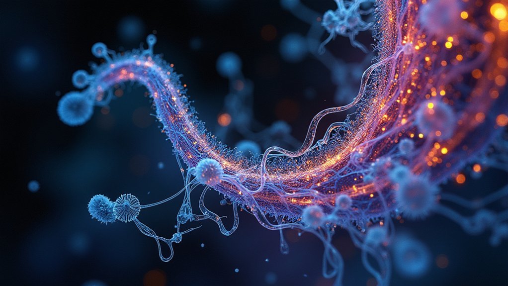
When observing living cells in their natural state, negative phase contrast imaging stands out as a powerful technique that illuminates cellular structures against a darker background.
You’ll see live cell dynamics with exceptional clarity as this method enhances the visibility of transparent biological specimens without staining.
This approach allows you to track real-time processes including cell migration, division, and interactions with surrounding environments. By manipulating phase shifts, you can detect subtle morphological changes that might otherwise remain invisible.
It’s particularly effective for smaller cells and microorganisms that conventional microscopy struggles to reveal.
Combining negative phase contrast with time-lapse microscopy creates powerful visualization opportunities, enabling you to monitor cellular responses to environmental changes or treatments over extended periods—all while maintaining cell viability.
Cellular Morphology Analysis Without Staining
Because traditional staining methods can alter cellular characteristics or even kill specimens, negative phase contrast imaging offers a revolutionary approach to morphological analysis.
You’ll observe transparent structures against a dark background, where phase shifts highlight cellular edges and internal components with remarkable clarity.
- Examine live cells in their natural state by leveraging differences in refractive indices to distinguish between cell types
- Identify morphological differences that indicate various functional states without disrupting cellular processes
- Detect subtle abnormalities that might signal disease development before they’re visible with conventional methods
- Track structural changes in real time, allowing you to observe how cells respond to environmental stimuli
This technique transforms your ability to study cellular morphology and potential disease identification while maintaining specimen integrity—something impossible with traditional staining approaches.
Monitoring Cell Division and Growth Patterns

While cells naturally proceed through their life cycles, negative phase contrast imaging uniquely captures these dynamic processes with exceptional clarity. You’ll observe mitosis and cytokinesis in real time as the technique highlights differences in refractive indices between dividing cells and their surrounding medium.
| Application | Benefit | Research Impact |
|---|---|---|
| Live cell imaging | No staining required | Preserves natural cell behavior |
| Growth pattern tracking | Reveals spatial organization | Informs developmental biology |
| Cell proliferation analysis | Quantifies division rates | Advances cancer therapy approaches |
This method lets you monitor morphological changes as cells proliferate under various conditions. You can track how different cell types—from stem cells to cancer lines—organize and multiply, providing essential insights into growth patterns. The non-invasive nature makes negative phase contrast imaging invaluable for longitudinal studies where preserving cell integrity matters.
Microbial Tracking in Environmental Samples
Environmental scientists rely on negative phase contrast imaging to reveal the hidden world of microorganisms that traditional microscopy often misses.
You’ll find this technique invaluable when tracking microbial populations in complex environmental samples, as it enhances visibility without staining that could alter their natural state.
- Notably improves detection of low-abundance microorganisms against darker backgrounds in soil and water matrices
- Enables real-time observation of motile microbes, revealing their movement patterns and behavioral responses
- Helps monitor microbial population dynamics as they respond to changing environmental conditions like nutrient availability
- Facilitates assessment of microorganisms’ contributions to biogeochemical cycles, including nitrogen fixation and organic matter decomposition
Clinical Applications in Diagnostic Microscopy
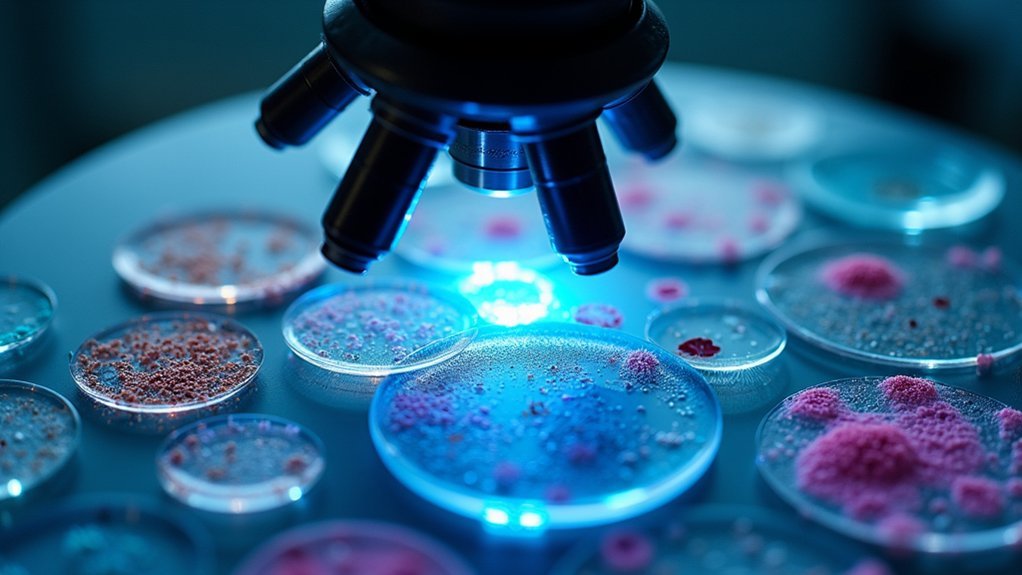
Negative phase contrast microscopy revolutionizes your clinical diagnostic capabilities through enhanced visualization of pathogens in bodily fluids without staining requirements.
You’ll find this technique particularly valuable when examining cytology specimens, where cellular details and abnormalities become readily apparent against the contrasting background.
The method’s ability to clearly delineate tissue margins during intraoperative assessment helps you achieve more precise surgical interventions and improve patient outcomes.
Pathogen Detection Analysis
Clinical laboratories rely extensively on negative phase contrast imaging for pathogen detection due to its remarkable ability to enhance visibility of microorganisms against darker backgrounds.
When examining clinical samples, you’ll find this technique particularly valuable for identifying transparent specimens like Mycobacterium tuberculosis in sputum samples without staining, preserving their natural morphology for accurate analysis.
- Visualize live pathogens with high-contrast images, maintaining their viability for further testing
- Examine complex biofilms and microbial communities with exceptional detail
- Detect low-contrast organisms that might be obscured by conventional staining methods
- Perform rapid pathogen identification and antibiotic susceptibility testing with greater accuracy
This imaging technique transforms nearly invisible microorganisms into clearly defined structures, greatly improving diagnostic capabilities while preserving the specimen’s integrity for thorough analysis.
Cytology Specimen Examination
In cytology diagnostics, you’ll find negative phase contrast imaging transforming the examination of cellular specimens across numerous medical specialties. This technique enhances the visibility of cellular components without staining, allowing you to clearly differentiate cells from surrounding matrices.
When diagnosing conditions including cancers and infections, you can identify abnormalities in cell morphology by observing differences in refractive indices. You’ll appreciate how this method reveals subtle cytoplasmic structures, organelles, and inclusions often missed with traditional bright-field microscopy.
The ability to observe live cells provides essential insights into cell behavior and interactions, markedly improving diagnostic accuracy.
Studies confirm that negative phase contrast increases the visualization of fine cellular details, giving you higher contrast in cytology specimens that helps pathologists make more precise evaluations.
Tissue Margin Assessment
When evaluating surgical excisions, you’ll find negative phase contrast imaging particularly valuable for tissue margin assessment. This technique enhances visualization of soft tissue structures without staining, allowing you to clearly differentiate between healthy and cancerous cells at the edges of excised specimens.
By revealing subtle differences in refractive indices, it helps identify tumor cell infiltration into surrounding tissues—critical information for surgical decision-making.
- Improves diagnostic accuracy by reducing false negatives, especially in breast cancer and skin cancer procedures
- Reveals detailed cellular arrangements and morphology in thin tissue sections
- Maintains natural tissue appearance by eliminating staining artifacts that might alter cellular features
- Provides real-time assessment of tissue margins during surgical procedures, potentially reducing the need for secondary surgeries
Study of Cell-to-Cell Interactions and Communication

Negative phase contrast imaging revolutionizes how researchers observe cellular communication by creating a stark visual contrast between cells and their surroundings.
You’ll appreciate how this technique highlights transparent objects that would otherwise remain invisible, allowing you to track live cell interactions in real-time without staining requirements.
When studying communication dynamics, you can clearly visualize subtle changes in shape as cells engage in adhesion and migration behaviors.
The enhanced brightness of cells against darker backgrounds makes it easier to observe the formation of specialized cellular structures like junctions and synapses.
This technique proves particularly valuable when examining challenging transparent specimens such as lymphocytes and epithelial cells, providing critical insights into immune responses and tissue repair processes.
Visualizing Structural Features in Transparent Specimens
Transparent biological specimens present significant challenges for conventional microscopy, yet negative phase contrast imaging transforms these invisible structures into clearly discernible elements.
You’ll observe remarkable detail in transparent specimens without staining or fixation, preserving their natural state for accurate biological assessments.
- Cellular components like organelles and membranes become visible through enhanced differences in refractive indices.
- Live cells can be studied in real-time, allowing you to track dynamic processes such as division and movement.
- The non-invasive method increases specimen brightness while darkening the background for ideal contrast.
- Fine structural features that would remain invisible under bright-field microscopy appear with striking clarity, revealing the complex architecture of transparent specimens.
Frequently Asked Questions
What Are the Uses of Phase Contrast Microscope?
You’ll use phase contrast microscopes to view living cells without staining, observe cellular structures, track cell movement, study bacterial motility, analyze blood cells, monitor cell division, and detect subtle morphological changes.
What Are the Objectives of a DIC Microscope?
A DIC microscope’s objectives let you observe living specimens without staining, visualize 3D structures, detect subtle refractive index differences, and enhance contrast in transparent samples, preserving cell viability for biological research.
What Is the Use of Inverted Phase Contrast Microscope?
You’ll use an inverted phase contrast microscope to observe living cells in culture dishes from below. It’s perfect for monitoring cell growth, division, and motility without disturbing adherent cells in larger containers.
What Are the Advantages and Disadvantages of a Phase Contrast Microscope?
You can observe live cells without staining, seeing dynamic processes in real-time with high contrast. However, you’ll face halo artifacts, lower resolution than fluorescence methods, and difficulties with thick specimens.
In Summary
Negative phase contrast imaging empowers you to view life’s microscopic processes as they unfold. You’ll witness cells dance, divide, and communicate without disrupting them through staining. It’s revolutionizing both research and clinical diagnostics by revealing what traditional methods can’t show. Whether you’re tracking microbes or analyzing cellular structures, this technique gives you unprecedented insight into the transparent world that’s invisible to conventional microscopy.


Leave a Reply