
Negative phase contrast imaging excels at visualizing cell motility, enhancing transparent microorganism visibility, monitoring cell division, examining unstained live cells, tracking intercellular interactions, analyzing bacterial biofilms, and evaluating clinical specimens. You’ll benefit from its ability to create high-contrast images where specimens appear bright against dark backgrounds without damaging stains. This technique preserves biological integrity while revealing critical cellular details that might otherwise remain invisible to conventional microscopy techniques.
Visualizing Cell Motility and Migration Patterns
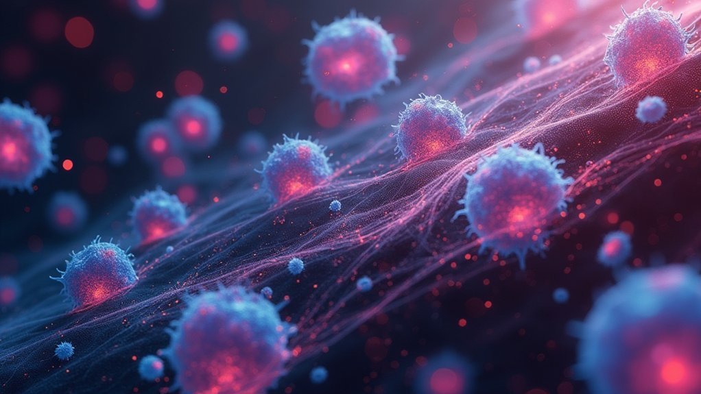
While traditional imaging techniques often struggle to capture dynamic cellular processes, negative phase contrast imaging revolutionizes how we observe cell movement.
Negative phase contrast imaging unveils the dynamic dance of cells that traditional techniques simply cannot capture.
You’ll see cell motility with exceptional clarity as this technique brightens cell bodies against darker backgrounds, making migration patterns immediately apparent.
You can track cells in real time without stains that might alter their natural behavior. This allows you to observe subtle changes in cell morphology as cells spread, interact, and respond to their environment.
The technique provides valuable quantitative data on speed and directionality of movement, which is particularly useful in cancer research where understanding cell migration offers insights into metastasis.
Enhancing Contrast in Transparent Microorganisms
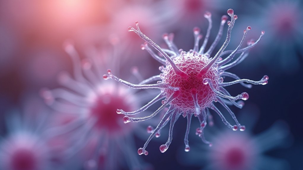
Because transparent microorganisms naturally absorb little light, they’re nearly invisible under conventional microscopy, but negative phase contrast imaging transforms these ghostly entities into clearly defined structures.
You’ll immediately notice how this technique enhances contrast by creating a high-contrast image where specimens appear bright against a darker background.
This method’s greatest advantage lies in its ability to reveal detailed features of live microorganisms without staining, preserving their natural state. The technique phase-shifts background light by -90°, making cell walls and internal structures clearly visible.
In microbiology, you’ll find this invaluable for studying bacteria and protozoa, allowing you to differentiate between species based on morphological characteristics. Additionally, the minimized halo effects provide clearer visualization of cellular interactions in aquatic environments, giving you unprecedented insights into microscopic life.
Monitoring Cellular Division Processes
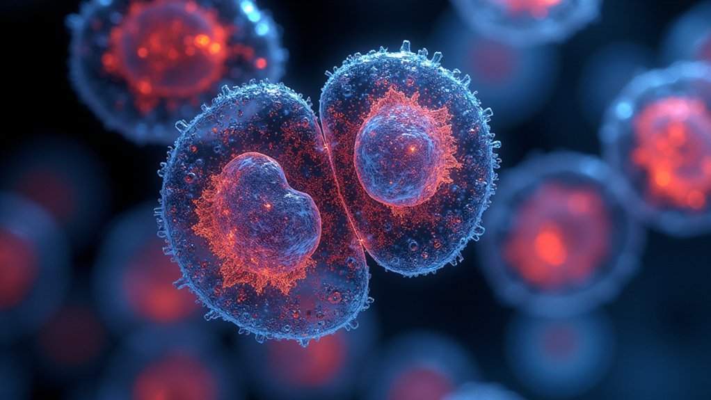
Since cellular division represents one of life’s most fundamental processes, negative phase contrast imaging offers researchers an unparalleled window into this dynamic event.
You’ll observe mitosis with remarkable clarity as this technique brightens cells against darker backgrounds, revealing subtle changes in cell morphology without disruptive staining.
The method excels at visualizing differences in refractive indices among cellular components, giving you detailed insights into mitotic spindle organization.
You can track chromosome alignment and separation in real time while eliminating background interference that might obscure vital details.
When studying cytokinesis, you’ll clearly see cleavage furrow formation and membrane invagination.
The enhanced contrast enables precise quantification of cell division phases, supporting significant cancer research by illuminating cell cycle regulation and potential therapeutic targets.
Examining Unstained Live Cell Structures
Beyond cell division analysis, negative phase contrast imaging truly shines when you’re examining living cellular structures in their native state. This technique transforms typically transparent biological specimens into clearly visible entities by shifting the background light phase, making cellular components appear bright against a darker field.
You’ll appreciate how light interference reveals delicate organelles and membranes without damaging stains. By detecting subtle differences in refractive index, you can assess specimen thickness and density with remarkable precision. In microbiology, this proves essential when you’re studying bacteria and protozoa morphology.
What sets negative phase contrast apart is your ability to observe dynamic processes in real time. You’ll witness cell division, movement, and other crucial functions while the specimen remains in its natural, unstained condition—preserving biological integrity throughout your observation.
Tracking Intercellular Interactions
When observing how cells communicate and interact with one another, negative phase contrast imaging offers unparalleled advantages for your research.
You’ll witness dynamic processes against darker backgrounds where cells appear brighter, making intercellular interactions clearly visible without disruptive staining procedures.
You can monitor cell migration and adhesion in real time, capturing the natural behavior of living specimens. This technique excels at revealing immune responses, such as interactions between immune cells and pathogens.
By observing fine details of cellular morphology as cells engage with one another, you’ll gain insights into tissue development and repair mechanisms.
The beauty of negative phase contrast lies in its ability to generate quantitative data on movement patterns and directional behaviors, enabling you to analyze complex intercellular signaling pathways with precision and accuracy.
Analyzing Bacterial Morphology and Biofilms
When you use negative phase contrast imaging, you’ll clearly see bacterial flagella movement patterns that reveal important motility behaviors without applying destructive stains.
You can assess cell wall integrity by observing the distinct contrast between cellular boundaries and their surroundings, enabling detection of structural abnormalities or damage.
This imaging technique also excels at biofilm structure analysis, showing the three-dimensional organization and revealing how bacteria interact within these complex communities.
Flagella Movement Visualization
Negative phase contrast imaging stands as an invaluable tool for microbiologists studying bacterial flagella movement. You’ll notice how this technique enhances flagella visibility without requiring stains, allowing you to observe their dynamic behavior in real time.
When analyzing bacterial motility, you can clearly see how these hair-like structures oscillate and rotate with remarkable detail. The improved resolution lets you examine how flagella interact with developing biofilms and surface adhesion processes.
This visualization technique reveals essential relationships between flagellar morphology and biofilm formation that standard imaging techniques often miss. You can also track how these structures influence antibiotic resistance in bacterial populations.
Cell Wall Integrity
Beyond flagella movement, the cellular architecture itself provides a window into bacterial health and behavior. Using negative phase contrast imaging, you’ll observe bacterial cell walls against a dark background, revealing structural details impossible to see with standard microscopy.
| Feature | Benefit |
|---|---|
| Real-time observation | Track biofilm formation dynamically |
| No staining required | View live cells in natural state |
| Enhanced contrast | Detect subtle morphological changes |
| Stress response analysis | Identify reactions to environmental stressors |
| Pathogenicity assessment | Correlate wall structure with virulence |
This technique excels when studying biofilms, where you can visualize the intricate spatial organization of microbial communities. You’ll easily assess cell wall integrity before and after antibiotic treatment, providing insights into resistance mechanisms. The method’s non-invasive nature preserves delicate bacterial structures while offering exceptional clarity for research applications.
Biofilm Structure Analysis
Through the power of negative phase contrast imaging, the complex architecture of bacterial biofilms becomes strikingly visible against darkened backgrounds.
You’ll observe dense bacterial clusters appearing darker, enhancing visibility and revealing intricate morphological characteristics within the biofilm structure.
This technique excels at distinguishing between bacterial cells and the surrounding extracellular matrix, providing clear insights into how microbial communities organize themselves.
You can track dynamic changes in bacterial adhesion and growth patterns over time, watching biofilm development unfold before your eyes.
When studying mixed species biofilms, the enhanced contrast helps you identify different bacteria based on their unique shapes.
This proves particularly valuable when evaluating treatment efficacy, as you’ll clearly see structural alterations in biofilms following antimicrobial interventions.
Assessing Clinical Specimens for Diagnostic Purposes
When examining clinical specimens for diagnostic purposes, medical professionals rely on negative phase contrast imaging to reveal critical cellular details without staining procedures.
You’ll find this technique particularly valuable when analyzing blood smears, where it helps differentiate various cell types and identify pathological changes with enhanced clarity.
In cytology examinations, you can visualize cancerous cells and assess their morphology more effectively, supporting early diagnosis and treatment planning.
The method excels at detecting infections by clearly revealing microorganisms in samples like urine or sputum without damaging the specimens.
Perhaps most impressively, negative phase contrast allows for real-time observation of live tissues, enabling you to monitor cellular structures as they respond to treatments—a significant advantage in modern clinical diagnostics.
Frequently Asked Questions
What Are the Uses of Phase Contrast Microscope?
You’ll use phase contrast microscopes to view live, unstained cells, observe cellular processes, examine blood samples, study transparent microorganisms, evaluate cell morphology, track cell motility, and monitor cellular interactions in real-time.
What Are the Objectives of a DIC Microscope?
DIC microscope objectives help you visualize transparent samples without staining. You’ll see enhanced contrast of cellular structures, 3D-like images, and fine details in live specimens through differential interference of polarized light beams.
What Is the Use of Inverted Phase Contrast Microscope?
You’ll use an inverted phase contrast microscope to observe living cells without staining. It’s perfect for monitoring cell cultures, examining morphology, and tracking cellular dynamics while easily adding reagents during long-term studies.
What Are the Advantages and Disadvantages of a Phase Contrast Microscope?
You’ll appreciate phase contrast microscopes for viewing living cells without staining. They enhance transparent specimens’ visibility, but you’ll face limitations with halo artifacts, thick specimens, and lower resolution compared to other techniques.
In Summary
Negative phase contrast imaging empowers you to see the invisible world of cells. You’ll discover detailed cell movements, observe transparent organisms, and witness division processes in real-time. It’s particularly valuable when you’re working with live specimens that can’t be stained. Whether you’re tracking bacterial interactions or making clinical diagnoses, this technique offers unparalleled clarity that conventional microscopy simply can’t match.


Leave a Reply