The 7 best multi-channel fluorescence techniques for stunning scientific images include ratiometric dual-channel imaging, spectral unmixing, FRET-based methods, multi-label immunofluorescence, live cell time-lapse imaging, fluorescence lifetime imaging microscopy (FLIM), and super-resolution multi-color microscopy. These approaches reveal complex cellular structures and interactions invisible to conventional microscopy, while eliminating artifacts and enhancing contrast. You’ll discover how these powerful techniques can transform your ordinary cell images into breathtaking scientific visualizations that capture dynamic biological processes.
Ratiometric Dual-Channel Imaging for Enhanced Contrast
While traditional fluorescence techniques often struggle with inconsistent signals, ratiometric dual-channel imaging offers a robust solution by measuring the relationship between two distinct fluorescence emissions.
You’ll find this approach eliminates artifacts from varying light intensities and photobleaching, ensuring your data accurately reflects cellular responses to stimuli.
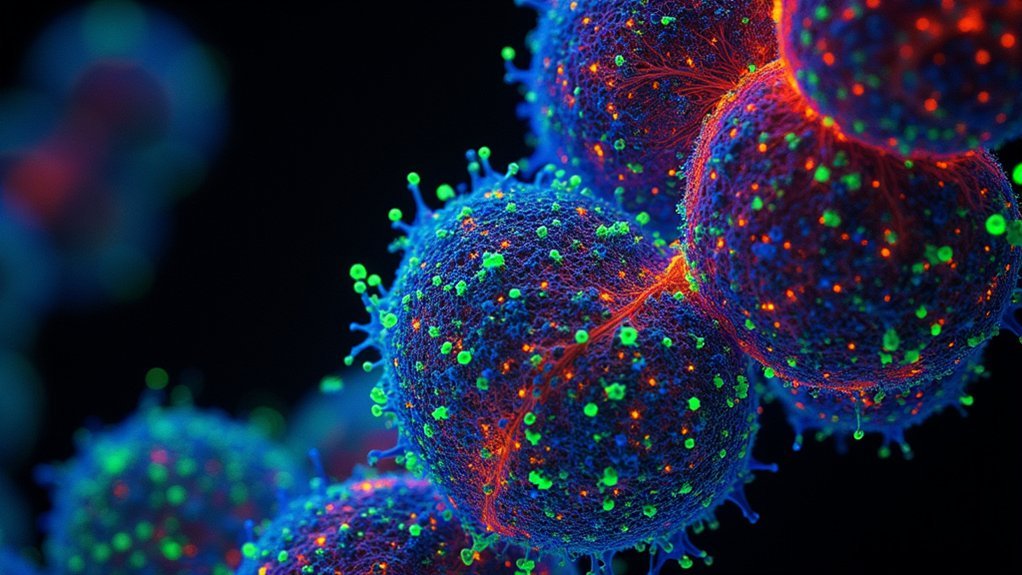
By capturing fluorescence images from two separate color channels, you’re able to visualize multiple cellular components simultaneously. This creates revealing multicolor representations that highlight spatial relationships between structures.
Dual-channel imaging unveils intricate cellular architecture by revealing spatial connections between components in vibrant multicolor detail.
When you divide the intensity values of these channels, you’ll achieve dramatically improved contrast that single-emission methods simply can’t match.
This technique proves particularly valuable when studying dynamic cellular processes like pH changes or signaling cascades, where reliable quantification makes the difference between ambiguous results and definitive insights.
Spectral Unmixing to Separate Overlapping Signals
When choosing spectral unmixing approaches for your fluorescence imaging, you’ll need to understand the difference between linear methods that assume proportional contributions from each fluorophore and non-linear techniques that account for more complex interactions.
Linear unmixing algorithms like non-negative least squares provide computational efficiency for most standard applications, while non-linear approaches offer improved accuracy for samples with significant scattering or autofluorescence.
Software platforms such as Zeiss ZEN, Leica LAS X, and open-source options like ImageJ with the PoissonNMF plugin offer varying capabilities for processing speed, user interface accessibility, and compatibility with your existing imaging systems.
Linear Versus Non-Linear Approaches
As researchers push the boundaries of multi-channel fluorescence imaging, the challenge of separating overlapping signals becomes increasingly critical. You’ll find two distinct methodologies at your disposal: linear unmixing, which uses mathematical algorithms to deconvolve signals based on known spectral profiles, and non-linear spectral unmixing, which analyzes complete emission spectra at each pixel.
| Approach | Strengths | Best Application |
|---|---|---|
| Linear | Computationally efficient | Well-separated fluorophores |
| Non-Linear | Superior with overlapping signals | Complex, crowded samples |
| Hybrid | Balances accuracy and speed | High-throughput screening |
When working with multichannel images containing closely emitting fluorophores, non-linear methods often deliver superior results by leveraging unique spectral fingerprints. You’ll need multispectral imaging systems to capture sufficient wavelength data for effective unmixing, but the improvement in image clarity makes this investment worthwhile.
Software Tools Comparison
Several leading software solutions now offer sophisticated spectral unmixing capabilities that can transform your multi-channel fluorescence imaging.
ImageJ and Fiji provide user-friendly plugins that effectively separate overlapping signals from multiple fluorescent dyes, allowing for precise quantification of distinct emissions.
For more advanced image analysis, MATLAB delivers powerful computational algorithms that handle complex unmixing calculations with exceptional accuracy.
Meanwhile, commercial platforms like Zeiss ZEN integrate spectral detectors with dedicated software to capture complete emission profiles, considerably reducing cross-talk between channels.
When working with challenging samples, consider ratiometric techniques available in these platforms to further enhance measurement reliability by normalizing overlapping signals.
Your choice between these tools should depend on your specific imaging requirements, budget constraints, and the complexity of your fluorophore combinations.
FRET-Based Techniques for Revealing Molecular Interactions
Because molecular interactions occur at nanometer scales, scientists rely on specialized methods to visualize these otherwise invisible processes. FRET technology stands out by detecting energy transfer between donor and acceptor fluorophores within 1-10 nanometers of each other—perfect for studying protein interactions and conformational changes.
When you implement FRET with multi-channel fluorescence microscopy, you’ll gain the ability to monitor multiple interaction pairs simultaneously. This approach excels in live cell applications, where you can track dynamic molecular events in real time.
For optimal results, consider ratiometric FRET measurements, which analyze the donor-to-acceptor emission ratio, effectively reducing artifacts from varying excitation intensities and photobleaching. This technique delivers reliable data on cellular processes like signaling pathways and membrane fusion events, even in complex biological environments.
Multi-Label Immunofluorescence for Structural Visualization
While single-color fluorescence provides basic information, multi-label immunofluorescence transforms your ability to visualize cellular architecture with remarkable detail.
By employing multiple fluorescent antibodies simultaneously, you’ll reveal complex biological interactions that would otherwise remain hidden.
When implementing this technique, select fluorescent dyes based on their spectral properties to minimize overlap. This careful selection guarantees each signal remains distinct in your composite image.
You’ll find ratiometric approaches particularly valuable, as they calculate intensity ratios between emissions to reduce artifacts and deliver more reliable protein localization data.
For colocalization studies, multi-label immunofluorescence excels by revealing spatial relationships between different proteins.
Your images will demonstrate how cellular structures interact, offering vital insights into biological functions through the powerful combination of targeted fluorescent markers and sophisticated imaging techniques.
Live Cell Multi-Channel Time-Lapse Imaging
Moving beyond static cellular architecture, live cell multi-channel time-lapse imaging captures the dynamic ballet of cellular processes as they unfold in real time.
To achieve thorough analysis, you’ll need 48-72 hour timelapses using systems like the Nikon Ti2 with Okolab cage incubators that maintain precise temperature and CO2 control.
Minimize phototoxicity by reducing exposure to excitation light and carefully planning your imaging frequency.
Combine phase contrast with fluorescent channels to enhance morphological insights and improve tracking. For robust visualization, use a combination of fluorescent dyes and fluorescent protein fusions, but avoid BFP variants due to their photobleaching susceptibility.
This approach creates a four-dimensional window into cellular dynamics, revealing intricate movements and interactions that static imaging simply can’t capture.
Fluorescence Lifetime Imaging Microscopy (FLIM)
The fluorescence lifetime signature represents a powerful dimension beyond simple intensity measurements, revealing essential biochemical information invisible to conventional fluorescence microscopy.
When you’re analyzing complex cellular environments, FLIM distinguishes between fluorophores with overlapping emission spectra but different lifetimes, enabling true multiple channels in crowded samples.
By measuring how long fluorescent dyes remain excited before emitting photons (typically nanoseconds), you’ll detect subtle changes in pH, viscosity, and molecular interactions.
This temporal dimension complements spatial information, making FLIM particularly valuable for protein interaction studies through FRET.
Modern FLIM systems using time-correlated single-photon counting deliver exceptional temporal resolution, allowing you to simultaneously track multiple cellular processes in living specimens—a capability traditional fluorescence microscopy simply can’t match.
Super-Resolution Multi-Color Microscopy Techniques
Breaking through the diffraction barrier, super-resolution microscopy techniques have revolutionized how you’ll visualize cellular structures at nanoscale resolution.
STED and PALM technologies achieve remarkable 20-50 nanometer precision by controlling fluorescence activation and deactivation in targeted molecules.
With multi-color microscopy capabilities, you’ll simultaneously track multiple cellular components within the same sample, providing unprecedented insights into complex biological interactions.
These techniques harness different emission wavelengths to create detailed composite images that reveal spatial relationships impossible to see with conventional microscopy.
For ideal results, you’ll need specialized equipment with environmental controls for temperature and CO2 levels.
The integration of ratiometric measurements across multiple fluorescence channels enhances your quantitative analysis reliability, making super-resolution imaging indispensable for advanced biological research requiring both spatial precision and molecular specificity.
Frequently Asked Questions
What Fluorescent Technique Can Improve Spatial Resolution?
You’ll improve spatial resolution by using multichannel microscopy, ratiometric techniques, and dual-channel imaging. With specialized filters, beam dividers, and high numerical aperture objectives, you’re capturing finer cellular details and enhancing measurement sensitivity.
What Are the Three Types of Fluorescence?
You’ll encounter three main types of fluorescence: intrinsic (natural cellular molecules), extrinsic (added dyes or probes), and fluorescent proteins (genetically encoded markers like GFP that cells produce themselves).
What Kind of Microscopy Uses Fluorescence and Images From Multiple Planes to Produce a 3D Image?
You’re looking for confocal microscopy, which uses fluorescence to capture images from multiple planes. It rejects out-of-focus light while scanning specific depths, allowing you to build detailed 3D reconstructions of biological samples.
What Is the Best Camera for Fluorescence Microscopy?
For fluorescence microscopy, you’ll want a high-sensitivity sCMOS camera with low noise, fast frame rates (30+ fps), at least 1.4 megapixels, and 12-bit+ dynamic range to capture weak fluorescent signals effectively.
In Summary
You’ve now explored seven powerful multi-channel fluorescence techniques that’ll transform your imaging capabilities. Whether you’re tracking molecular interactions with FRET or resolving intricate cellular structures with super-resolution methods, these approaches offer unprecedented visualization power. By mastering these techniques, you’ll generate stunning images that reveal biological processes with exceptional clarity and detail. Remember, the right technique depends on your specific research question and available equipment.


Leave a Reply