
Temperature fluctuations ruin your imaging results by causing optical components to expand or contract, disrupting critical alignments and changing refractive indexes. Your imaging plates experience increased dark noise and reduced sensitivity, especially above 35°C, compressing dynamic range and reducing contrast between tissue densities. These thermal changes directly impact diagnostic accuracy, making it harder to distinguish subtle details. Maintaining stable, cool storage environments below 35°C is essential for preserving the image quality your diagnoses depend on.
The Science Behind Thermal Expansion in Imaging Systems

When temperature changes occur in an environment, imaging systems experience subtle but significant physical reactions that can compromise image quality.
Temperature fluctuations cause materials in your optical components to expand or contract, disrupting carefully calibrated alignments essential for sharp imaging.
Thermal changes silently sabotage precision—as components expand and contract, your perfect optical alignments slowly drift into failure.
This thermal expansion isn’t just about physical dimensions changing—it also alters the refractive index of optical materials. As temperatures shift, light bends differently through lenses and sensors, causing wavelength shifts and focusing errors that blur your results.
For high-precision imaging, even minor temperature variations can be devastating. The repeated heating and cooling creates thermal cycling stress that eventually leads to material fatigue and potential failure of critical components.
That’s why professional imaging setups maintain strict temperature controls—they’re preserving both image quality and equipment longevity.
How Temperature Affects Image Plate Sensitivity
When you store CR image plates at temperatures above 35°C, you’ll notice sensitivity threshold shifts that compromise diagnostic accuracy.
These elevated temperatures also cause contrast range compression, making it harder to distinguish between subtle anatomical differences in your radiographic images.
Dark noise levels increase dramatically in both Fuji and AGFA systems as temperatures rise, with measurements showing significant noise elevation that directly impacts your imaging quality and clinical reliability.
Sensitivity Threshold Shifts
Although often overlooked in clinical settings, temperature fluctuations greatly alter the sensitivity thresholds of computed radiographic (CR) image plates. When temperatures exceed 35°C, you’ll notice a significant increase in dark noise levels—particularly severe in Fuji systems above 40°C and consistently problematic in AGFA systems across all tested temperature ranges.
These temperature-induced threshold shifts manifest in three critical ways:
- Higher average pixel values in regions of interest
- Decreased image clarity and diagnostic reliability
- Compromised adherence to radiographic imaging standards
Your image plates require storage below 35°C to maintain ideal sensitivity thresholds.
Without proper temperature control, diagnostic accuracy suffers as dark noise degrades image quality.
Implementing strict thermal management protocols guarantees your radiographic imaging maintains the precision required for reliable clinical assessment.
Contrast Range Compression
Temperature’s impact extends beyond sensitivity thresholds into contrast range compression—a phenomenon that fundamentally alters diagnostic image quality.
When your imaging plates experience temperature fluctuations, particularly above 40°C for Fuji CR systems, the ability to distinguish between different tissue densities diminishes.
These temperature changes compress the dynamic range of your images by elevating dark noise levels, effectively reducing the contrast between structures.
You’ll notice this as altered average pixel values in regions of interest, making subtle pathologies harder to detect. AGFA plates are especially vulnerable, showing increased noise across all tested temperature ranges.
To preserve ideal contrast, store your imaging plates below 35°C.
This simple protocol enhancement guarantees your diagnostic images maintain the clarity and distinction necessary for accurate interpretation, preventing temperature-induced contrast compression from compromising clinical decisions.
Noise Level Elevation
Despite careful calibration efforts, your imaging plates remain vulnerable to temperature-induced noise elevation that can undermine diagnostic accuracy.
Research reveals that proper temperature control is critical for maintaining ideal CR image quality.
When your storage area exceeds recommended temperatures, dark noise levels increase dramatically:
- Fuji CR plates show substantial noise increases above 40°C
- AGFA plates demonstrate sensitivity at all tested temperatures
- Average pixel values in regions of interest rise measurably with temperature
You’ll achieve best results by keeping storage temperatures below 35°C.
This simple practice preserves image plate sensitivity and prevents the degradation of diagnostic information.
Temperature fluctuations directly impact your images’ clarity and can compromise the subtle details needed for accurate interpretation.
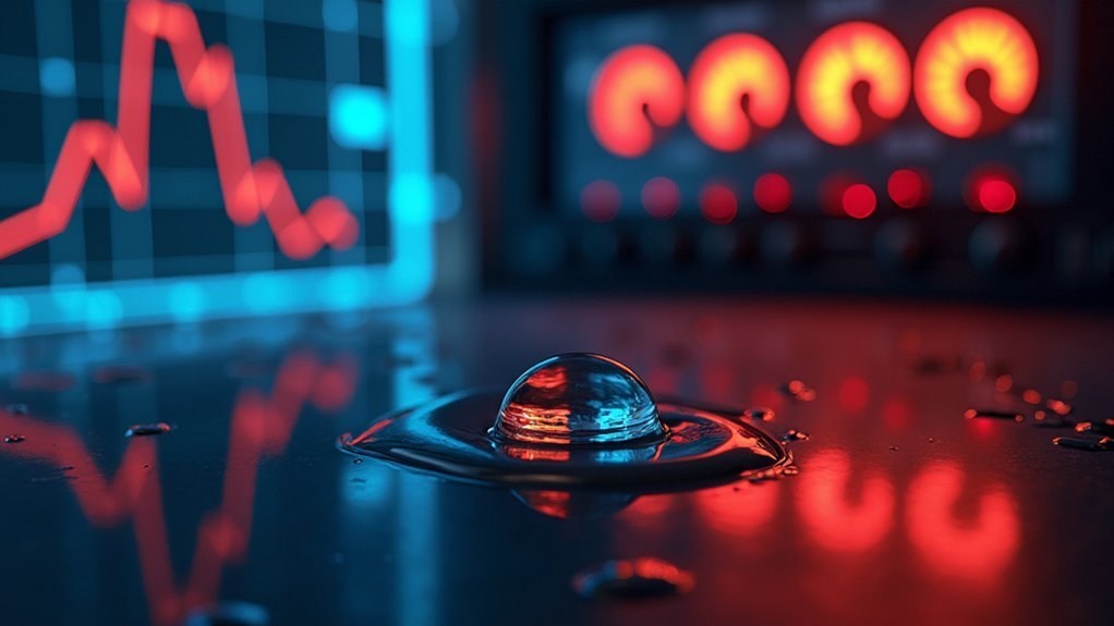
Dark Noise: The Silent Quality Killer in Medical Imaging
Precision in medical diagnostics hinges on factors that often remain invisible to practitioners. Dark noise, the electronic interference that occurs even without light exposure, can considerably compromise your imaging results when temperatures aren’t properly controlled.
| Temperature Range | Fuji CR Systems | AGFA Systems |
|---|---|---|
| Below 35°C | Minimal impact | Noticeable |
| 35-40°C | Slight increase | Moderate |
| Above 40°C | Major | Severe |
| Storage Impact | Manageable | Critical |
| Image Quality | Preserved | Compromised |
When your imaging equipment experiences temperature fluctuations, particularly above 35°C, dark noise levels rise dramatically. This invisible quality killer affects diagnostic accuracy by reducing image clarity. You’ll need to maintain storage temperatures below 35°C to guarantee peak performance and uphold imaging standards that your diagnostic work demands.
Optimal Storage Conditions for CR Image Plates
You’ll need to maintain CR image plates below 35°C to prevent dark noise from compromising image quality, as studies with Fuji systems show significant degradation above 40°C.
Your storage facility should feature consistent temperature controls, given that AGFA plates demonstrated increased noise across all tested temperature ranges.
Implementing regular monitoring systems and dedicated storage cabinets with climate control can effectively minimize these thermal threshold effects while preserving the diagnostic value of your radiographic images.
Thermal Threshold Effects
When considering the performance of computed radiography (CR) systems, temperature control emerges as a critical factor in maintaining image quality.
Research reveals that specific thermal thresholds considerably impact your imaging results, with storage temperatures above 40°C causing substantial dark noise increases in Fuji CR plates, while AGFA plates show sensitivity at all tested temperatures.
To preserve ideal image clarity and diagnostic accuracy, you’ll need to:
- Maintain storage environments below 35°C to minimize dark noise effects
- Monitor temperature fluctuations regularly, especially in warmer climates
- Implement cooling solutions in areas where equipment is stored
These thermal thresholds aren’t merely recommendations—they represent critical boundaries beyond which your imaging quality degrades measurably, affecting diagnostic reliability and potentially compromising patient care.
Noise Prevention Strategies
To effectively prevent dark noise in CR image plates, ideal storage conditions must be consistently maintained.
You’ll need to store your CR plates at temperatures below 35°C to preserve image quality and diagnostic accuracy. Research on both Fuji and AGFA systems demonstrates significant image degradation when this threshold is exceeded, with particularly severe effects above 40°C.
Monitor your storage environment regularly, as even temporary temperature spikes can introduce unwanted noise that compromises pixel value consistency across regions of interest.
Implement a dedicated storage solution with temperature regulation to maintain stable conditions year-round.
Remember that each manufacturer’s systems respond differently to thermal stress—AGFA plates showed sensitivity at all tested temperatures, indicating they may require even stricter controls than the 35°C general guideline suggests.
Comparing System Responses: Fuji vs. AGFA Temperature Tolerance
Although both systems require careful temperature management, Fuji and AGFA CR imaging systems demonstrate particularly different tolerances to temperature fluctuations.
While Fuji plates show significant dark noise increase only above 40°C, AGFA plates exhibit increased noise at all tested temperatures, making them more sensitive to even minor temperature changes.
Temperature sensitivity varies significantly – Fuji systems remain stable until 40°C while AGFA plates show sensitivity to even slight temperature shifts.
For ideal performance with your imaging equipment:
- Store all image plates below 35°C to minimize dark noise impact
- Monitor Fuji systems closely when approaching the 40°C threshold
- Exercise extra caution with AGFA systems as they’re affected at all temperature variations
Understanding these temperature tolerances helps you maintain diagnostic accuracy and prevent image quality degradation that could compromise patient care and diagnostic capabilities.
Temperature Thresholds That Trigger Image Degradation
Specific temperature thresholds directly impact the quality of diagnostic images across different CR systems.
When you store Fuji CR image plates above 40°C, you’ll notice a significant spike in dark noise levels, immediately compromising image quality and diagnostic accuracy.
AGFA systems show even less resilience, with noise increases occurring at all tested temperature points.
To preserve peak imaging performance, you should maintain storage environments below 35°C for both systems.
These temperature thresholds aren’t merely suggestions—they represent critical boundaries where image degradation becomes clinically significant.
The dark noise measurements serve as reliable indicators of when your images will start losing clarity and detail.
Practical Strategies for Temperature-Controlled Storage
You’ll need dedicated climate-controlled storage areas that maintain temperatures below 35°C to protect your computed radiographic image plates from dark noise effects.
Consider installing automated temperature monitoring systems that alert staff when conditions approach critical thresholds, preventing the quality degradation documented in both Fuji and AGFA CR systems.
Proper insulation and reliable HVAC systems represent essential investments that will safeguard your imaging equipment’s sensitivity and guarantee consistent diagnostic reliability.
Storage Environmental Considerations
When implementing temperature-controlled storage for radiographic imaging equipment, your primary goal should be maintaining consistent temperatures below 35°C to preserve image quality.
Beyond temperature monitoring, you’ll need thorough environmental controls that address related factors affecting your CR plates and systems.
- Position storage units away from heat-generating equipment – even nearby devices can create localized temperature pockets that exceed the critical 35-40°C threshold where Fuji and AGFA systems experience increased dark noise.
- Install temperature monitoring systems with automated alerts – receive notifications before conditions reach damaging levels.
- Consider humidity control alongside temperature – excessive moisture can compound temperature-related degradation and affect the material stability of your imaging plates.
Remember that temperature fluctuations, even temporary ones, can misalign optical components and permanently compromise diagnostic accuracy.
Temperature Monitoring Solutions
Proper monitoring forms the backbone of any effective temperature control strategy for radiographic storage. By implementing dedicated temperature monitoring solutions, you’ll considerably reduce the risk of dark noise and image degradation caused by fluctuations in ambient temperature.
Consider deploying data logging devices that track temperature in real-time, allowing you to make prompt adjustments before damage occurs. Regular calibration of these systems guarantees accuracy, maintaining storage conditions below the recommended 35°C threshold.
Establish standard operating procedures for routine temperature checks and document any variations to identify patterns that may compromise imaging quality.
For ideal results, invest in advanced technologies like modular climate chambers that enhance environmental stability. These measures provide consistent conditions that preserve your imaging materials’ integrity and improve diagnostic reliability.
Real-World Implications of Poor Temperature Management

Although often overlooked in clinical settings, temperature fluctuations can severely compromise diagnostic accuracy and patient care outcomes. Your imaging equipment and samples are constantly battling against thermal changes that degrade results and potentially lead to misdiagnosis.
- Increased dark noise levels occur when temperature exceeds 40°C in Fuji systems and at all temperatures in AGFA systems, directly reducing contrast and detail in radiographic images.
- Microscopy data becomes unreliable as even slight temperature variations alter enzymatic reactions and protein diffusion rates.
- Sample integrity deteriorates through evaporation and condensation, while thermal drift causes focus loss and misalignment.
Maintaining temperatures below 35°C isn’t just a recommendation—it’s essential for preserving image quality and ensuring accurate diagnostic information for your patients.
The Cost of Heat: Quantifying Image Quality Loss
The financial impact of temperature-related image degradation extends far beyond equipment maintenance costs.
Temperature-related image degradation silently erodes your bottom line while compromising diagnostic integrity.
When your CR system operates above ideal temperature thresholds, you’re sacrificing diagnostic accuracy with every degree increase.
Studies of Fuji CR systems reveal dark noise spikes dramatically above 40°C, while AGFA systems show noise increases at all tested temperatures. This translates to concrete losses: even minor temperature fluctuations of a few degrees result in substantial variations in average pixel values.
Maintaining storage environments below 35°C isn’t just a technical recommendation—it’s a financial imperative.
Each substandard image potentially requires rescans, wasting resources and extending patient wait times. More concerning is the risk of missed diagnoses when subtle pathological features become obscured by temperature-induced noise, potentially leading to costly medical errors.
Beyond Storage: Environmental Controls During Imaging
While storage conditions play an essential role in preserving imaging quality, environmental factors during the actual imaging process demand equally rigorous attention. Your results suffer when temperature fluctuations cause thermal drift, component misalignment, and altered biological reactions.
To guarantee reliable imaging outcomes:
- Implement environmental control systems like heated lids or microfluidic platforms to prevent evaporation and maintain thermal stability.
- Monitor temperature continuously throughout your imaging session, as even minor deviations can compromise focus and clarity, especially in sensitive techniques like TIRF.
- Calibrate your equipment regularly to account for seasonal variations that might affect your imaging setup’s baseline performance.
Best Practices From Clinical Settings for Temperature Management
Clinical environments have pioneered robust temperature management protocols that laboratory researchers can adopt for improved imaging outcomes. You’ll find that leading facilities maintain imaging plate storage consistently below 35°C to minimize dark noise effects that degrade image quality and compromise diagnostic accuracy.
You should implement regular temperature monitoring systems throughout your imaging suite, not just in storage areas. This prevents the optical misalignments and focus issues that thermal fluctuations cause.
Consider installing specialized temperature control systems like those used in hospitals—advanced heating elements and incubators that stabilize environmental conditions during critical examinations.
For best results, incorporate materials with low thermal sensitivity and design your systems with thermal compensation features. These adaptations greatly reduce thermal drift and guarantee your imaging data remains reliable and consistent.
Future Directions in Temperature-Resistant Imaging Technology
As researchers push the boundaries of imaging technology, next-generation systems are incorporating unprecedented thermal precision with innovations like VAHEAT, which maintains stability within 0.1°C during critical imaging procedures.
You’ll soon benefit from these emerging developments that promise to transform your imaging results:
- Temperature-stable crystal materials engineered specifically to minimize thermal expansion, ensuring consistent optical properties regardless of environmental conditions.
- Programmable temperature profiles that you can customize for sensitive experiments, particularly useful in your biological and medical imaging applications.
- New industry standards for temperature stability that will guide protocol development, helping you achieve more reliable and reproducible imaging results.
These advancements represent the collaborative success of engineers and scientists working to develop materials that perform flawlessly despite temperature variations.
Frequently Asked Questions
How Does Temperature Affect Results?
Temperature affects your results by causing misalignment in optical systems, increasing image noise, altering biological reactions, damaging optical materials, and reducing focus quality. These changes greatly compromise your imaging accuracy and reliability.
Why Are Results Less Accurate at Higher Temperatures?
Higher temperatures increase dark noise, alter refractive indices, and cause thermal cycling in your equipment. You’ll see degraded image quality, wavelength shifts, and material fatigue—all making your imaging results less reliable and accurate.
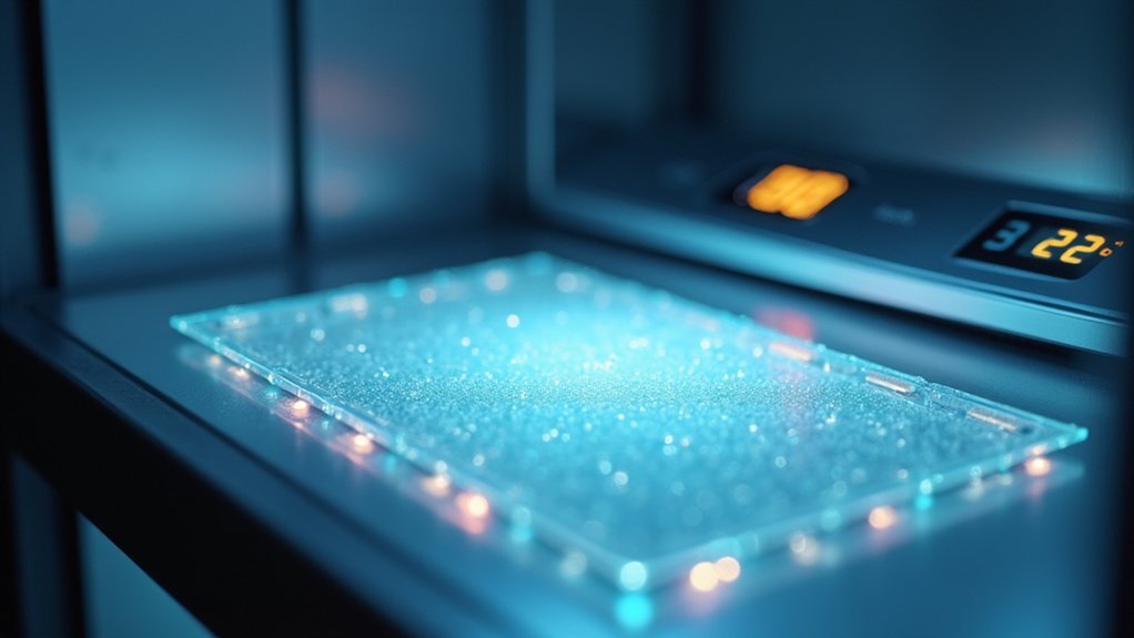
What Temperature Should a CT Scan Room Be?
You should maintain your CT scan room between 20°C to 22°C (68°F to 72°F). This temperature range guarantees ideal equipment performance, prevents thermal noise, and provides comfort for your patients during imaging procedures.
Why Does Temperature Fluctuate so Much?
Temperature fluctuates due to your building’s HVAC cycling, outdoor weather changes, equipment heat generation, door openings, and varying occupancy. These factors create thermal gradients that your environmental controls can’t always compensate for perfectly.
In Summary
You’ve seen how temperature fluctuations create thermal expansion, sensitivity shifts, and increased dark noise—all compromising your imaging quality. By maintaining consistent temperatures for your CR image plates and equipment, you’ll achieve clearer diagnostics and extend system life. Whether you’re using Fuji or AGFA systems, proper environmental controls aren’t optional—they’re essential. Remember, temperature stability isn’t just about equipment protection; it’s about ensuring reliable patient care.


Leave a Reply