For crystal-clear live specimen videos, position your key light at a 45-degree angle to enhance textures while using diffusion materials like white sheets to soften harsh light. Add a fill light at lower intensity opposite your key light to reduce shadows without flattening depth. Implement backlighting to separate specimens from backgrounds and maintain a color temperature of 5000K-6500K for accurate reproduction. These fundamental techniques form just the foundation of professional-quality microscopy illumination.
Numeric List of 9 Second-Level Headings
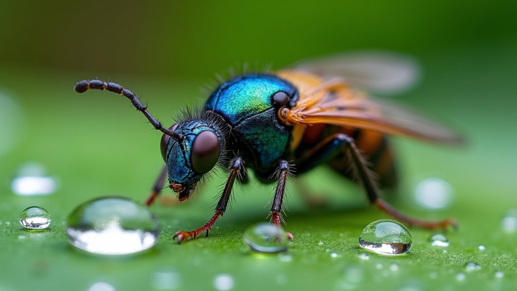
While planning your live specimen video production, you’ll need to address these nine essential lighting aspects:
- Key Light Positioning at 45-Degrees
- Diffusion Techniques for Natural Appearances
- Backlighting Methods for Specimen Separation
- Camera Distance Optimization
- Light Temperature Combinations
- Avoiding Lens Flare
- Shadow Management for Detail Enhancement
- Color Fidelity Preservation
- Background-Subject Contrast Techniques
Make sure you experiment with each heading to maximize image quality in your footage.
When working with delicate specimens, you’ll want to use a lot of diffusion to soften harsh lighting.
Position your key light carefully to create those dynamic shadows that highlight important details.
Remember that proper lighting isn’t just about brightness—it’s about creating depth, enhancing natural colors, and separating your specimen from its background for truly professional results.
Understanding Light Quality for Microscope Specimens
You’ll find that mastering diffusion techniques transforms harsh light into soft illumination that reveals delicate specimen details without creating distracting shadows.
The direction of your light sources—particularly angling your key light and properly positioning fill lights—creates the depth and dimension needed to accurately represent three-dimensional structures under the microscope.
Balancing color temperature guarantees your specimens appear natural and consistent throughout your video sequence, preventing the misleading color shifts that can compromise scientific accuracy.
Diffusion Techniques Matter
When capturing live specimens under a microscope, the difference between average and exceptional footage often comes down to how you manage your light. Proper diffusion transforms harsh illumination into soft, even light that reveals intricate specimen details while minimizing distracting shadows.
You’ll achieve better clarity by increasing your light source size. Don’t invest in expensive equipment—simply bounce light off walls or white sheets to create natural diffusion that enhances your footage quality.
| Diffusion Method | Effect on Clarity | Cost |
|---|---|---|
| Softbox | Excellent detail | High |
| White sheet | Good shadow reduction | Low |
| Bounce cards | Enhanced color accuracy | Low |
| Paper diffusers | Reduced glare | Minimal |
| Distance adjustment | Controls intensity | Free |
Experiment with different materials and distances to find the perfect diffusion for your specific specimens. You’ll immediately notice improved color accuracy and detail visibility in your microscope videos.
Light Direction Effects
Light direction represents the unsung hero in microscope videography that transforms ordinary footage into engaging scientific documentation.
When you position your light source at an angle rather than directly overhead, you’ll reveal textures and structures that might otherwise remain hidden from view.
Try side lighting to enhance the three-dimensional appearance of your specimens. This technique creates subtle shadows that accentuate contours and surface features, giving viewers a better sense of depth and form.
For transparent or semi-transparent subjects, backlighting works wonders by allowing light to pass through the specimen, highlighting internal structures and creating natural contrast.
You’ll need to experiment with different angles and intensities as each specimen responds uniquely to light direction.
Remember that diffused sources often provide more even illumination while still benefiting from directional effects.
Color Temperature Balance
Although often overlooked, color temperature dramatically affects how accurately your specimens appear in microscope videos. For ideal results, aim for 5000K-6500K to replicate natural daylight. This range reveals true specimen colors without introducing the yellowish tint of warmer temperatures (below 5000K) or the bluish cast of cooler ones (above 6500K).
Invest in color-corrected LED lights to maintain consistent color temperature throughout filming sessions. This reduces post-production color correction work while ensuring scientific accuracy.
When setting up your lighting, incorporate diffusion materials to soften harsh illumination, eliminate glare, and create even lighting across your entire specimen.
Don’t forget to harmonize your primary light source with ambient lighting in your workspace. This balanced approach prevents unwanted color casts that could misrepresent your live specimens.
Key Light Positioning for Maximum Detail Visibility
Mastering the position of your key light serves as the foundation for capturing exceptional live specimen videos.
Place your key light at a 45-degree angle to your specimen, slightly above eye level, to reveal textures while avoiding harsh shadows that can obscure important details.
Attach a softbox to your key light to diffuse illumination, reducing glare and enhancing the visibility of fine features. This softer light creates a more natural appearance while maintaining depth.
Balance is critical—adjust intensity until your specimen is well-lit without overexposing delicate structures.
Complete your setup with a fill light positioned opposite your key light at lower power. This secondary light gently fills shadows without flattening the specimen’s three-dimensional qualities, ensuring viewers can appreciate every intricate detail.
Diffusion Techniques to Eliminate Harsh Reflections
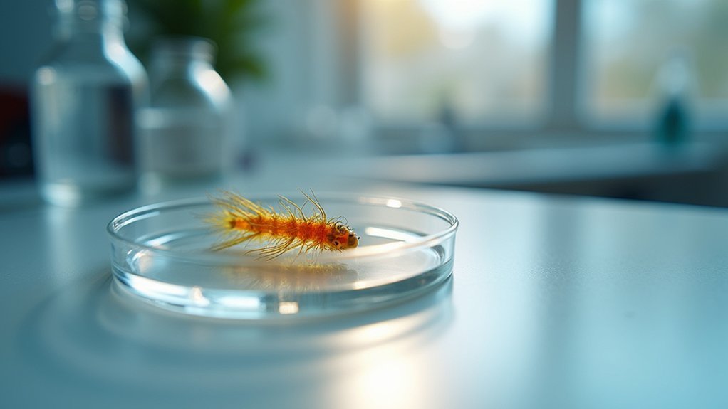
When capturing live specimens, harsh reflections can obscure critical details and diminish video quality. You’ll achieve clearer results by implementing effective diffusion techniques.
Try using larger softboxes or hanging sheer curtains to spread light evenly across your subject. For enhanced diffusion, bounce light off white surfaces to create natural illumination without direct harshness.
Light diffusion transforms harsh illumination into gentle, even coverage—revealing specimen details otherwise lost to glare and reflection.
A dual-layer diffusion setup can dramatically reduce unwanted glare—position your key light at an angle rather than directly above to minimize distracting reflections.
Remember to experiment with different distances between your light source and diffusion material for best results.
- Feel the excitement of capturing previously unseen details
- Experience the satisfaction of professional-quality specimen footage
- Enjoy the confidence of controlling every aspect of your lighting
- Marvel at the transformation diffusion makes to your video quality
- Delight in sharing crystal-clear observations with colleagues
Creating Depth With Strategic Shadow Management
While strategic lighting creates specimen depth, you’ll need to master shadow balance techniques by positioning your key light between 10-11 o’clock and a softer fill light at 9 o’clock.
These depth-enhancing light positions transform flat-looking specimens into three-dimensional subjects that reveal texture and structural details viewers might otherwise miss.
You can achieve contrast without harshness by experimenting with different lighting intensities, maintaining shadows that accentuate features while avoiding overly dark areas that obscure important details.
Shadow Balance Techniques
The artistry of shadow balance transforms flat, clinical videos into dynamic visual narratives with remarkable depth. Position your key light between 10 and 11 o’clock to create natural shadows that contour facial features while ensuring clear visibility of your specimen.
Complement this with a fill light at 9 o’clock to soften harsh contrasts without eliminating the shadows that create dimension.
Effective shadow management requires experimenting with light distances and angles. By deliberately incorporating a darker side, you’ll enhance three-dimensionality and prevent the flat, unprofessional look that plagues amateur videos.
- Dramatic side lighting that reveals texture and detail
- Subtle shadow shifts that guide the viewer’s eye
- Mysterious partial illumination that creates visual interest
- Bold contrast that emphasizes important features
- Gentle shadow patterns that add sophistication and depth
Depth-Enhancing Light Positions
Strategic positioning of your lighting setup creates the illusion of depth that transforms ordinary specimen videos into compelling visual narratives.
Position your key light at the 10 to 11 o’clock angle to cast natural shadows that define facial features and textures, avoiding the flat appearance that undermines dimension.
Add a fill light at 9 o’clock to soften these shadows without eliminating them completely—maintaining the contrast that communicates depth to viewers.
For maximum three-dimensionality, incorporate an edge light directly opposite your subject (at 180°) to create separation from the background.
Experiment with light-to-subject distance to fine-tune shadow intensity.
Remember that larger light sources produce softer, more flattering shadows.
These precise placements guarantee your specimens appear with the depth and dimension that makes scientific observations truly valuable.
Contrast Without Harshness
Creating depth in specimen videos requires mastering the delicate balance between contrast and harshness. Position your key light between 10 and 11 o’clock to create natural shadows that enhance your specimen’s features while maintaining clarity.
Add a fill light at 9 o’clock to soften shadows without flattening the subject’s dimensionality.
To achieve perfect contrast:
- Experiment with light distance—closer placement intensifies shadows, while greater distance creates gradual, more forgiving shifts
- Use larger light sources or diffusers for softer shadows that preserve detail
- Keep one side slightly darker to accentuate natural contours
- Adjust fill light intensity to control the shadow-to-highlight ratio
- Test different diffusion materials to find the perfect balance between definition and softness
Remember that shadows aren’t your enemy—they’re essential tools for creating the depth that makes your specimens appear three-dimensional and lifelike.
Three-Point Lighting Adaptations for Microscopy
Professional three-point lighting techniques can be effectively adapted for microscopy work, enhancing the clarity and detail of your live specimen videos. Position your key light at a 45-degree angle to avoid glare while providing primary illumination. Add a fill light to soften shadows and maintain detail visibility. Complete the setup with a back light angled from behind to create separation, highlighting specimen edges and adding depth.
| Light Type | Positioning | Purpose |
|---|---|---|
| Key Light | 45° angle | Primary illumination |
| Fill Light | Opposite key | Shadow reduction |
| Back Light | Behind specimen | Edge definition |
| Diffusers | On key/fill lights | Soften harsh lighting |
You’ll need to adjust intensity and positioning for different specimens. Use diffusers on your key and fill lights to prevent harsh shadows that might obscure important microscopic details.
Background Lighting Considerations for Specimen Contrast
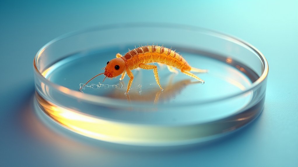
While your specimen remains the star of the show, the background lighting plays an essential supporting role in creating effective microscopy videos. A well-lit background that’s not too bright prevents distractions and maintains focus on your specimen.
Opt for darker backgrounds to enhance contrast, making your specimen pop, but watch for grain in low-light situations.
Position practical lights strategically to add dimension without overwhelming your subject. Always balance background illumination with your key light for a harmonious composition that keeps attention where it belongs.
- Dark backgrounds dramatically reveal delicate specimen structures
- Colored backgrounds evoke different emotional responses from viewers
- Perfectly balanced lighting creates professional, trustworthy results
- LED accents add excitement and visual interest to otherwise clinical footage
- Proper contrast transforms ordinary specimens into mesmerizing subjects
DIY Solutions for Professional-Quality Microscope Illumination
You don’t need expensive equipment to achieve stellar microscope lighting. With a few household items, you can transform your microscope videos from amateur to professional quality.
Let’s start with diffusion—simply stretch a white sheet over your light source to spread illumination evenly and eliminate harsh shadows.
Another one of my favorite techniques is bouncing light off nearby walls or surfaces. This creates natural, indirect lighting that enhances specimen details without introducing glare.
Position your key light at a 45-degree angle to create depth, then use a fill light (like a desk lamp) at lower intensity on the opposite side.
If you need to use additional illumination, consider arranging practical lights in the background to add depth while maintaining focus on your specimen.
Troubleshooting Common Lighting Issues in Live Specimen Capture
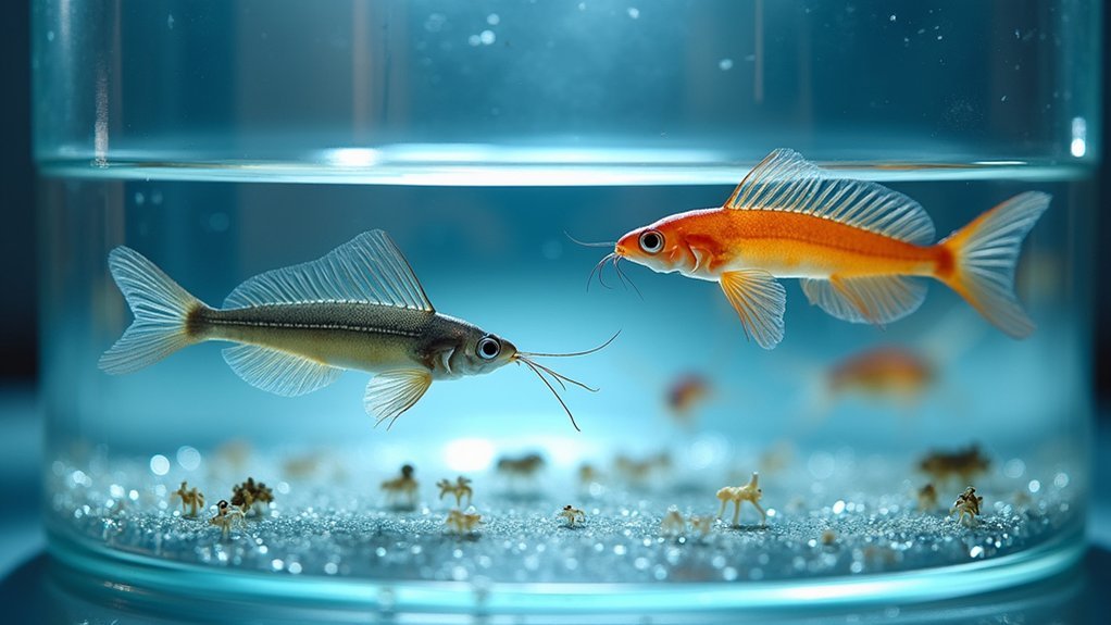
Even with careful setup, live specimen videos often encounter lighting challenges that can frustrate both beginners and experienced microscopists.
Lighting challenges plague microscopy videos regardless of experience level, turning exciting discoveries into exercises in frustration.
When you notice glare obscuring important details, try repositioning your key light at an angle—the first one you choose isn’t always ideal. Remember, distance matters; move your light source farther away if you’re getting distracting hotspots on your specimen.
- Unexplained color shifts that make identification impossible
- Frustrating reflections that hide vital structural details
- Maddening shadows that obscure the very features you’re trying to document
- Distracting background elements competing for attention
- Disappointing loss of texture and depth that fails to capture your specimen’s true nature
One common oversight is neglecting background illumination, which can dramatically improve contrast without overpowering your subject.
Frequently Asked Questions
How to Get Perfect Lighting for Videos?
Position your key light at 10-11 o’clock and add a fill light at 9 o’clock. Use diffusion for softness, bounce light off walls, and experiment with butterfly or edge lighting for depth.
What Is the 3 Lighting Rule?
The 3 lighting rule requires you to use three light sources: a key light as your primary source, a fill light to soften shadows, and a back light to highlight edges and create depth.
What Type of Lighting Is Best for Video?
For video, you’ll want diffused, three-point lighting with a key light, fill light, and backlight. Soft lighting from larger sources creates flattering illumination while reducing harsh shadows on your subjects.
How to Get Good Lighting for Makeup Videos?
For great makeup videos, you’ll need a key light at 10-11 o’clock, a diffuser to soften harsh shadows, and a fill light at 9 o’clock. Choose light backgrounds and add depth with practical background lighting.
In Summary
Perfect lighting isn’t just about brightness—it’s about strategy. You’ll see dramatic improvements when you implement proper diffusion, positioning, and shadow management. Don’t let poor illumination obscure your specimens’ details. Whether you’re using professional equipment or DIY solutions, remember that light is your most powerful tool for revealing microscopic structures. Master these techniques and you’ll consistently produce crystal-clear, publication-worthy videos of your live specimens.




Leave a Reply