
For crystal-clear specimen photos, set up proper Köhler illumination first, then adjust your condenser height for even lighting. Use aperture diaphragm settings between f/8-f/16 to balance resolution and contrast. Keep color temperature around 5500K and use polarized light filters to reduce glare. Start with 4X magnification before increasing power. For transparent specimens, consider phase contrast techniques. Maintain ISO between 100-400 with shutter speeds of 1/60-1/125s. These fundamentals will transform your microscopy results.
Second-Level Headings for “What Transmitted Light Settings Give Crystal-Clear Specimen Photos?”
When setting up your microscope for transmitted light photography, how you configure each component dramatically affects image quality. To capture crystal-clear specimen images, you’ll want to optimize several key settings.
First, adjust your condenser with a polarized light filter to reduce glare and greatly enhance contrast in crystal images.
Next, fine-tune your diaphragm opening to control light intensity—this increases depth of field for sharper definition. Begin with lower magnification (4X) for initial focusing before shifting to higher powers for detailed examination.
For professional-quality results, implement focus stacking by capturing multiple images at slightly different focal planes and combining them into one perfectly clear composite.
Finally, verify your camera is properly mounted at the correct angle to prevent vignetting and capture the full field of view without distortion.
Understanding Köhler Illumination for Optimal Specimen Contrast
Mastering Köhler illumination stands as the foundation of professional microscopy, transforming ordinary specimen images into striking, high-contrast visualizations.
You’ll achieve uniform lighting across your specimen by properly aligning the light source, condenser, and objectives. This setup guarantees light rays enter the specimen plane parallel, minimizing distortion and maximizing image quality.
To implement this technique, adjust your condenser height and diaphragm for ideal brightness without glare or shadows. This precision is especially important when examining birefringent materials under polarized light, where every detail matters.
Proper condenser alignment transforms polarized microscopy, revealing critical details in birefringent specimens that would otherwise remain invisible.
Don’t neglect regular calibration of your optical components—misalignments can dramatically reduce specimen visibility.
Configuring Your Microscope’s Condenser for Precise Light Control
Three critical adjustments to your microscope’s condenser can dramatically transform image quality in transmitted light photography.
First, adjust the condenser height to achieve uniform illumination across your entire specimen—this prevents uneven brightness that can mask important details.
Next, manipulate the condenser aperture diaphragm to balance light intensity and contrast, revealing fine structures that would otherwise remain invisible.
Finally, confirm you properly center the condenser to maintain a consistent light path and eliminate vignetting.
- Use a high numerical aperture setting when photographing minute structures for maximum resolution
- Regularly clean the condenser lens to prevent dust from degrading image quality
- Experiment with different aperture settings to find the ideal balance between contrast and clarity
Remember that meticulous condenser configuration is often the difference between mediocre and spectacular specimen photographs.
Aperture Diaphragm Settings: Balancing Resolution and Contrast
The aperture diaphragm stands as your primary tool for achieving the perfect balance between resolution and contrast in transmitted light microscopy.
When photographing crystals, you’ll need to make strategic adjustments based on your specimen’s characteristics. A smaller aperture increases depth of field and enhances image clarity, though it reduces light intensity. Conversely, opening the diaphragm wider improves contrast but sacrifices some resolution.
For ideal crystal photography, aim for f-stop settings between f/8 and f/16, which provide the perfect balance for most specimens.
Remember to adjust your aperture according to your objective lens—higher magnifications typically require smaller openings to maintain detail.
Field Diaphragm Adjustment Techniques for Even Illumination
While the aperture diaphragm controls contrast and resolution, your field diaphragm plays an equally essential role in achieving professional-quality crystal photographs. Proper adjustment guarantees even illumination across your entire specimen.
For best results, close the field diaphragm slightly until its edges are just visible in your viewfinder. This simple technique enhances contrast and reduces stray light that can diminish image clarity.
- Position the diaphragm to control light intensity without overexposing delicate specimens
- Pair field diaphragm adjustments with your condenser lens for uniform brightness
- Recalibrate settings when specimen thickness changes to maintain consistent photographic results
Remember that mastering field diaphragm adjustment techniques is critical for balanced illumination. By carefully controlling light distribution, you’ll capture the fine details and subtle variations in your specimens with remarkable clarity.
Color Temperature Considerations in Transmitted Light Microscopy
Choosing the right color temperature considerably impacts the quality and accuracy of your transmitted light microscopy photos.
The ideal range falls between 4500K and 6500K, with 5500K providing the best neutral white balance for maximum specimen clarity.
For optimal microscopy imaging, seek the 5500K sweet spot where specimens reveal their true nature with perfect neutrality and clarity.
You’ll achieve more consistent results with LED light sources, which maintain stable color temperature compared to traditional bulbs that shift as they warm up.
If your specimens appear yellowish, your light is too warm (below 4500K); if they’ve a bluish tint, it’s too cool (above 6500K).
Different wavelengths can either enhance or obscure specific features in your samples.
To fine-tune visual contrast, consider using color filters that match your specimen’s specific requirements.
This targeted approach will enhance detail and produce vibrant, accurate microscopy images.
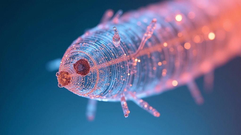
Polarization Techniques to Enhance Birefringent Crystal Structures

Capturing stunning images of birefringent crystal structures requires proper polarization techniques to reveal their unique optical properties.
You’ll need two polarizer filters—position one above the condenser and the second above the eyepiece or camera.
Begin with a low magnification objective (4X) to locate your crystal specimens before moving to higher magnifications for detailed examination.
- Adjust the orientation of your polarizer filters to maximize contrast, ensuring the background appears dark while crystal colors remain vibrant.
- Use a mechanical stage to rotate specimens precisely, finding ideal angles that showcase their optical characteristics.
- Apply focus stacking to combine multiple images at different focal depths for superior clarity in your final photographs.
The quality of your birefringent crystal images depends on these polarized light settings and your ability to manipulate them effectively.
Light Intensity Management for Different Specimen Densities
Effective light intensity control serves as the foundation for capturing detailed transmitted light photographs across varying specimen densities.
You’ll need to adjust the light intensity based on your specimen’s thickness and refractive properties—denser materials typically require lower light levels to prevent overexposure and preserve detail.
Utilize your polarized light microscope’s diaphragm to control light entrance, making incremental adjustments for maximum clarity.
When working with highly birefringent specimens, a neutral density filter helps reduce brightness without disrupting color balance.
Don’t hesitate to adjust the orientation of your light source, as different angles can dramatically enhance crystal structure visibility.
Monitor brightness and contrast through your camera’s live view while fine-tuning transmitted light settings.
This real-time feedback guarantees you’ll capture the specimen’s intricate details with precision across different density levels.
Phase Contrast Settings for Transparent and Translucent Samples
When transparent and translucent specimens fail to produce sufficient contrast in brightfield microscopy, phase contrast settings become your essential tool for revealing their hidden details.
Phase contrast microscopy transforms invisible phase shifts into visible intensity variations by utilizing a specialized phase plate that creates optical path differences between diffracted and undiffracted light.
Phase contrast illumination reveals the invisible, converting subtle specimen phase shifts into striking visual contrasts through optical manipulation.
For ideal clarity in your specimen images:
- Correctly align your condenser diaphragm with the phase annulus of your objective—misalignment noticeably reduces contrast and obscures fine structures
- Use a monochromatic light source to enhance the phase contrast effect and improve image quality
- Confirm proper setup of phase contrast components, as even minor adjustments can dramatically affect your ability to observe transparent specimens
Differential Interference Contrast for Three-Dimensional Detail
Differential Interference Contrast (DIC) microscopy takes your imaging capabilities beyond phase contrast by generating stunning three-dimensional representations of transparent specimens.
This technique employs polarized light and a Nomarski prism to split light into two beams that interact differently with your sample, converting phase shifts into visible contrast enhancement.
You’ll find DIC particularly valuable when examining biological samples without staining.
Cell membranes and organelles pop with detail as the system detects minute differences in refraction index throughout the specimen. By adjusting your focus and polarizer angles, you can fine-tune the three-dimensional appearance to highlight specific structures.
Materials scientists benefit equally when analyzing anisotropic materials like polymers, where DIC reveals microstructural details critical for understanding material properties and behavior.
Camera Exposure Settings for Accurate Specimen Representation
To capture the true essence of specimens under transmitted light, mastering your camera’s exposure settings becomes essential.
Keep your ISO low (100-400) to minimize noise and enhance clarity in crystal images. Select a mid-range aperture (f/8-f/11) to achieve greater depth of field, ensuring more of your specimen remains in focus. Use shutter speeds between 1/60-1/125 seconds to balance light exposure without introducing motion blur.
- Switch to manual mode for precise control, allowing you to adjust settings based on your specific specimen’s lighting conditions.
- Consider bracketed exposures by taking multiple shots at different exposure levels.
- Remember that proper exposure settings work together to reveal intricate details that might otherwise be lost in transmitted light photography.
Post-Processing Workflows to Preserve Specimen Detail
After capturing your specimens under transmitted light, proper post-processing becomes essential for preserving the intricate details that make each crystal unique.
Begin by applying Raw Development Settings in your editing software to maintain maximum detail when working with images from your light microscope or polarized light microscope.
For birefringent materials, select ProPhoto RGB as your working color space to guarantee vibrant color representation of crystal structures.
Experiment with different rendering methods—Method B typically yields superior results for crystal photographs by preserving fine details.
Save your work as DNG files to maintain high-quality output without compression losses.
Organize your image stacks using filename templates in your software preferences, allowing for more efficient detail preservation as you compare and refine multiple captures of the same specimen.
Frequently Asked Questions
What Provides Light to See Specimens?
You’ll use a light source positioned beneath your specimen stage to provide illumination. This transmitted light passes through your sample, making transparent specimens visible and allowing you to see structural details clearly.
Which Microscope Can Increase the Brightness of Images of a Transparent Specimen?
You’ll get brighter images of transparent specimens using a microscope with LED illumination, polarizing capabilities, plan achromatic objectives, adjustable diaphragm, and a high numerical aperture condenser to focus more light onto your sample.
What Uses Transmitted or Reflected Light to Obtain an Image?
You’ll use transmitted light for transparent specimens where light passes through them, while you’ll need reflected light for opaque samples where illumination bounces off the surface back to your microscope’s objective lens.
What Supplies Light to Illuminate the Specimen Being Viewed?
You’ll find that light sources, such as LED or halogen lamps, supply illumination for your specimen. They’re positioned beneath the stage in transmitted light microscopy, sending light upward through your sample for viewing.
In Summary
You’ll find that mastering transmitted light microscopy isn’t about fixed settings but understanding how to adjust for each specimen. Perfect your Köhler illumination, dial in your aperture and field diaphragms, and experiment with contrast techniques for your specific samples. Remember, the best results come from balancing resolution against contrast while considering your camera’s capabilities. With practice, you’ll develop intuition for creating crystal-clear specimen photos every time.


Leave a Reply