Time-lapse microscopy requires stable environmental controls (37°C, 5% CO2) to maintain cell viability during extended imaging. You’ll need to minimize phototoxicity by using lower illumination intensity, red-spectrum fluorophores, and shorter exposure times. Select appropriate sampling frequencies based on the cellular processes you’re studying, and implement reliable autofocus systems to compensate for drift. Advanced image processing tools help analyze cell movements and morphological changes. The following techniques will transform your ability to capture dynamic cellular events with precision.
Fundamental Principles of Time-Lapse Microscopy
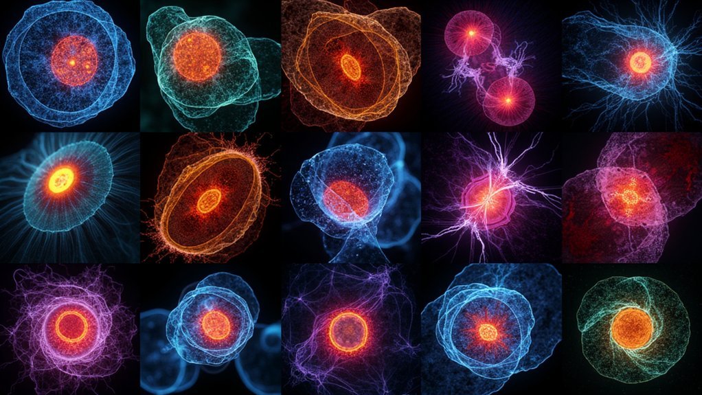
While static images provide snapshots of cellular structures, time-lapse microscopy reveals the dynamic nature of living cells by capturing sequential images at predetermined intervals. This technique transforms hours or days of cellular processes into condensed visual sequences lasting just minutes.
When planning your time-lapse imaging experiment, you’ll need to determine appropriate temporal resolution by adjusting capture frequency—lower rates accelerate perceived motion, while higher rates slow complex events for detailed analysis.
For successful live-cell imaging, you must maintain stable environmental conditions throughout your experiment. Control temperature, CO2 levels, and humidity carefully to guarantee cell viability during extended imaging periods.
Select appropriate microscopy methods—phase contrast or fluorescence—based on your specific research questions. Remember that proper focus maintenance and timely filter adjustments are essential for capturing accurate cellular dynamics.
Selecting the Right Equipment for Cell Viability
When selecting equipment for time-lapse imaging, you’ll need reliable temperature control systems that maintain cells at their ideal temperature (typically 37°C for mammalian cells) throughout your experiment.
You can minimize phototoxic effects by choosing microscopes with sensitive detectors that require less illumination intensity and filters that block harmful wavelengths.
An integrated approach that combines specialized culture dishes, appropriate light sources, and software-controlled exposure timing will greatly extend cell viability during your imaging sessions.
Minimize Phototoxic Effects
Since phototoxicity represents one of the greatest challenges in live cell imaging, selecting appropriate equipment becomes essential for maintaining cell viability throughout your experiments.
When conducting fluorescence imaging, you’ll need to address several factors to reduce damage to your live cells.
- Choose sensitive detectors that require minimal light intensity, reducing exposure while maintaining image quality.
- Opt for red-spectrum fluorescent probes as these longer wavelengths cause less cellular damage.
- Implement strict illumination controls—turn off light sources between acquisitions and use the lowest effective laser power.
- Select appropriate imaging media that maintains stable osmolarity and pH, as poor media conditions can amplify phototoxic effects.
Don’t forget to utilize autofocusing features to keep cells in ideal focus without unnecessary light exposure during long-term experiments.
Temperature Control Systems
Although often overlooked in imaging setup planning, temperature control represents a critical factor for successful live cell imaging experiments. For most mammalian cells, you’ll need to maintain a consistent 37°C environment to accurately mimic physiological conditions.
When selecting temperature control systems for live cells, consider options like heated stages, incubation chambers, or custom environmental control units. The best systems offer real-time monitoring capabilities that prevent thermal fluctuations which can stress cells and compromise your results.
Pay attention to the thermal conductivity of your imaging platform—materials with high thermal stability guarantee more consistent temperatures during long-term time-lapse imaging.
Investing in quality temperature regulation equipment considerably improves cell viability and experimental reproducibility, eliminating artifacts caused by temperature shifts that could otherwise invalidate your findings.
Environmental Control Systems for Extended Imaging
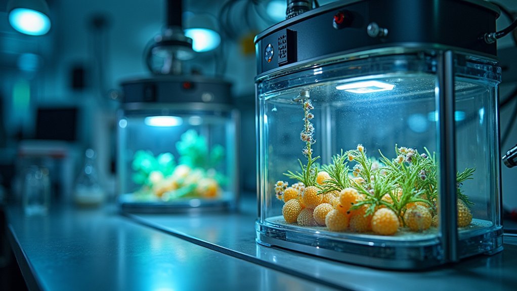
Because live cells require specific physiological conditions to thrive, environmental control systems form the backbone of successful time-lapse imaging experiments.
When you’re monitoring cellular processes over hours or days, maintaining stable conditions becomes critical for reliable results.
Your imaging setup should include these essential components:
- Temperature regulation – Keep mammalian cells at 37°C to guarantee normal metabolic functions
- CO2 control – Maintain approximately 5% CO2 to stabilize pH in cell culture media
- Humidity management – Prevent media evaporation that alters osmolarity and stresses cells
- Automated monitoring – Use systems that make real-time adjustments to minimize environmental fluctuations
For shorter experiments, using larger volumes of imaging media can help buffer against environmental changes that might compromise your cellular health and experimental outcomes.
Optimizing Illumination to Minimize Phototoxicity
While maintaining stable environmental conditions preserves cellular health, even the most perfectly controlled chamber won’t save cells from the invisible threat of phototoxicity. When imaging live cells, you’ll need to strategically reduce light-induced damage.
Start by using the lowest illumination intensity that still provides adequate signal-to-noise ratio. Select fluorescent probes in the red spectrum, which require less excitation energy and minimize cellular harm. Implement shuttering mechanisms to block the light source between acquisitions, considerably reducing cumulative exposure.
Shorter exposure times during image capture will help maintain cell viability while still documenting dynamic processes.
Finally, invest in sensitive detectors that allow you to capture high-quality images with minimal light levels. These practices will extend your cells’ viability throughout long-term time-lapse experiments while preserving image quality.
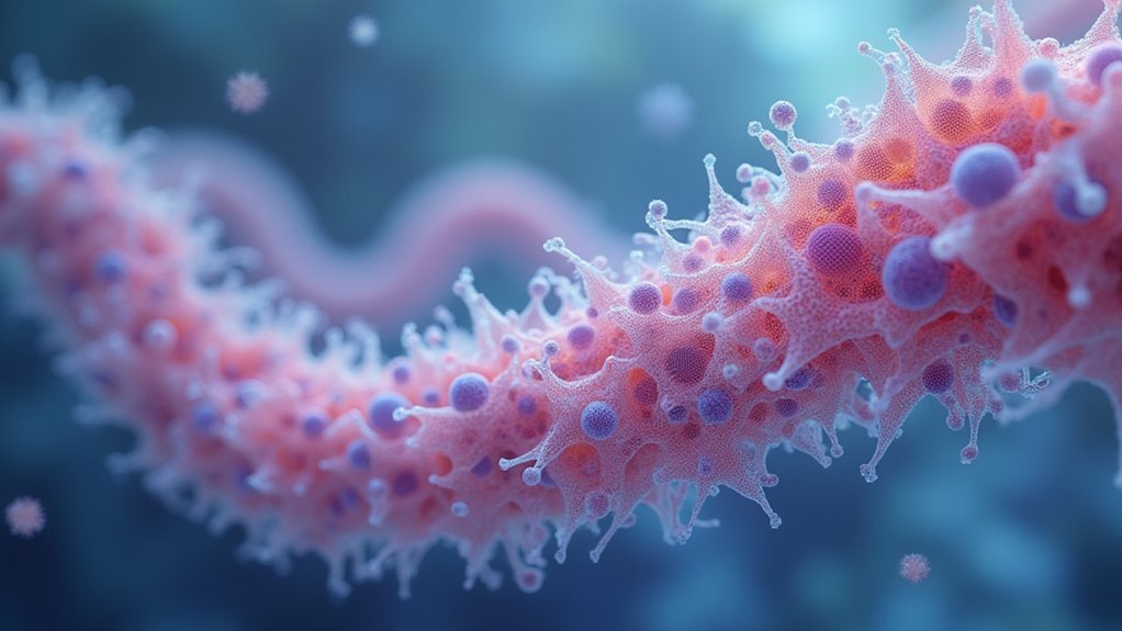
Fluorescent Labeling Strategies for Dynamic Cell Processes
Since cellular processes occur in a complex, ever-changing landscape, your choice of fluorescent labels will determine what you can observe and for how long.
Select bright, specific fluorophores like GFP variants that minimally disrupt cellular functions when tracking dynamic biological processes.
When designing your imaging techniques for live cells, consider:
- Opt for red-spectrum fluorescent proteins to reduce phototoxicity during extended imaging sessions
- Carefully optimize fluorophore concentrations and exposure times to prevent cellular stress
- Choose stable markers with low photobleaching rates for long-term studies spanning days
- Combine multiple fluorescent proteins with advanced imaging techniques like multiphoton microscopy to track several processes simultaneously
This strategic approach will guarantee you capture accurate, thorough data on cellular dynamics without compromising cell viability.
Image Acquisition Parameters and Focus Stability
When setting up your time-lapse experiment, you’ll need to select the ideal sampling frequency to balance temporal resolution against phototoxicity and data storage requirements.
Your choice of autofocus system—whether hardware-based methods like infrared reflection or software-based algorithms that analyze image contrast—will directly impact focus reliability during extended imaging sessions.
To maintain crisp images throughout your experiment, you should implement drift compensation methods such as reference point tracking or predictive focusing algorithms that actively correct for mechanical and thermal fluctuations.
Optimal Sampling Frequency
Determining the right sampling frequency stands as one of the most critical decisions in time-lapse microscopy design. Your ideal sampling frequency should capture cellular dynamics without causing unnecessary stress to live cells. Effective time-lapse protocols balance data acquisition with cellular health.
When configuring your imaging system, consider these essential factors:
- Match your sampling rate to cellular dynamics – faster processes require 1 frame per second while slower events may need only 1 frame every few minutes.
- Monitor for signs of phototoxicity, which increases with higher sampling frequencies.
- Utilize autofocusing features to maintain focus stability throughout your experiment.
- Maintain consistent environmental conditions (temperature, humidity) to support both focus stability and natural cellular behavior.
Autofocus System Selection
Maintaining precise focus throughout extended time-lapse experiments represents perhaps the greatest technical challenge researchers face when imaging live cells.
You’ll need a reliable autofocus system to compensate for inevitable drift caused by thermal fluctuations and sample movement.
Invest in a high-quality autofocus that employs advanced algorithms for rapid, accurate focal plane detection.
Systems utilizing laser scanning or structured illumination techniques deliver superior precision for high-resolution applications.
Regular calibration is essential, particularly in environments with variable temperature and humidity.
Your choice of autofocus system directly impacts imaging experiment outcomes.
More sophisticated systems maintain stability while minimizing unnecessary exposure, reducing photodamage risk to delicate specimens.
Remember that proper autofocus selection and calibration can mean the difference between capturing meaningful cellular dynamics and wasting days on blurry, unusable data.
Drift Compensation Methods
Three vital drift compensation strategies can dramatically improve your time-lapse imaging results.
When tracking live cells over long time periods, even minor focus shifts can blur essential cellular dynamics. You’ll need reliable methods to maintain focus stability throughout your experiment:
- Implement automated focusing systems that dynamically adjust to counteract temperature or vibration-induced drift.
- Establish stable mounting systems and minimize external vibrations at the hardware level.
- Capture multiple fields of view across larger imaging areas to better assess and respond to drift patterns.
- Perform regular calibration of both your imaging system and environmental controls.
These drift compensation approaches guarantee your time-lapse experiments produce sharp, analyzable data rather than blurry, unusable images—vital when tracking subtle cellular processes that unfold over hours or days.
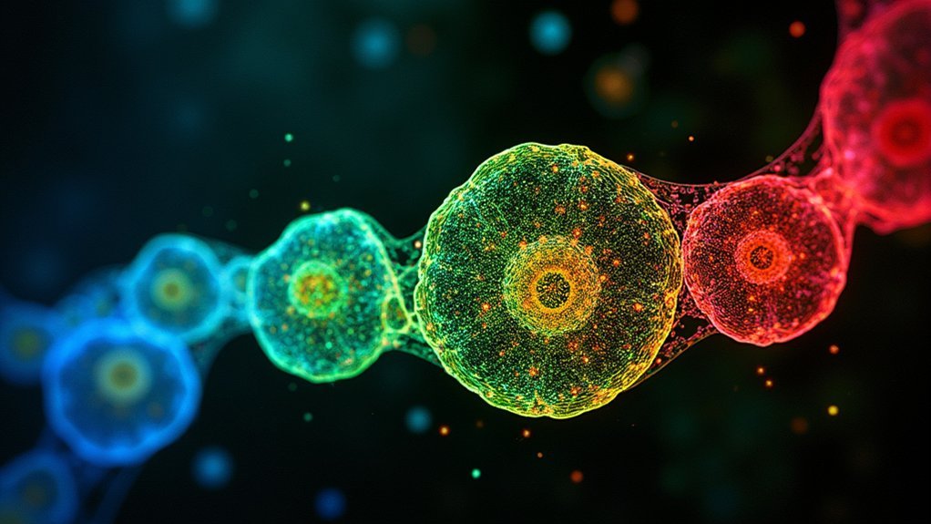
Data Processing and Analysis of Time-Lapse Sequences
Once time-lapse imaging data is collected, researchers face the crucial challenge of effectively processing and analyzing the vast sequences generated during live cell experiments.
You’ll need specialized software tools that align, enhance, and filter images to reduce noise and improve clarity, allowing for more accurate visualization of cellular dynamics.
Quantitative analysis of time-lapse sequences typically involves tracking cell movements, measuring fluorescent imaging intensity changes, and analyzing morphological transformations over time.
Automated image analysis software can dramatically accelerate the processing of large datasets, freeing you to focus on interpretation rather than manual handling.
Frequently Asked Questions
What Microscopy Techniques Can Be Used in Live-Cell Imaging?
You can use widefield, confocal, spinning-disc confocal, TIRF, multiphoton, and FCS microscopy techniques for live-cell imaging. CARS microscopy also offers chemical specificity for advanced studies like lipid dynamics.
What Is the Time-Lapse Imaging Technique?
Time-lapse imaging lets you capture sequential images at set intervals over time. You’ll see dynamic cellular processes unfold as if accelerated, with adjustable frame rates that compress hours or days into viewable sequences.
What Techniques Would Allow You to Visualize Living Cells Over a Period of Time?
You can visualize living cells over time using time-lapse microscopy, fluorescent protein labeling, widefield or confocal imaging, and environmental chambers that maintain ideal conditions while minimizing phototoxicity during extended observation periods.
What Technique Allows Living Cells to Be Examined?
You can examine living cells using time-lapse microscopy, which captures images at regular intervals. You’ll need fluorescent labeling, environmental controls, and specialized microscopes like widefield or confocal systems to visualize cellular processes effectively.
In Summary
As you’ve explored these time-lapse techniques, you’ve learned that successful live cell imaging requires careful balance between temporal resolution and cellular health. Remember, you’ll need appropriate equipment, strict environmental control, and optimized illumination to capture meaningful data. Your choice of fluorescent labels and acquisition parameters will determine the quality of your results. Master these fundamentals, and you’ll transform fleeting cellular events into powerful visual narratives.


Leave a Reply