Safe staining methods are critical for specimens as they preserve cellular integrity while ensuring accurate diagnostic results. You’ll protect both the specimen’s morphology and laboratory personnel from toxic chemical exposure. Proper techniques prevent cellular distortion, enhance visualization of structures, and reduce environmental impact from hazardous waste. By implementing low-toxicity alternatives and standardized protocols, you maintain specimen viability and comply with safety regulations. Discover how these methods can transform both the quality and sustainability of your laboratory work.
13 Second-Level Headings
When organizing your documentation about safe staining methods, you’ll need clear second-level headings to guide readers through specific procedures and protocols.
Structure your content with sections like “Contamination Risk Prevention” and “Personal Protective Equipment Requirements” to highlight safety priorities.
Include headings focused on “Chemical Exposure Minimization” that detail proper handling of reagents and disposal protocols.
Don’t forget sections addressing “Reliable Staining Results” and “Specimen Integrity Preservation” to emphasize how safety measures directly impact accurate diagnostics.
Create distinct headings for “Pathogenic Organisms Identification” to explain how proper staining procedures enhance visualization of critical structures.
Well-organized documentation guarantees laboratory personnel can quickly reference safety protocols, reducing workplace hazards while maintaining specimen quality.
Meticulously structured safety protocols protect both laboratory staff and the integrity of diagnostic specimens.
Your headings should create a logical flow that reinforces the connection between safety practices and diagnostic excellence.
Preserving Specimen Integrity During Staining Processes
You’ll need to balance chemical exposure with proper fixation techniques to prevent structural damage during the staining process.
Heat or methanol fixation preserves cellular morphology and guarantees specimens won’t wash off during subsequent steps, directly contributing to diagnostic accuracy.
Your attention to timing and selection of appropriate fixatives will enhance visualization of cellular structures while maintaining the specimen’s natural state, especially when working with delicate structures like capsules.
Preventing Structural Damage
Preserving the structural integrity of specimens throughout the staining process requires meticulous attention to fixation techniques and timing.
When you’re handling delicate samples, proper heat or chemical fixation prevents cellular shrinkage and distortion that can compromise cell morphology. Be vigilant about over-fixation, as it can destroy structural details needed for accurate visualization.
You’ll need to control both concentration and exposure time of staining solutions carefully. Extended contact with stains can degrade cellular integrity, leaving you with misleading results.
Don’t rush the air drying phase before staining—incomplete drying causes samples to wash away during the procedure.
Safe staining methods aren’t just about protecting yourself—they’re essential for maintaining specimen quality.
Maintaining Diagnostic Accuracy
To maintain diagnostic accuracy, every step of the staining process must protect specimen integrity. When you’re working with biological samples, safe staining methods aren’t just about preventing contamination—they’re essential for preserving cellular morphology that leads to correct interpretations.
Use appropriate fixation techniques, such as methanol for BSL2 organisms, to create fixed smears that accurately represent the original specimen. Always employ positive controls to validate your staining results and confirm the identification of specific structures.
Proper labeling and standardized preparation methods will reduce errors that could compromise diagnostic outcomes.
Remember that reproducibility is the cornerstone of reliable diagnostics. By following safety protocols and using well-standardized stains, you’ll guarantee consistent results that can be trusted across different tests and laboratories.
Reducing Toxic Chemical Exposure for Laboratory Personnel
Laboratory personnel face significant health risks when working with traditional staining chemicals, making exposure reduction a critical priority in modern lab settings.
You’ll minimize these risks by implementing thorough safety protocols that include proper ventilation systems and personal protective equipment when handling hazardous staining chemicals.
- Replace toxic chemicals like formaldehyde with low-toxicity alternatives to prevent both acute and chronic health issues.
- Install fume hoods and verify they’re regularly maintained to effectively manage airborne contaminants.
- Provide regular training on best practices for handling, storage, and disposal of hazardous materials.
Maintaining Cell Structure and Morphology
While safe handling practices protect laboratory staff, proper staining techniques guarantee specimen integrity throughout the preparation process.
You’ll find that safe staining methods preserve cell structure and morphology by minimizing exposure to harsh chemicals that could damage cellular components.
When you employ proper fixation techniques, such as methanol fixation, you help maintain the overall shape and integrity of your specimens.
Following appropriate staining protocols guarantees that membranes and organelles remain visible and accurately represent their natural state during microscopic examination.
Always maintain a sterile environment to prevent contamination that could lead to morphological changes and misinterpretation.
Remember to document your process systematically—this attention to detail guarantees that cell structures remain identifiable for future reference and comparative analysis.
Environmental Impact of Traditional vs. Safe Staining Methods
Although widely used for decades, traditional staining methods carry significant environmental burdens that extend far beyond the laboratory walls.
When you utilize formaldehyde and xylene in your staining protocols, you’re generating hazardous waste that requires costly disposal procedures and specialized safety equipment.
By switching to non-toxic alternatives like water-based dyes, you’ll dramatically reduce your lab’s environmental footprint while enhancing occupational safety.
- Hazardous chemicals in traditional stains require strict regulatory compliance for disposal, increasing operational costs and environmental liability.
- Safe staining methods minimize chemical exposure risks, creating healthier work environments for your laboratory staff.
- Non-toxic alternatives reduce the need for extensive safety protocols and equipment, improving both sustainability and cost-effectiveness.
Your choice of staining methods directly impacts both environmental sustainability and the health of those working in your laboratory.
Non-Toxic Alternatives to Common Histological Dyes
As research priorities shift toward sustainability and safety, you’ll find a growing arsenal of non-toxic alternatives replacing traditional histological dyes in modern laboratories.
Natural dyes like saffron offer effective tissue staining without the harmful effects associated with synthetic options, while preserving specimen integrity.
You can now choose fluorescent dyes such as SYBR Green for nucleic acid visualization with minimal cytotoxicity to living cells.
Plant-based alternatives like beetroot extract demonstrate comparable staining capabilities while considerably reducing environmental impact.
Some advanced imaging technologies even eliminate staining requirements altogether.
The development of these non-toxic alternatives emphasizes biocompatibility and environmental safety without compromising effectiveness.
Proper Handling and Disposal of Staining Reagents
Because staining reagents often contain hazardous chemicals, proper handling protocols are essential for laboratory safety.
You’ll need to wear personal protective equipment and use fume hoods when working with these substances to minimize exposure risks. Label all staining reagents clearly with hazard symbols and store them according to compatibility guidelines.
When disposing of chemical waste, you must follow established waste disposal protocols to comply with environmental regulations:
- Segregate chemical waste from biological waste in designated containers
- Neutralize or deactivate used staining reagents before disposal when applicable
- Document all hazardous materials disposal according to facility requirements
Regular training for laboratory personnel on safe handling practices isn’t optional—it’s critical for preventing incidents and maintaining safety standards while working with potentially dangerous staining compounds.
Effect of Safe Staining on Image Quality and Contrast
While many researchers assume safer staining methods might compromise image quality, the opposite often proves true. Safe staining methods actually preserve cellular structures’ integrity, enhancing morphological details visible under microscopy.
When you employ proper fixation techniques like methanol fixation, you’ll prevent cellular distortion and shrinkage that typically diminish contrast and clarity. Using certified stains that meet industry standards guarantees consistent visualization of specimens, reducing variability that affects diagnostic interpretation reliability.
You’ll find that avoiding harmful chemicals during staining minimizes sample degradation, allowing for better visualization of cellular components.
Additionally, safe staining practices maintain specimen viability, enabling you to observe dynamic processes with improved contrast between living cells and their surrounding environment. The result is clearer imaging that reveals subtle structural details previously obscured by harsh chemical agents.
Long-Term Preservation of Stained Biological Samples
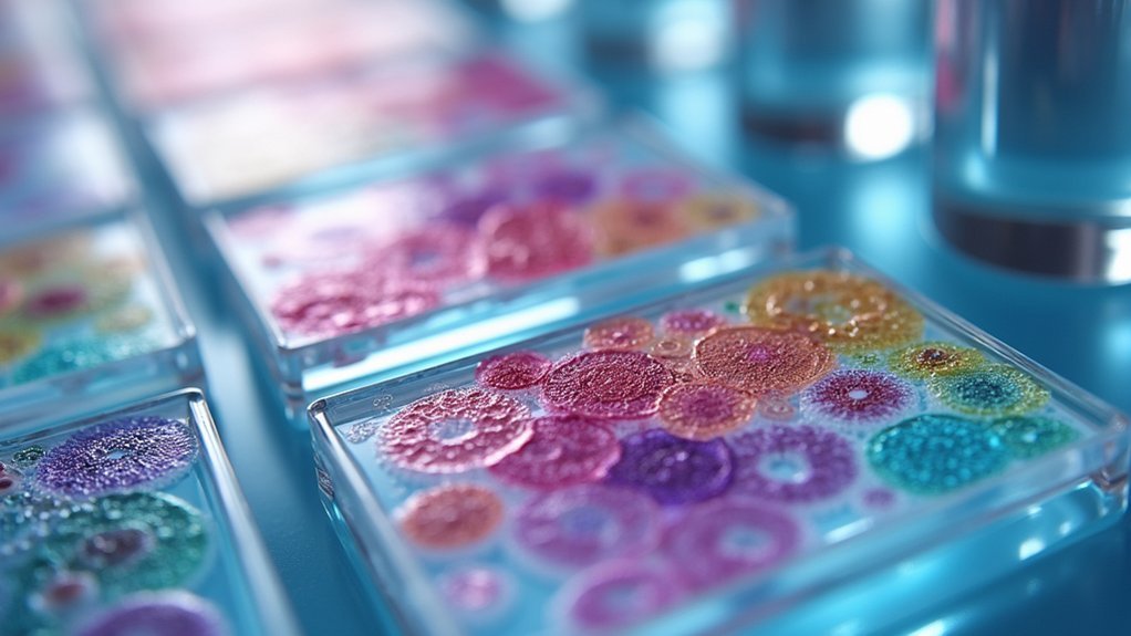
When preserving stained biological samples, you’ll need to take into account archival quality factors including stable dyes and non-toxic fixing agents like methanol or formaldehyde.
You can prevent degradation by storing specimens in controlled environments away from light and moisture, ideally at lower temperatures.
Implementing standardized staining protocols and regular quality checks will guarantee your samples remain viable for future analysis and reference without losing diagnostic clarity.
Archival Quality Considerations
To achieve truly archival-quality preservation of stained biological specimens, you’ll need to contemplate multiple factors beyond basic staining techniques. High-quality stains that resist fading are essential for maintaining structural integrity over decades.
You must control environmental conditions meticulously—temperature fluctuations, humidity levels, and light exposure can all compromise long-term preservation efforts.
- Implement standardized staining protocols to guarantee reproducibility when samples are examined years later.
- Store specimens in specialized containers that buffer against environmental changes.
- Establish regular assessment schedules to identify early signs of degradation.
Remember that proper initial fixation forms the foundation of archival quality. If you notice deterioration during monitoring, don’t hesitate to reflect on re-staining rather than losing valuable specimens.
Your attention to these preservation details now will protect research investments for future generations.
Preventing Sample Degradation
Preventing sample degradation extends beyond archival considerations to address the specific chemical and physical factors that threaten specimen integrity over time.
When you implement safe staining methods, you’ll protect cellular structures from harmful chemical reactions that can distort your observations and compromise research validity.
Proper fixation techniques using methanol or heat guarantee your specimens maintain their natural morphology throughout storage periods.
For long-term preservation, you’ll need to use standardized stains and reagents consistently to prevent degradation of delicate biological features.
Choosing environmentally safe stains not only protects lab personnel but also safeguards your specimens from adverse reactions during extended storage.
Regulatory Compliance in Laboratory Staining Procedures
Although many laboratory professionals focus primarily on scientific outcomes, regulatory compliance forms the backbone of safe staining practices in modern laboratories.
You’ll need to implement thorough safety data sheets for all staining reagents and establish proper chemical handling protocols to meet OSHA and CDC standards. Your lab must also follow RCRA guidelines for waste disposal to minimize environmental impact while maintaining detailed documentation per Good Laboratory Practices.
- Regular training for lab personnel on safe staining methods is mandated by regulatory bodies
- Proper implementation of safety data sheets provides critical information for emergency responses
- Accurate record-keeping of all staining procedures guarantees traceability and accountability
Maintaining occupational safety through regulatory compliance isn’t just about avoiding penalties—it’s about creating a culture of responsibility that protects your team and promotes research integrity.
Advanced Techniques for Minimizing Specimen Damage
You’ll find that selecting low-toxicity reagents dramatically reduces specimen degradation while maintaining diagnostic accuracy.
Sequential staining protocols allow for controlled reagent exposure, preventing the harsh effects of simultaneous chemical interactions on delicate cellular structures.
Microfluidic delivery systems offer precise control over stain application, ensuring minimal specimen stress through regulated flow rates and reduced reagent volumes.
Low-Toxicity Reagent Selection
When selecting fixatives and stains for specimen preparation, choosing low-toxicity reagents like methanol and formaldehyde can dramatically reduce cellular damage while maintaining structural integrity.
You’ll preserve morphology for accurate visualization during staining processes, while minimizing harm to delicate cellular structures.
Consider implementing these safer alternatives in your laboratory:
- Less harmful stains such as nigrosin for negative staining, which prevents interference with capsules and membranes
- Advanced fluorescent dyes that enhance imaging quality while reducing exposure risks for both specimens and staff
- Low-toxicity fixatives that minimize artifacts and improve diagnostic reliability
Sequential Staining Protocols
Beyond selecting safer reagents, the implementation of precise sequential staining protocols greatly reduces specimen damage during preparation. You’ll find that carefully applying stains and reagents in a specific order preserves delicate specimens while enhancing visualization of cellular structures.
Start with proper fixation techniques, such as methanol fixation for BSL2 organisms, to maintain cell morphology before staining. Standardize your staining durations and temperatures to prevent overexposure that leads to specimen degradation.
Don’t skip using positive and negative controls—they validate your process and help identify potential damage immediately.
Optimize your staining solutions by adjusting concentrations and pH levels to minimize cellular stress. These advanced approaches considerably decrease damage risks while ensuring high-quality imaging results that accurately represent your specimen’s natural state.
Microfluidic Delivery Systems
Three revolutionary advances in microfluidic technology have transformed specimen staining approaches for research laboratories.
You’ll find these systems utilize precision-engineered channels to control staining solutions with remarkable accuracy, preserving structural integrity while reducing specimen damage. The controlled environment minimizes shear stress and turbulence that typically compromise delicate samples during traditional staining methods.
- Automated handling of reagents dramatically improves reproducibility and consistency across experiments, eliminating variability from manual processing.
- Real-time monitoring capabilities optimize staining reagents dynamically, adapting to each specimen’s unique requirements.
- Lower concentration solutions can be effectively utilized, reducing both hazardous material exposure and waste generation.
Safe Staining Methods for Live Cell Imaging
Although conventional staining techniques often sacrifice cell viability, modern crucial stains now allow researchers to observe living specimens without causing cellular damage.
When you’re conducting live cell imaging, vital stains offer selective absorption by living cells while excluding others, ensuring accurate cellular morphology studies.
You’ll find non-toxic stains like New Methylene Blue particularly significant for supravital staining that reveals cellular functions without compromising viability.
Avoid heat fixation, which denatures proteins and disrupts cellular integrity. Instead, use low-concentration chemical fixatives to preserve structures during imaging.
Don’t forget to maintain your safety by using personal protective equipment when handling staining chemicals.
These safe staining methods enable you to capture dynamic cellular processes in real-time while keeping both specimens and researchers protected throughout the imaging process.
Risk Assessment for Different Staining Protocols
When implementing any staining protocol, you’ll need to conduct thorough risk assessments to identify potential hazards specific to each technique.
Consider the hazardous chemicals involved, including carcinogens like formaldehyde and xylene, which require strict handling protocols and personal protective equipment to minimize exposure risks.
- Evaluate ventilation requirements for volatile stains, guaranteeing proper airflow to protect laboratory personnel from inhalation hazards.
- Document proper waste disposal procedures that comply with regulatory guidelines to prevent environmental contamination.
- Develop emergency response procedures for accidental spills or exposures, including eyewash station locations and first aid protocols.
Regular review of your staining protocols against current safety standards guarantees continuous improvement.
Don’t overlook training requirements—all staff should understand specific risks associated with the staining methods they’re using.
Frequently Asked Questions
Why Is It Important to Stain Specimens?
You stain specimens to enhance cellular visibility, reveal structures, differentiate microorganisms, and identify key features. It improves microscopic examination, aids accurate diagnosis, and helps you classify bacteria for proper treatment selection.
What Is the Importance of Staining Methods?
Staining methods are important because they help you visualize structures that wouldn’t be visible otherwise. They enhance contrast, reveal cellular details, and allow you to distinguish between different types of microorganisms and tissue components.
What Is the Most Critical Stain to Identify Bacteria?
The Gram stain is your most critical bacterial identification tool. It differentiates bacteria as Gram-positive (purple) or Gram-negative (pink), providing essential information about cell wall structure that guides appropriate antibiotic treatment decisions.
Why Do Some Specimens Need to Be Stained?
You’ll need to stain some specimens because most microorganisms are nearly transparent. Staining enhances contrast, reveals cellular structures, differentiates between cell types, and highlights specific components essential for proper identification and classification.
In Summary
You’ll find that safe staining methods aren’t just procedural requirements—they’re essential safeguards for specimen integrity, researcher health, and environmental protection. By adopting safer techniques, you’re preserving cellular morphology while reducing toxic exposures. Remember that regulatory compliance isn’t optional in modern laboratory practice. As you develop your protocols, always prioritize safety without compromising the quality of your scientific observations and results.



Leave a Reply