Stage drift during live cell imaging is prevented through thermal stabilization systems that maintain consistent temperatures, hardware autofocus technologies like Perfect Focus Solution, and environmental chambers controlling temperature and humidity. You’ll also need proper mechanical stability through rigid materials and flat surfaces. Software-based correction algorithms provide real-time adjustments during extended imaging sessions. Implement regular calibration and select photostable fluorophores to minimize drift effects. The most successful approaches combine multiple stabilization strategies for best results.
Understanding the Causes of Stage Drift in Microscopy
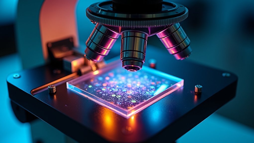
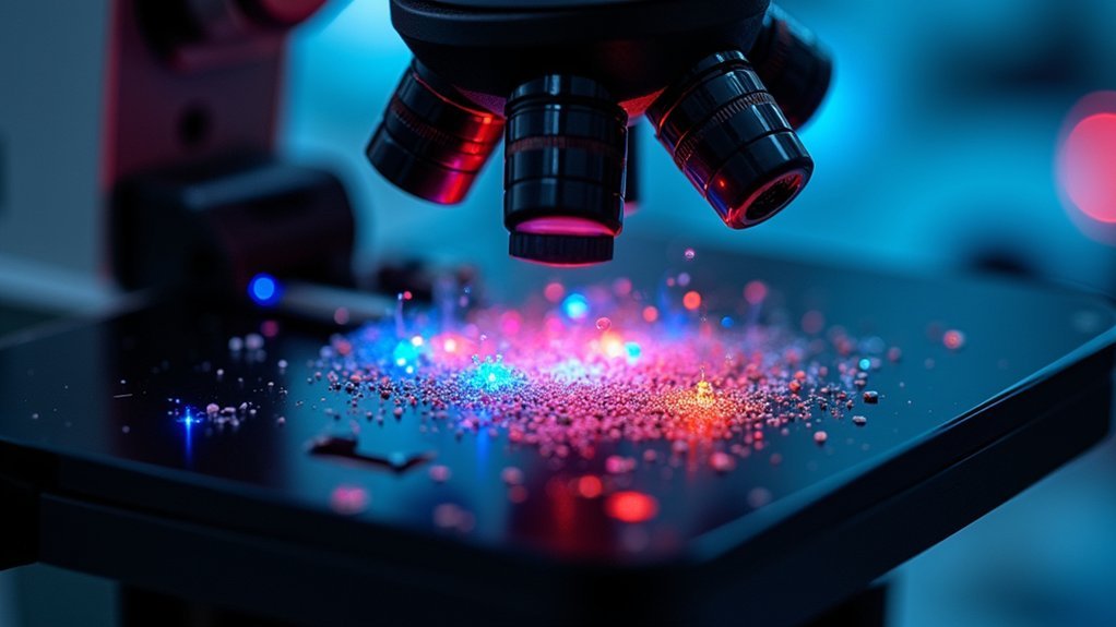
When conducting live cell imaging experiments, you’ll likely encounter stage drift, a persistent challenge that compromises image quality and data reliability. This phenomenon stems from several key factors.
Thermal fluctuations cause microscope components to expand or contract, leading to focus drift that disrupts your imaging plane. Environmental conditions like mechanical vibrations from footsteps or nearby equipment create subtle stage movements that accumulate over time.
Thermal variations and subtle vibrations gradually undermine imaging stability, compromising your experimental integrity over time.
In inverted microscopes, the weight of optical components can gradually shift the stage position, especially during extended live cell imaging sessions.
You’ll also notice that changes in the refractive index of your imaging medium, caused by temperature or concentration fluctuations, create perceived drift. Unstable microscope positioning on uneven surfaces further compounds these issues by introducing additional instability in the optical path.
Thermal Stabilization Systems for Drift Prevention
You’ll find thermal stabilization systems essential for maintaining consistent temperature and preventing focus drift during live cell imaging.
Modern systems employ Peltier elements or heated stages that keep mammalian cells at an ideal 37°C while compensating for expansion and contraction of materials that can cause mechanical drift.
These sophisticated temperature control mechanisms often incorporate feedback loops that continuously monitor conditions, automatically adjusting to guarantee your samples remain perfectly in focus throughout extended imaging sessions.
Temperature Control Mechanisms
Since living cells require precise environmental conditions to function normally, temperature control represents one of the most critical factors in preventing stage drift during live cell imaging.
Effective thermal stabilization systems typically employ Peltier elements or heated stages to maintain the ideal 37°C environment your cells need to thrive while preventing thermal-induced drift.
When implementing temperature control for your live cell imaging setup, focus on:
- Continuous temperature monitoring with real-time feedback systems that dynamically adjust settings
- Uniform heat distribution across your imaging area using specialized materials that minimize heat loss
- Rapid thermal equilibration capabilities to quickly stabilize your sample after any environmental disruption
Even slight temperature fluctuations can cause focus drift, so precision in your thermal stabilization system isn’t just beneficial—it’s essential for accurate results.
Focus Drift Compensation
Although temperature fluctuations remain a primary culprit in stage drift, implementing dedicated focus drift compensation technologies provides an essential safety net for your live cell imaging experiments.
Thermal stabilization systems equipped with Peltier elements or heating blankets create a consistent environment around your specimen, minimizing the expansion and contraction that typically cause focus drift.
Look for systems that incorporate feedback mechanisms to continuously monitor and adjust temperature conditions.
Advanced setups featuring real-time focus correction technologies, such as Continuous Optical Feedback (COF), dynamically compensate for subtle focus shifts as they occur.
These systems work in the background, automatically making micro-adjustments that maintain crisp image clarity throughout extended imaging sessions.
Hardware Autofocus Technologies: The Perfect Focus Solution
The Perfect Focus Solution (PFS) represents one of the most advanced hardware autofocus systems available for combating stage drift during live cell imaging.
Using Continuous Optical Feedback technology, this system constantly adjusts focus in real-time, eliminating drift during extended fluorescence imaging sessions without manual intervention.
You’ll benefit from these key advantages:
- High precision focus maintenance within 1/3 of the objective’s focal depth using an internal linear CCD detector
- Infrared LED technology that detects focus position without interfering with your observations or causing photobleaching
- Automatic focus correction during stage movements, enabling seamless time-lapse imaging
The PFS also features Anchor Focus Position capability, allowing you to retain selected Z-positions for rapid acquisition, greatly enhancing your imaging workflow efficiency.
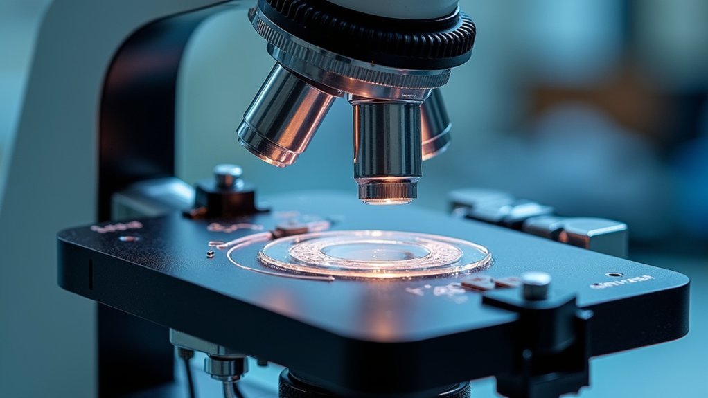
Environmental Control Chambers in Drift Reduction
Maintaining stable environmental conditions represents one of the most effective strategies for minimizing stage drift during live cell imaging. When you’re using oil immersion objectives, even slight temperature fluctuations can cause significant drift as materials expand or contract.
Environmental chambers regulate temperature, humidity, and CO2 levels, creating a stable ecosystem that prevents media evaporation and maintains pH—critical factors that, when unstable, can trigger cell movement and subsequent drift.
Environmental stability prevents cell movement by maintaining crucial parameters that would otherwise contribute to unwanted microscopy drift.
These systems often integrate with both hardware and software components, providing real-time monitoring capabilities that allow you to make immediate adjustments.
Ensure your microplate is properly seated to achieve thermal equilibrium before beginning extended imaging sessions.
Modern chambers with adequate storage capacity for data logging help you track environmental parameters over time, making troubleshooting easier when drift occurs despite precautions.
Mechanical Stability: Stage Design and Material Considerations
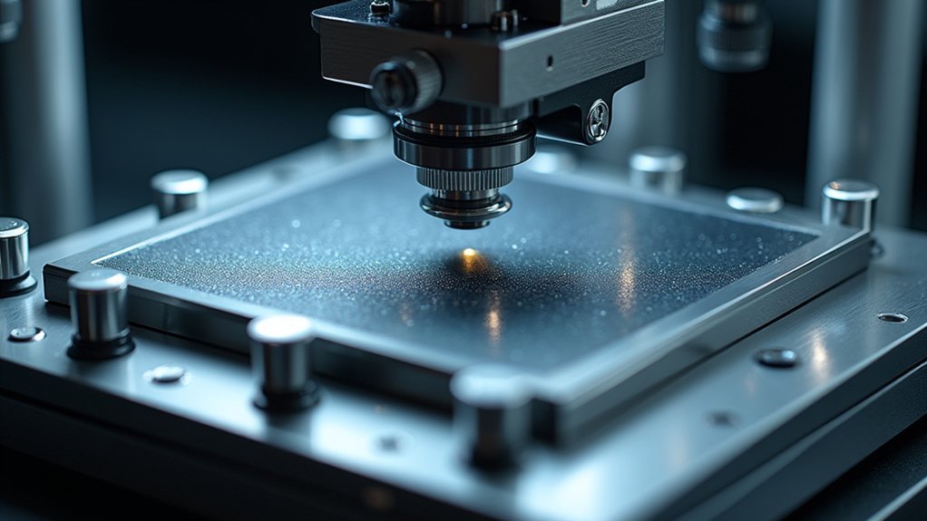
Superior mechanical stability begins with thoughtful stage design and materials selection, which form the foundation of drift-free live cell imaging. Your imaging system’s performance depends critically on the stage’s ability to resist vibrations and maintain position during extended observations.
- Rigid materials matter – Aluminum and carbon fiber components provide the structural integrity needed to prevent flexing and minimize thermal expansion that can cause drift.
- Distribute weight properly – Large, flat stage surfaces evenly support your specimens and equipment, reducing the risk of tipping or subtle shifts that compromise image quality.
- Incorporate vibration dampening – Strategic use of dampening materials isolates your imaging platform from environmental noise, while precision motorized stages allow for adjustments without introducing instability.
Regular calibration of stage components prevents performance degradation over time.
Vibration Isolation Techniques for Precise Imaging
Three critical vibration isolation techniques can make the difference between blurry images and crystal-clear cellular details during live cell imaging. You’ll need to implement these systems to maintain consistent focus during extended observation periods.
| Technique | Mechanism | Benefits |
|---|---|---|
| Passive Isolation | Heavy platforms, rubber mounts | Absorbs vibrations without power requirements |
| Active Isolation | Sensors and real-time actuators | Dynamically counteracts detected vibrations |
| Air Tables | Pneumatic isolation systems | Provides floating suspension for equipment |
| Optical Tables | Honeycomb core with damping materials | Minimizes transmission of environmental vibrations |
| Combined Approaches | Multi-layered isolation strategies | Offers thorough protection against various vibration types |
Don’t overlook regular calibration alongside these isolation methods—it’s essential for maintaining focusing precision during critical experiments where even microscopic movement can compromise your results.
Software-Based Drift Correction Algorithms
While physical stabilization methods provide the foundation for drift control, intelligent software algorithms now serve as the frontline defense against stage drift during live cell imaging.
These solutions analyze real-time image data to continuously adjust focus and stage position, compensating for movement without requiring hardware modifications.
Modern drift correction leverages machine learning to enhance precision, analyzing pixel intensity changes across frames to identify and correct focal plane shifts autonomously.
- You’ll benefit from algorithms that integrate with existing imaging systems, streamlining your workflow during extended observations.
- You can reduce manual intervention during long-term imaging sessions as the software makes automatic adjustments.
- You should regularly calibrate your imaging system to maximize the effectiveness of these algorithms.
Focus Maintenance During Long-Term Time-Lapse Experiments
When conducting live cell imaging over extended periods, maintaining precise focus becomes essential for capturing meaningful biological processes without data loss.
Focus drift can compromise your image quality and data integrity, making automatic correction systems invaluable.
The TE2000-PFS microscope’s Continuous Optical Feedback technology makes real-time adjustments with precision better than 1/3 of the objective’s focal depth.
You’ll benefit from automatic focus correction during stage movements, which eliminates manual adjustments and reduces photobleaching, ultimately enhancing specimen viability.
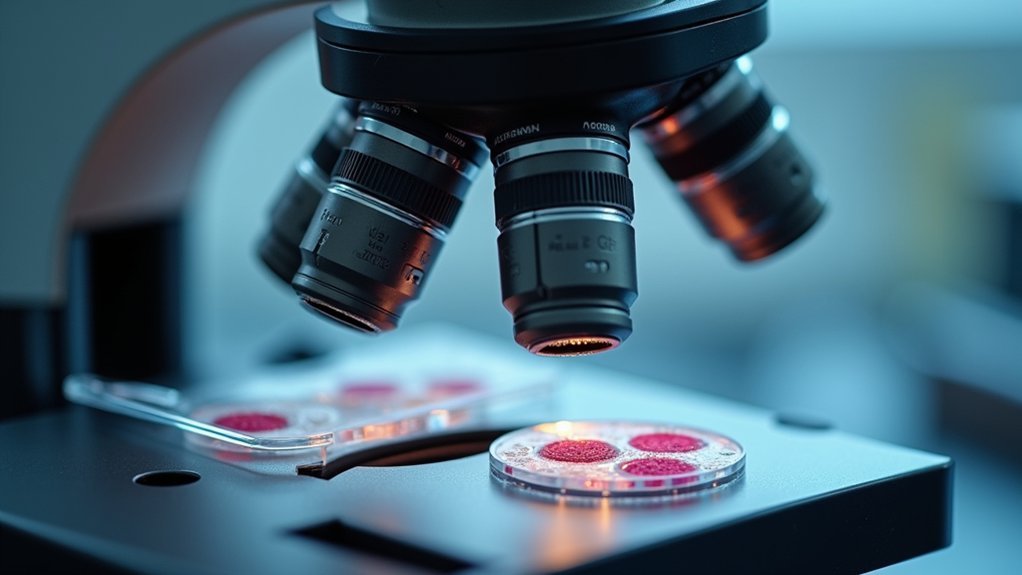
Specialized Sample Mounting for Drift Minimization
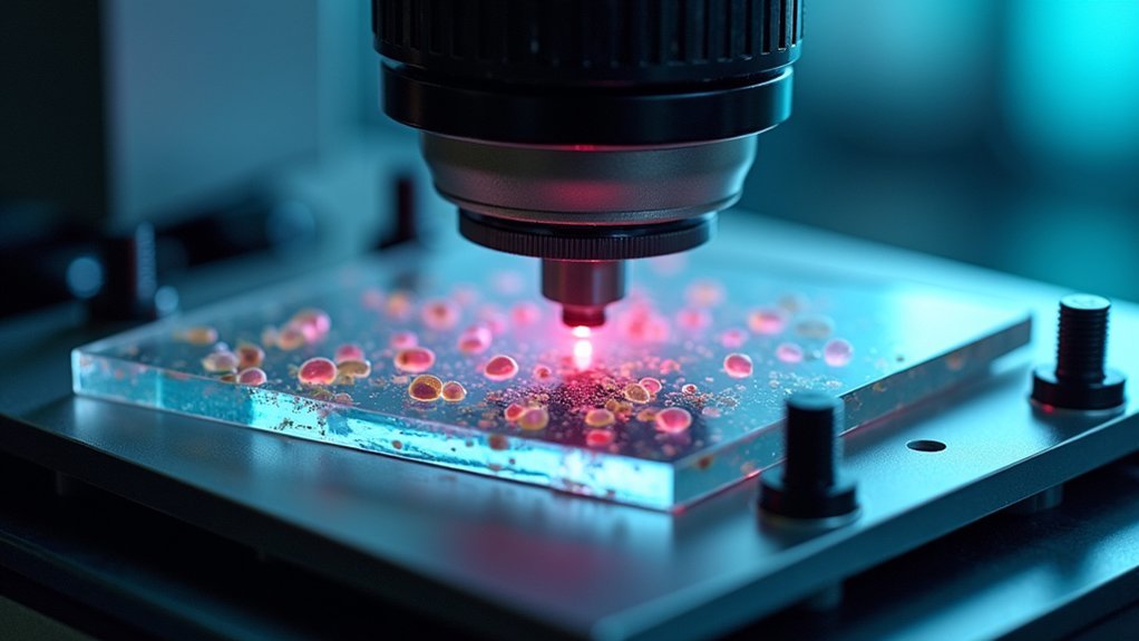
Proper sample mounting represents one of the most critical yet often overlooked factors in preventing stage drift during live cell imaging.
Sample mounting isn’t just a detail—it’s foundational to minimizing drift and ensuring reliable live cell data acquisition.
You’ll find that customized culture dishes and high-precision mechanical stages with fine adjustment controls greatly reduce unwanted movement during extended observation periods.
Temperature-controlled stages and humidity chambers work together to maintain ideal conditions for your specimens, preventing thermal expansion and media evaporation that typically contribute to drift.
For maximum stability, consider:
- Adhesive or vacuum-based mounting techniques that secure samples against external vibrations
- Specialized culture dishes designed specifically for microscopy applications
- Combined temperature and humidity control systems that prevent both thermal drift and osmolarity changes
These mounting strategies will help you achieve the stable imaging platform necessary for capturing high-quality time-lapse sequences with minimal positional variability.
Real-Time Feedback Systems in Modern Microscopes
Real-time feedback systems in modern microscopes implement laser-based focus locks that continuously monitor your sample’s position to instantly correct any drift.
You’ll find these systems utilize piezoelectric adjustment mechanisms to make sub-nanometer corrections that maintain perfect focus despite environmental fluctuations.
Sophisticated drift compensation algorithms process positional data and preemptively adjust the stage, ensuring your live cell imaging captures cellular dynamics without the frustration of focus drift.
Laser-Based Focus Locks
While traditional microscopy struggles with focal plane maintenance, laser-based focus locks have revolutionized live cell imaging by virtually eliminating stage drift.
These systems use Continuous Optical Feedback (COF) technology combined with infrared LEDs that deliver real-time adjustments without interfering with your specimens.
The impressive focusing precision—less than 1/3 of the objective’s focal depth—is achieved through internal linear CCD detectors that constantly monitor the focal point.
You’ll find these advantages particularly valuable:
- Markedly reduced photobleaching through minimized manual focus adjustments
- Enhanced specimen viability during extended time-lapse imaging sessions
- Compatibility with multiple observation methods including DIC and TIRF
Piezoelectric Adjustment Mechanisms
The heart of stage drift prevention lies in piezoelectric adjustment mechanisms, which represent the next evolution in focus maintenance technology. These systems continuously monitor your specimen’s focal point and make rapid, precise adjustments—often at precisions less than 1/3 of the objective’s focal depth—ensuring your cells remain perfectly in focus.
| Feature | Benefit | Application |
|---|---|---|
| Real-time feedback | Eliminates manual intervention | Long-term imaging |
| Continuous Optical Feedback | Maintains ideal focus | Dynamic observations |
| Sub-micron precision | Enhances image quality | High-resolution studies |
| Rapid response time | Compensates for sudden movements | Temperature-sensitive experiments |
| Integrated automation | Reduces photobleaching | Extended live cell viability |
You’ll find these mechanisms particularly valuable during lengthy imaging sessions, where they considerably improve specimen viability by reducing the need for repeated refocusing that can damage delicate cells.
Drift Compensation Algorithms
Modern microscopy has been revolutionized by drift compensation algorithms that function as the brain behind real-time focus maintenance.
These sophisticated systems utilize Continuous Optical Feedback (COF) technology to automatically adjust focus in real-time, ensuring your specimens remain crystal clear throughout extended imaging sessions.
When you’re tracking live cell dynamics, these algorithms work tirelessly to:
- Detect focal shifts using internal linear CCD detectors with precision less than 1/3 of the objective’s focal depth
- Continuously monitor and correct focus, greatly reducing photobleaching and improving specimen viability
- Adapt to environmental changes and sample movements without interrupting your critical observations
Optimizing Imaging Parameters to Combat Drift Effects
Successful live cell imaging depends critically on strategically enhancing your imaging parameters to counteract stage drift.
Start by ensuring proper thermal equilibration of your microplates before imaging to minimize expansion-related drift. You’ll need regular calibration of your imaging system to maintain precise focus adjustments during extended sessions.
Consider implementing hardware autofocus systems to reduce manual adjustments that often introduce drift during stage movements.
Select high signal-to-noise ratio, photostable fluorophores that allow for shorter exposure times, reducing phototoxicity while maintaining image quality.
Don’t overlook environmental factors—consistently monitor and adjust CO₂ levels and humidity to keep cells in ideal condition.
These stable conditions help prevent cellular responses that could contribute to positional changes and subsequent drift during your imaging experiments.
Frequently Asked Questions
What Are the Challenges of Live Cell Imaging?
You’ll face challenges like stage drift from thermal changes and vibrations, focal shifts due to refractive index variations, and environmental factors affecting media stability. Maintaining cell viability while capturing quality images is also difficult.
What Is the Stage Control of a Microscope?
The stage control of a microscope is your precision movement system that lets you position your sample exactly where you need it. It’s often motorized with feedback mechanisms for stable, accurate positioning during imaging.
In Summary
To stop stage drift during live cell imaging, you’ll need a multi-tiered approach. Invest in thermal stabilization systems and proper environmental chambers to minimize temperature fluctuations. Choose microscopes with hardware autofocus technologies like Perfect Focus. Don’t overlook mechanical stability through robust stage design and specialized sample mounting. Finally, optimize your imaging parameters and utilize real-time feedback systems to maintain focus throughout your long-term experiments.


Leave a Reply