
Microscope imaging software revolutionizes your workflow by automating acquisition tasks and eliminating separate hardware controls. You’ll benefit from consistent imaging conditions, advanced analysis tools, and precise measurements with minimal human error. Digital documentation organizes images with timestamps and annotations, while enabling remote collaboration for faster decision-making. High-throughput capabilities process thousands of samples simultaneously, reducing experiment duration dramatically. Discover how these integrated solutions transform microscopy from basic visualization to extensive quantitative analysis.
Why Use Software For Your Microscope Image Workflow?
While traditional microscopy relies heavily on manual processes, implementing specialized software transforms your imaging workflow into a streamlined, efficient system.
Modern imaging software automates acquisition and analysis tasks that would otherwise consume hours of valuable research time, dramatically increasing your throughput.
You’ll gain precise control over microscope settings and cameras through integrated solutions, ensuring consistent imaging conditions across experiments.
Advanced analysis tools eliminate subjective interpretation by providing accurate, quantifiable data from your specimens.
Digital documentation features organize your images and metadata systematically, supporting traceability requirements in both research and industry.
Additionally, you’ll benefit from real-time remote capabilities that enable instant collaboration with colleagues, regardless of their location—allowing your team to make critical decisions faster and more effectively.
Seamless Integration of Microscope Hardware Components
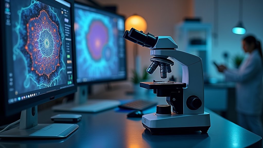
Because modern research demands both precision and efficiency, today’s microscope imaging software excels at unifying diverse hardware components into cohesive systems. You’ll find that these platforms eliminate the frustration of managing separate controls for your microscope and camera, creating a streamlined workflow that enhances productivity.
| Component | Integration Benefit |
|---|---|
| Cameras | Automatic exposure and calibration |
| Stage controls | Precise positioning and tracking |
| Light sources | Optimized illumination settings |
| Specialized modules | Application-specific functions |
This integration doesn’t just simplify your microscope imaging process—it fundamentally transforms it. When your hardware components communicate seamlessly, you’ll experience improved data sharing capabilities and collaboration opportunities. The customizable nature of these platforms allows you to configure your setup according to specific research requirements, whether you’re focused on life sciences, materials analysis, or industrial applications.
Automated Image Acquisition for Consistent Results

When researchers demand reproducible results, automated image acquisition becomes essential to their microscopy workflow.
By implementing software that guarantees consistent imaging conditions, you’ll minimize variability across multiple users and sessions, enhancing the repeatability of your research outcomes.
The system’s auto and manual exposure adjustments provide optimized settings for clear, reliable images while automating complex documentation processes.
This traceability supports compliance requirements and guarantees reproducibility of your work.
Integration with digital cameras enables instant image capture and processing, streamlining your workflow and reducing manual intervention time.
The thumbnail gallery feature lets you quickly review and select acquired images, making data management more efficient.
You’ll spend less time wrestling with equipment settings and more time analyzing meaningful results.
Enhanced Image Analysis With Digital Processing Tools
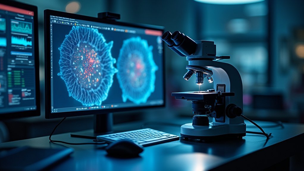
Modern microscopy extends beyond acquisition into powerful digital processing capabilities. Your digital microscope software now offers automated analysis tools that dramatically reduce human error while improving measurement accuracy.
These advanced algorithms process high-resolution images rapidly, letting you identify and quantify cellular structures or particles in a fraction of the time manual methods require.
High-performance algorithms transform digital microscopy with rapid identification and quantification capabilities far exceeding traditional manual analysis.
You’ll benefit from streamlined workflows through integrated software solutions that maintain consistent documentation and traceable records—essential for compliance and quality control.
Features like automated particle counting and EDS chemical composition analysis provide thorough insights into your samples without additional steps.
When working with complex biological samples, you can precisely reconstruct sample volumes in three dimensions, visualizing interactions that would otherwise remain hidden in traditional microscopy approaches.
Multi-Scale Visualization and Correlative Microscopy
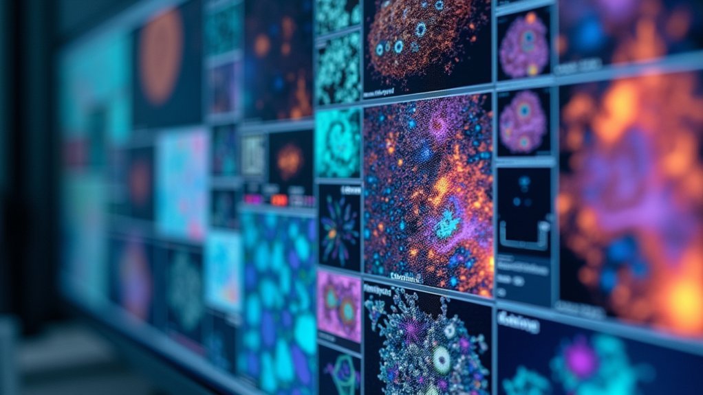
Modern correlative microscopy software lets you integrate diverse imaging data seamlessly, creating extensive visualizations across multiple scales.
You’ll find tools that unify light and electron microscopy datasets, revealing structural relationships impossible to see with a single technique.
These platforms enable multi-modal sample exploration, allowing you to trace cellular processes from macro to nano levels while maintaining spatial context throughout your analysis.
Seamless Data Integration
Because biological research often requires examining specimens across multiple scales, today’s microscope software solutions excel at integrating diverse imaging data into cohesive visualizations.
You’ll benefit from tools that combine widefield, laser scanning, and FIB-SEM microscopy in correlative cryogenic workflows, markedly enhancing imaging accuracy.
Software like Atlas 5 provides customizable modular structures for detailed multi-modal imaging across ZEISS SEM, XRM, and FIB-SEM platforms.
These solutions enable high-contrast volume imaging with precise reconstruction capabilities, ensuring seamless data integration across different microscopy techniques.
When analyzing complex biological samples, you can automate the imaging of hundreds of sections through correlative array tomography, creating unified 3D datasets.
This integration also supports real-time collaboration, allowing your team to share findings efficiently and accelerate research progress.
Multi-Modal Sample Exploration
While traditional microscopy limits researchers to single scales and modalities, today’s multi-modal sample exploration tools transform how you’ll interact with complex specimens.
You’ll benefit from correlative cryo workflows that seamlessly integrate widefield, laser scanning, and focused ion beam techniques to precisely localize fluorescent macromolecules.
The Atlas 5 system enables you to create detailed, multi-scale images customized to your specific research needs. During image acquisition, correlative array tomography automates the imaging of hundreds of biological sections, combining them into thorough 3D datasets.
With SmartPI and nanoscale analysis software, you can automatically count and analyze up to 200,000 particles with elemental precision. Digital microscope cameras and integrated imaging software further streamline your workflow, ensuring high-resolution capture while maintaining efficiency throughout your multi-modal exploration process.
Streamlined Data Management and Documentation
As research and quality control workflows become increasingly complex, digital documentation through microscope image software emerges as a critical solution for maintaining data integrity.
Your extensive software package transforms how you manage microscopy data by creating clear, organized records with timestamps and annotations that track changes accurately over time.
You’ll benefit from automated analysis tools that maintain consistency across different users and inspections, greatly reducing human error and variability.
The centralized digital records enable seamless collaboration among team members, enhancing decision-making while ensuring compliance during audits.
Remote Collaboration and Real-Time Monitoring Capabilities
Modern microscopy transcends physical barriers through powerful remote collaboration features.
You’ll gain immediate access to high-resolution microscope images from anywhere on your network, enabling seamless teamwork regardless of location.
With real-time streaming capabilities, you can instantly share detailed visuals with colleagues for immediate consultation and review.
Collaborate instantly through high-resolution visual streams, enabling real-time feedback regardless of physical location.
This connectivity accelerates decision-making processes in both research and industrial environments, as team members can provide instant feedback and contribute to problem-solving discussions without delay.
The software’s automated measurement and reporting tools further enhance collaboration by standardizing data collection and presentation.
You’ll notice improved productivity as your team members simultaneously access, analyze, and discuss microscope data, making smarter, data-driven decisions while maintaining consistent workflow across distributed locations.
High-Throughput Screening and Batch Processing
Because research efficiency directly impacts discovery timelines, high-throughput screening capabilities transform your microscopy workflow.
When you implement software with batch processing features, you’ll analyze thousands of samples simultaneously instead of tediously processing them one by one.
High-throughput screening software like OpenHiCAMM integrates robotics with image processing, allowing you to track and analyze images across multiple slides automatically.
You’ll notice immediate benefits: experiments that once took weeks can be completed in days or even hours.
Beyond speed, you’ll gain consistency. Automated acquisition and processing deliver reproducible results across experiments, minimizing variability and human error.
This reliability translates to more accurate data and stronger research conclusions.
Advanced Particle Analysis and Quantification

While traditional microscopy might limit your analysis capabilities, advanced particle analysis software revolutionizes how you quantify and characterize microscopic structures.
You’ll gain unprecedented control over your research with tools that automate the analysis of up to 200,000 particles, eliminating human error and ensuring consistent results.
Advanced particle analysis offers three key advantages:
- Chemical composition determination through Energy Dispersive Spectroscopy (EDS) analysis, providing deeper material insights
- Flexible offline processing that allows you to evaluate data using multiple methods without continuous online processing
- Customizable analysis parameters, including the ability to exclude irrelevant particles from your measurements and statistics
This technology streamlines your workflow while dramatically improving accuracy, giving you more reliable data for your research and development projects.
Customizable Reporting and Results Presentation
Modern microscopy software empowers you to transform complex image data into streamlined, visually compelling presentations tailored to your specific audience.
You’ll find intuitive tools for creating custom data visualizations that highlight key findings while maintaining scientific rigor and accuracy.
These reporting capabilities enable you to generate publication-ready outputs with precise formatting requirements, saving valuable time in your manuscript preparation workflow.
Streamlined Data Visualization
Once microscopic images have been captured and processed, your ability to effectively communicate findings becomes paramount. Advanced visualization tools integrated with Microscope Cameras transform complex data sets into understandable visuals that tell compelling stories about your research.
Streamlined data visualization offers you significant advantages:
- Interactive exploration – Manipulate your data dynamically to uncover hidden patterns and relationships that might otherwise remain obscured in static presentations.
- Template automation – Save hours of formatting time while maintaining consistency across multiple projects using pre-designed report templates.
- Integration capabilities – Seamlessly combine high-resolution images with analytical data, creating thorough visualizations that enhance understanding and facilitate better decision-making.
These visualization features guarantee your microscopic findings aren’t just stored but effectively communicated to stakeholders, colleagues, and regulatory bodies.
Tailored Publication Outputs
Publishing your microscopic discoveries effectively requires more than just collecting quality data—it demands presentation tools that meet diverse publication standards.
Modern microscope software empowers you to create tailored publication outputs that align perfectly with your research objectives.
You’ll benefit from customizable report templates that incorporate relevant metrics, annotations, and visualizations specific to your work. The software’s automated analysis capabilities guarantee your results maintain consistency when integrated into final reports.
Your high-resolution imaging can be properly annotated and scaled, providing essential context for reviewers and readers.
Perhaps most valuable is the flexibility to export your findings in multiple formats, facilitating seamless collaboration with colleagues and enhancing your work’s impact.
This adaptability transforms raw microscopic data into compelling visual narratives ready for publication in any scientific forum.
Frequently Asked Questions
What Is Microscope Software?
Microscope software is a program you’ll use to control your microscope and camera, capture images, make adjustments, and perform analysis. It’ll streamline your workflow through automated features and documentation capabilities.
Why Is Microscope Technology Important?
Microscope technology is essential because it lets you visualize microscopic structures, advancing your research capabilities. You’ll discover cellular details invisible to the naked eye, supporting breakthroughs in medicine, biology, and materials science.
How to Present Microscope Images?
To present microscope images effectively, you’ll need high-resolution digital cameras, image analysis software for annotations, organized thumbnail galleries, consistent exposure adjustments, and proper digital documentation with timestamps for context and traceability.
Why Is the Microscope an Important Tool?
The microscope’s your window to the invisible world. It lets you visualize cellular structures, analyze materials, and diagnose diseases. You’ll gain insights that are essential for advancing biological, medical, and materials research.
In Summary
You’ve seen how microscope software transforms your imaging workflow. It’s not just about integration and automation—it’s about accessing deeper analysis and collaboration. Whether you’re processing thousands of images or extracting precise measurements, software expands what’s possible with your microscope. By implementing these digital tools, you’ll save time, increase accuracy, and ultimately make more meaningful discoveries from your microscopy data.


Leave a Reply