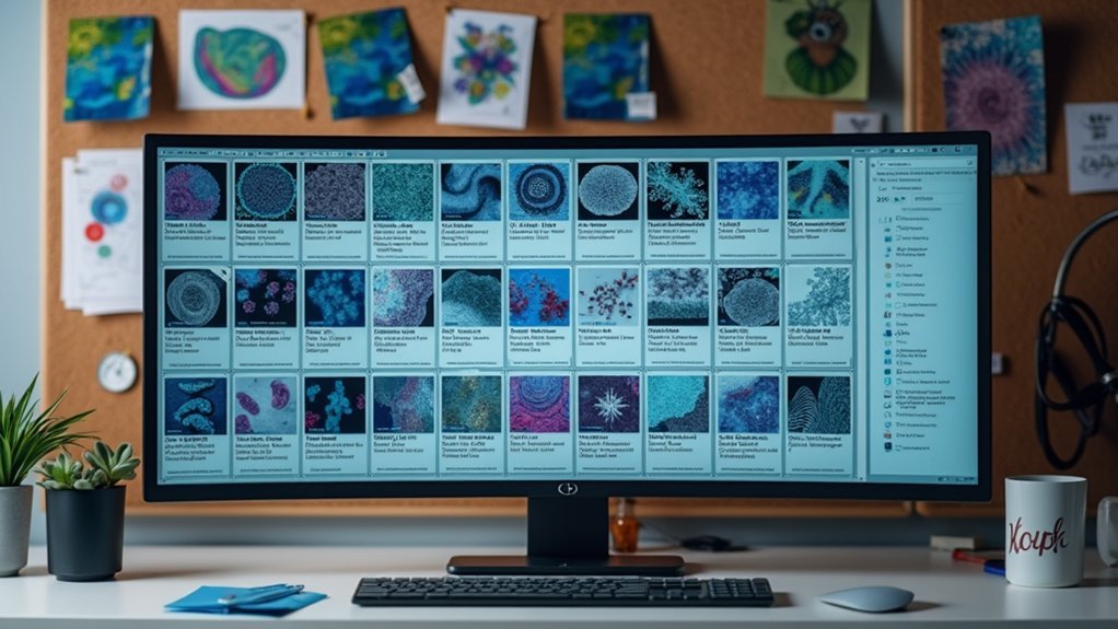
When naming scientific images, you’ll want to: use lowercase for researcher names, include lab identifiers as prefixes, add descriptive metadata tags, implement version numbers (e.g., “_v01”), incorporate instrument-specific codes, add resolution indicators, specify sample preparation methods, use microscopy technique identifiers, apply sequential numbering with leading zeros, and integrate standardized metadata tags. These practices prevent confusion, streamline collaboration, and guarantee proper tracking of your research materials. The following standards will transform your image management system.
Use Descriptive Metadata Tags for Experiment Details

Metadata serves as the backbone of effective scientific image organization. When tagging your images, always include descriptive metadata tags with key experiment details such as the experiment name, conditions, and tested variables. This practice dramatically improves identification and retrieval efficiency within databases.
To enhance searchability, incorporate relevant contextual information like dates, your initials, and project codes. Using controlled vocabulary consistently will reduce ambiguity when categorizing images across different experiments. These tags also help track modifications and versioning, guaranteeing you can always reference the correct iteration of your data.
For maximum interoperability between systems, adopt standardized metadata formats like Dublin Core or Exif. These established frameworks guarantee your scientific images remain accessible and properly contextualized across various platforms.
Establish Instrument-Specific Identifiers
Including instrument identifiers in your file names provides immediate context about how an image was captured.
You’ll benefit from a consistent coding system that incorporates microscope resolution details, such as “SEM_1500x” for scanning electron microscopy at 1500x magnification.
Don’t forget to add essential equipment parameters like “CONFOCAL_488nm_60%laser” to document specific imaging conditions that affect interpretation and reproducibility.
Instrument Prefix Coding System
The foundation of any robust scientific image filing system begins with a consistent instrument prefix coding system. By assigning unique identifiers to each scientific instrument in your lab, you’ll streamline file organization and enhance searchability.
Implement standardized abbreviations like “SEM” for scanning electron microscopes or “TEM” for transmission electron microscopes at the beginning of each filename.
- Create a thorough list of instrument prefixes for all equipment in your facility
- Document your coding system in a shared readme file accessible to all team members
- Train new researchers on your prefix conventions during onboarding
- Review your system annually to incorporate new technologies
- Use three-letter codes whenever possible for consistency (e.g., AFM, NMR, XRD)
This approach guarantees everyone can quickly identify image sources and maintain organizational integrity across projects.
Microscope Resolution Indicators
Precision in documenting resolution capabilities transforms ordinary file names into informative research assets. When establishing your file naming conventions, include both microscope type and resolution parameters—for example, “Confocal_1024x1024” or “TEM_300kV”—to instantly communicate imaging specifications.
Adding resolution indicators like “Image01_512x512” enables quick sorting and identification of image quality levels across your datasets. This practice prevents confusion when sharing data with colleagues and streamlines retrieval in collaborative environments.
For ideal organization, pair these indicators with a good format for date and instrument prefixes. Your microscope resolution indicators should follow a consistent pattern across all files.
Document these standards in a readme.txt file to make certain all team members adhere to the same conventions, maintaining clarity and efficiency throughout your research workflow.
Equipment-Specific Parameter Notations
Effective scientific documentation hinges on your ability to encode equipment details directly into file names. When implementing a file naming convention for scientific images, incorporate equipment-specific metadata to guarantee traceability across different instruments and experimental conditions.
- Add instrument model abbreviations (like “FTIR” or “SEM”) at the beginning of filenames.
- Include unique identifiers for wavelength settings, magnification levels, or detector types.
- Document calibration dates or operational modes to provide context for data interpretation.
- Use standardized abbreviations for common parameters to maintain brevity.
- Create a master reference spreadsheet linking filename codes to detailed equipment information.
Implement Date and Time Formatting Using ISO Standards
When organizing scientific image files, consistent date and time formatting becomes essential for efficient data management and retrieval. Adopt the ISO 8601 format for date designations by using YYYY-MM-DD in your file names. This standard guarantees chronological sorting and eliminates ambiguity across international collaborations.
For images where timing is critical, extend your naming convention to include HH-MM-SS, creating complete timestamps like 2023-11-15_14-30-22. Always maintain four-digit years and two-digit months and days for consistency, even for single-digit values (e.g., 2023-01-05 rather than 2023-1-5).
This standardized approach helps you track experimental progressions, manage version control, and guarantees your data remains properly organized across different systems and software platforms.
Structure Filenames by Magnification Levels and Settings

When structuring scientific image filenames, you’ll want to categorize them by resolution scales to quickly identify the level of detail captured.
Separate optical microscopy images (typically 4x-100x) from electron microscopy files (often denoted in thousands, like 5000x) to prevent confusion between these fundamentally different imaging techniques.
Always include the objective magnification settings in your filename (e.g., “40x_BrightField” or “1000x_TEM”) to provide immediate context about how the image was acquired.
Categorize by Resolution Scales
Scientific images captured at varying magnifications require systematic naming to maintain order in your research collections. When categorizing by resolution scales, include the magnification level directly in your filename (e.g., “10x” or “1000x”) to instantly identify the scale of observation.
Establish a standardized prefix for each magnification level and add the imaging technique used, such as “brightfield” or “fluorescence,” to create an informative naming structure.
- Use consistent magnification notation (e.g., “40x_brightfield_001”)
- Include imaging settings in every filename (e.g., “100x_fluorescence_sample3”)
- Implement a sequential numbering system for multiple images of the same type
- Group related images with standardized prefixes (e.g., “EM_1000x_tissue_005”)
- Maintain consistency between resolution scale indicators across all research projects
Separate Optical From Electron
Distinguishing between optical and electron microscopy images requires clear structural differences in your file naming conventions. For good practices, always prefix filenames with “Optical_” or “Electron_” to quickly identify the imaging technique used.
For optical images, incorporate magnification levels like “100x” or “400x” directly in the filename to indicate resolution detail. With electron microscopy files, include the accelerating voltage (e.g., “20kV”) to document critical imaging parameters.
Combine these elements with your sample identifier for thorough context: “Optical_SampleA_100x.jpg” or “Electron_SampleB_20kV.tif”. This systematic approach guarantees your image collection remains organized and searchable.
Maintaining consistent file naming across your entire project makes comparing images at different magnifications and settings straightforward, saving valuable time during analysis and publication preparation.
Denote Objective Magnification Settings
Three essential components define effective magnification notation in scientific image filenames. By clearly indicating the objective power, microscopy type, and optical settings, you’ll create a system that enables quick identification and efficient file sorting.
Your naming convention should consistently position magnification information—whether you place “10X” at the beginning or end of your file naming structure matters less than doing it uniformly across your dataset.
- Use standard abbreviations (10X, 40X, 100X) for instant recognition
- Pair magnification with microscopy type (LM-40X or 40X-EM)
- Include special conditions like “oil” or “dry” when relevant
- Implement a consistent format for version numbers alongside magnification
- Consider creating a lab-specific magnification code system for complex imaging techniques
Include Sample Preparation Methods in Naming Scheme

When developing your file naming standards, don’t overlook the critical role sample preparation methods play in experimental context. Incorporating preparation techniques directly into filenames—such as “SamplePrep-MethodA-SampleID” or specific methods like “FreezeDrying”—provides essential information for reproducibility and accurate interpretation.
Sample preparation method details embedded in filenames enhance reproducibility and prevent critical context loss.
This good practice allows colleagues to immediately understand how samples were handled prior to imaging, reducing misinterpretation risks.
You’ll find that standardized preparation method inclusion facilitates efficient sorting and filtering within research databases, making it easier to compare results across similar techniques.
For streamlined file naming standards, consider using concise codes for common methods (e.g., “SPF” for “Sample Preparation Freeze-dried”) while maintaining clarity. This approach guarantees critical experimental conditions remain documented without creating unwieldy filenames.
Create Consistent Versioning for Edited Images
As scientific images undergo multiple processing stages, implementing a robust versioning system becomes crucial for maintaining data integrity.
You’ll need to establish clear naming conventions that track modifications throughout your workflow. Incorporate version numbers directly into filenames by adding “_v01” for initial versions and incrementing for subsequent edits.
When you’ve adjusted an image, add standardized modification codes like “-e” for edited or “-c” for cropped alongside the version number.
- Use simple version indicators (avoid “_v001” in favor of “_v01”)
- Apply modification codes consistently (e.g., “-e” for edited images)
- Document all changes in a master spreadsheet for reference
- Maintain a consistent format across all image files
- Make certain all team members understand your versioning system
This consistent format guarantees everyone can quickly identify the most current edited versions while preserving modification history.
Develop Standardized Abbreviations for Cell Types

Beyond tracking image versions, the actual subject of your scientific imagery requires careful labeling. Implementing standardized abbreviations for cell types enhances clarity in your file naming system and facilitates seamless communication across the scientific community.
Choose intuitive, widely recognized abbreviations that allow for efficient sorting and searching in collaborative environments. Document these in a README file to guarantee consistency among team members.
| Abbreviation | Cell Type | Origin | Usage |
|---|---|---|---|
| HEK | Human Embryonic Kidney | Human | HEK293_GFP_20230615 |
| PC12 | Pheochromocytoma | Rat | PC12_NGF_48h_20230521 |
| HUVEC | Umbilical Vein Endothelial | Human | HUVEC_Angiogenesis_v2 |
| MEF | Mouse Embryonic Fibroblast | Mouse | MEF_p53KO_IF_20230412 |
Consistent abbreviations prevent confusion, minimize errors in data management, and improve workflow efficiency when handling multiple cell types in your research.
Organize Filenames With Microscopy Technique Identifiers
Incorporating microscopy technique identifiers at the beginning of your filenames provides immediate visual cues about the imaging method used.
You’ll find it helpful to maintain a documented list of standardized abbreviations for each technique (CLSM for confocal laser scanning microscopy, SEM for scanning electron microscopy) to guarantee consistency across research teams.
Cross-reference these identifiers with specific imaging parameters in your metadata to create a robust system that facilitates efficient image retrieval and analysis.
Microscopy Technique Naming Prefixes
When organizing scientific image files, microscopy technique identifiers serve as essential prefixes that immediately communicate the imaging method used. By including standardized prefixes like “EM” for Electron Microscopy or “FL” for Fluorescence Microscopy, you’ll guarantee clarity and consistency across your research datasets.
Consistent use of these identifiers offers numerous benefits:
- Enables efficient sorting and searching of images by technique
- Facilitates immediate identification without opening files
- Improves collaboration by establishing shared protocols
- Reduces risk of mislabeling in large datasets
- Enhances overall data organization in research projects
This standardized approach is particularly valuable when handling diverse microscopy data or when collaborating with other researchers.
Your team will understand the context of each image instantly, streamlining analysis and preventing confusion during your scientific workflow.
Document Instrument-Specific Abbreviations
Building upon microscopy technique naming prefixes, an extensive documentation system for instrument-specific abbreviations provides the foundation for consistent file organization.
To document instrument-specific abbreviations effectively, create a centralized glossary in your project’s readme file. It’s a good idea to standardize these identifiers across your team to guarantee files remain searchable and properly categorized. Include both the instrument model and technique parameters in your naming convention.
| Abbreviation | Full Description |
|---|---|
| ZA700-SEM | Zeiss Auriga 700 Scanning Electron Microscope |
| LCS-CLSM | Leica Confocal System – Confocal Laser Scanning Microscopy |
| N80-TEM-200x | Nikon 80i Transmission Electron Microscopy at 200x magnification |
Cross-Reference Imaging Parameters
Effective microscopy file management depends on properly cross-referencing imaging parameters within your filenames. When organizing microscopy images, use consistent format that includes technique identifiers like “confocal,” “SEM,” or “TEM” to immediately clarify the method used.
Avoid spaces in file names; instead, use underscores or hyphens to separate important elements while maintaining a logical sequence.
- Include magnification levels directly in filename (e.g., SEM_40x_Sample3)
- Standardize alphanumeric characters for consistent sorting (01, 02 rather than 1, 2)
- Position technique identifiers at the beginning for easier filtering
- Incorporate unique identifiers for differentiating similar images
- Document your abbreviation system in a README file for team reference
This standardized approach guarantees images are easily retrievable and contextually rich, even years after collection.
Format Researcher Names and Lab Identifiers Consistently
Three critical elements of scientific image file naming are researcher names, lab identifiers, and their consistent formatting. When organizing your files, standardize researcher names by placing the last name first, followed by the first name (e.g., “smith-john-experiment.jpg”).
Always use lowercase letters for both researcher names and lab identifiers to prevent retrieval issues. The format makes sure all team members can locate files regardless of search method.
Stick with lowercase for researcher names and lab identifiers to ensure everyone can find files, no matter how they search.
Include lab identifiers as prefixes (e.g., “labA_smith-john_experiment_v01.jpg”) to immediately signify ownership and context. Document standardized abbreviations for labs in a readme.txt file for reference.
Each researcher or lab should have a unique identifier to avoid confusion during collaborations. This consistency is especially important when tracking the first letter of each component in complex naming structures.
Design Sequential Numbering Systems for Image Series

Sequential numbering systems provide the backbone of effective image series management in scientific research. When organizing experimental images, implement a consistent approach using leading zeros for clarity (e.g., 001, 002) to maintain uniform file lengths and guarantee proper sorting in directories. Your system should include descriptive prefixes that identify the context, such as “experiment1_001.jpg,” making image identification immediate.
- Use three-digit numbering (001-999) to accommodate future additions
- Include category identifiers before sequence numbers (e.g., treatment1_001.jpg)
- Maintain consistent digit length with leading zeros (01 instead of 1)
- Guarantee sequence reflects the intended documentation order
- Incorporate relevant metadata tags alongside numbers for enhanced context
This naming best practice prevents confusion during analysis and streamlines collaboration, especially in projects with hundreds of related images requiring systematic organization.
Frequently Asked Questions
What Is the Best Naming Convention for Images?
You’ll improve image visibility by using lowercase names with hyphens, starting with destination page, followed by sequence number, relevant keywords, and business name. Don’t exceed 60 characters or use unusual characters to guarantee compatibility.
What Is a Good Naming Convention for Files?
For good file naming, you’ll want to include your project name, initials, and date (YYYYMMDD). Use hyphens instead of spaces, add descriptive keywords, implement version control (v01), and keep names under 50 characters.
What Is the Standard Photo File Name?
Standard photo file names should include descriptive keywords, use lowercase letters with hyphens, incorporate date (YYYYMMDD), and remain under 60 characters. You’ll want to add version numbers (-v01) for tracking edits when necessary.
What Are the Three Points When Naming Image Files?
When naming image files, you’ll want to use descriptive keywords first, replace spaces with hyphens, and keep names short but meaningful. This improves searchability while ensuring technical compatibility across different systems.
In Summary
You’ll find that adopting these file naming standards drastically improves your lab’s workflow efficiency and data management. When you’re consistent with descriptive metadata, standardized formats, and logical organization, you’re building a foundation for seamless collaboration and reproducible science. Remember, well-named files don’t just save time—they’re essential for maintaining research integrity and ensuring your valuable scientific images remain accessible for years to come.


Leave a Reply