
For microscope image annotation, you’ll find several excellent online tools to enhance your research workflow. ImageJ/FIJI offers extensive scientific analysis capabilities, while Cell Profiler excels at batch processing. Labelbox and V7 Darwin provide AI-assisted annotation features, and CVAT delivers precise polygon and keypoint marking. SuperAnnotate streamlines complex projects with advanced management tools, and all platforms offer free trials. Explore these options to discover which interface best matches your specific microscopy needs.
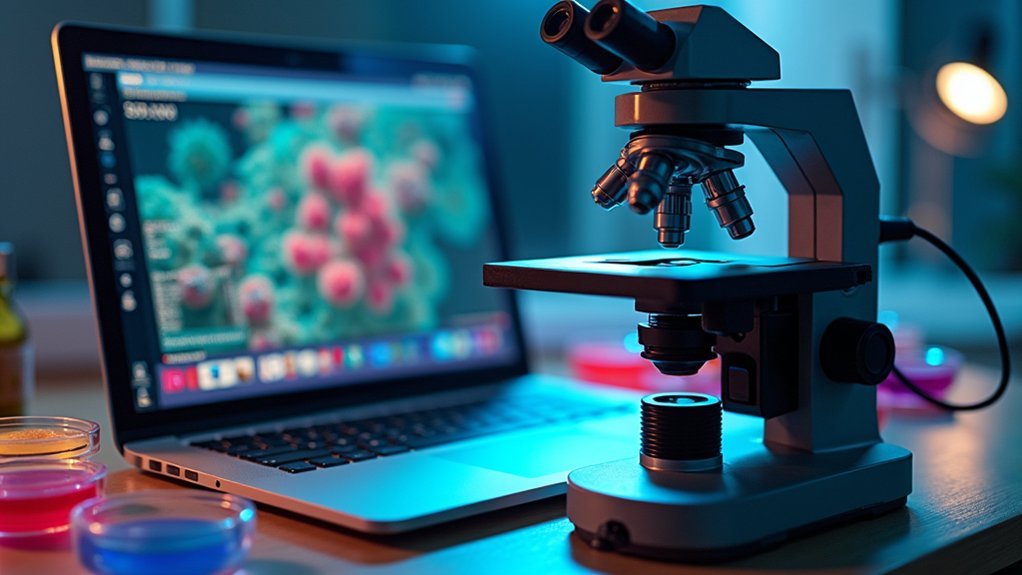
7 Best Online Tools For Microscope Image Annotation
When working with microscopic imagery, you’ll need reliable annotation tools to analyze and categorize your findings effectively. ImageJ/Fiji offers beginner-friendly interfaces perfect for your first annotation projects, while open-source platforms like CVAT and Labelme enable collaborative work on complex microscopy datasets.
Effective microscopy analysis demands reliable annotation tools – from beginner-friendly ImageJ to collaborative platforms like CVAT for complex datasets.
For larger research initiatives, consider SuperAnnotate or Dataloop with their advanced project management features and AI-assisted labeling that dramatically improve efficiency. These microscopy annotation tools support various export formats including COCO and JSON, ensuring seamless integration with your machine learning workflows.
Most platforms offer free trials, allowing you to test their user-friendly interfaces before committing.
Whether you’re annotating individual cells or tissue samples, you’ll find options ranging from simple open-source solutions to thorough commercial platforms tailored to your specific research needs.
ImageJ/FIJI: Comprehensive Platform for Scientific Image Analysis
Although many annotation tools exist, ImageJ/FIJI stands out as the gold standard for scientific microscopy analysis. This powerful open-source software provides you with essential capabilities for processing microscopy images across various formats.
You’ll find it particularly valuable for manipulating fluorescence images through adjustments to brightness, contrast, cropping, and labeling.
Key advantages of ImageJ/FIJI include:
- Seamless handling of 3D stacks from confocal microscopy, enabling thorough quantitative analysis of three-dimensional datasets
- Pre-bundled plugins that expand functionality for specialized applications in neuroscience and beyond
- Compatibility with numerous image formats, making it adaptable to different microscopy systems
- Access to a robust online community offering extensive documentation, tutorials, and troubleshooting resources
Cell Profiler: Automated Processing for High-Volume Microscopy Data

Cell Profiler excels at handling thousands of microscope images through its powerful batch processing capabilities, saving you countless hours of manual work.
You’ll find the software’s custom analysis pipelines particularly valuable when extracting specific cellular measurements across large datasets. These pipelines can be tailored to your exact research needs, enabling automated detection of features like fluorescence intensity, morphology, and cell population dynamics.
Streamlined Batch Processing
For researchers drowning in microscopy data, batch processing represents a critical lifeline. Cell Profiler’s streamlined batch capabilities let you process thousands of microscopy images simultaneously through automated pipelines, dramatically reducing analysis time.
You’ll appreciate the intuitive workflow that transforms image annotation from tedious manual work into an efficient, reproducible process.
With Cell Profiler’s batch processing, you can:
- Design customizable image processing workflows once and apply them across entire datasets
- Extract quantitative measurements like fluorescence intensity and cell density automatically
- Analyze time-lapse movies to track cellular changes across multiple timepoints
- Process complex datasets with consistent parameters, ensuring reliable results
This high-throughput analysis approach means you’ll spend less time on repetitive tasks and more time interpreting meaningful biological insights from your microscopy data.
Custom Analysis Pipelines
Beyond batch processing, the true power of Cell Profiler emerges through its custom analysis pipelines. You’ll create tailored workflows that transform your microscopy data into valuable quantitative measurements with remarkable efficiency.
Whether you’re performing cell segmentation, feature extraction, or tracking cellular changes over time, this open-source software adapts to your specific research requirements.
Cell Profiler excels with diverse image types, supporting both fluorescence imaging and brightfield techniques. Built-in modules let you quantify antibody fluorescence intensity and cell density—critical metrics for thorough image annotation.
Labelbox: AI-Assisted Microscopic Annotation Solutions
Labelbox’s AI-assisted annotation tools will dramatically speed up your cell analysis workflows by automating repetitive labeling tasks.
You’ll find the polygon precision capabilities especially valuable when marking irregular cellular structures that require exact boundary definitions.
These advanced annotation features let you capture even the most minute details in your microscopic samples while maintaining scientific accuracy.
AI Accelerates Cell Analysis
While traditional microscope image annotation remains time-consuming, Labelbox has revolutionized this process with AI-assisted labeling tools specifically designed for cellular analysis.
You’ll experience dramatic efficiency gains as machine learning algorithms pre-label your microscopy images, allowing your team to focus on refinement rather than starting from scratch.
Labelbox enhances your cell analysis workflow through:
- Advanced annotation options including polygons and bounding boxes tailored for microscopic structures
- Customizable interface that simplifies complex annotation tasks for non-technical team members
- Collaborative features enabling multiple researchers to annotate simultaneously for faster project completion
- Seamless API integration that incorporates AI-assisted labeling into your existing microscopy workflow
The platform’s intelligent approach to cell analysis means you’ll get more accurate annotations in less time, ultimately accelerating your research outcomes.
Polygon Precision Capabilities
Among Labelbox’s standout features, the polygon precision tools offer unparalleled accuracy when annotating complex microscopic structures. You’ll find that these advanced polygon annotation capabilities enable you to delineate intricate details within your microscope images with remarkable precision.
The AI-assisted labeling function accelerates your workflow by suggesting polygon shapes based on existing patterns, dramatically reducing the time you spend on manual annotations.
Labelbox’s customizable interface lets you streamline data labeling processes, making it easier to manage large datasets efficiently.
What’s more, the platform supports team collaboration, allowing multiple researchers to contribute to polygon annotations simultaneously. This collaborative approach proves especially valuable when working on extensive microscopy projects where accuracy and efficiency are paramount to successful AI model training.
V7 Darwin: Medical-Grade Image Annotation for Research Applications

For researchers handling sensitive microscopy data, V7 Darwin stands out as a specialized annotation platform designed with regulatory compliance at its core. This solution supports FDA compliance while handling complex medical image annotations, including ultra-high-resolution and multi-spectral formats essential for advanced research applications.
What makes V7 Darwin particularly valuable for microscopy work:
- User-friendly automation features accessible to non-technical researchers, reducing annotation time dramatically.
- Composable workflows that adapt to complex, multi-stage research protocols.
- Seamless integration of autoML model training directly into your dataset management pipeline.
- Regulatory adherence (FDA, CE, HIPAA) that protects sensitive research data while enabling collaborative work.
You’ll appreciate how Darwin streamlines the entire AI lifecycle, from initial annotations through model improvement, without requiring coding expertise.
CVAT: Open-Source Annotation Tool for Detailed Microscopic Features
When budgets are tight but annotation needs remain complex, CVAT offers a compelling alternative to commercial platforms. This Intel-developed open-source tool excels at both image annotation and video annotations of microscopic features. You’ll find its semi-automatic annotation capabilities particularly useful for accelerating tedious microscopy work while maintaining precision.
| Feature | Benefit | Use Case |
|---|---|---|
| Polygon annotation | Precise outlining | Cell membrane tracing |
| Keypoints | Structure marking | Organelle identification |
| Collaborative features | Team efficiency | Multi-researcher projects |
CVAT’s user-friendly interface accommodates both beginners and experts, making it accessible across skill levels. The platform’s support for varied annotation types—including bounding boxes, polygons, and keypoints—ensures you can accurately capture even the most intricate microscopic structures in your research datasets.
SuperAnnotate: Streamlined Workflow for Complex Microscopy Projects

SuperAnnotate stands out as an extensive solution specifically engineered for tackling intricate microscopy imaging projects. Its platform streamlines your annotation workflow with powerful tools designed for scientific image analysis.
- Comprehensive annotation capabilities – Supports both object detection and semantic segmentation, critical for precise cellular and subcellular structure identification.
- AI-assisted labeling – Leverages machine learning to accelerate your annotation process, reducing manual effort when working with complex datasets.
- Advanced project management tools – Includes analytics and filtering features that help you monitor progress and organize large microscopy projects efficiently.
- Risk-free evaluation – Take advantage of their 14-day free trial to test SuperAnnotate’s capabilities before integrating it into your research workflow.
Frequently Asked Questions
Which Tool Is Used for Annotating Images?
For annotating images, you can use ImageJ/Fiji, Labelme, CVAT, VoTT, or SuperAnnotate. Each tool offers specific features like arrows, polygons, bounding boxes, and collaborative options for your annotation needs.
What Is the Best Annotation Tool for OCR?
For OCR annotation, you’ll find Tesseract is highly regarded for its accuracy and open-source flexibility. You can also consider commercial options like ABBYY FineReader or cloud services from Amazon Textract, Google Cloud Vision, or Microsoft Azure.
Does Google Have an Annotation Tool?
Google doesn’t have a dedicated annotation tool, but you can use Google Slides, Drawings, Docs, or Photos for basic image annotations. For more sophisticated OCR annotations, you’ll need specialized third-party tools.
How Do You Annotate a Picture Online?
You can annotate pictures online by choosing a web-based tool like MakeSense.AI or VGG Image Annotator, uploading your image, and using their tools to add bounding boxes, polygons, or labels.
In Summary
You’ve now explored seven powerful tools to enhance your microscope image annotation workflow. Whether you’re analyzing complex cellular structures with ImageJ or leveraging AI assistance through Labelbox, there’s a solution to match your specific research needs. Don’t hesitate to experiment with multiple platforms as your projects evolve. The right annotation tool won’t just save you time—it’ll reveal deeper insights in your microscopy work.


Leave a Reply