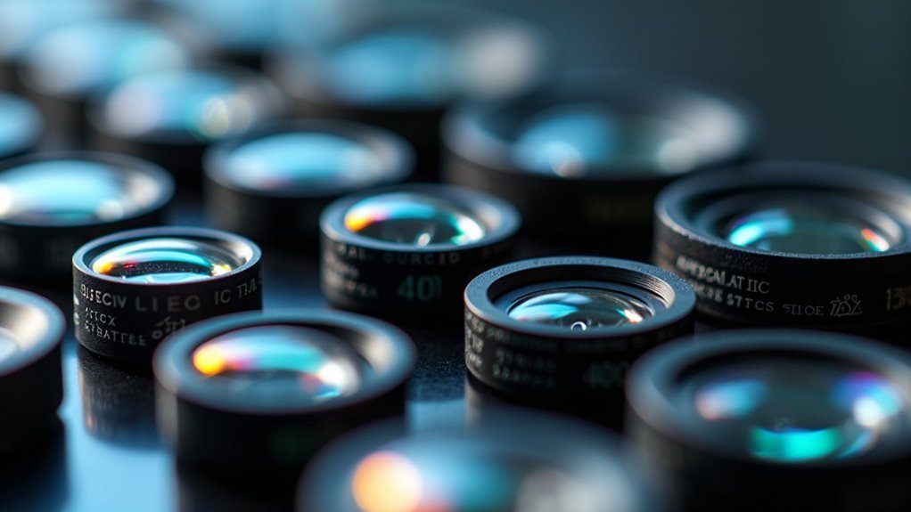
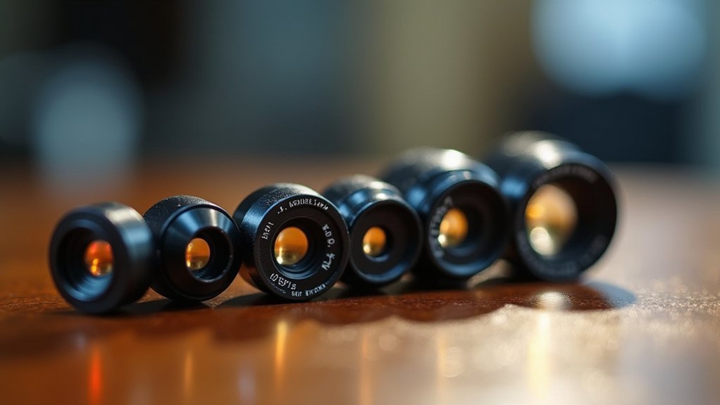
Microscope objectives range from low to high magnification to suit various needs. You’ll find 1X-2X for wide views of large specimens, 4X for scanning and navigation, 10X for routine examination, 20X for balanced detail, 40X for cellular structures, 60X for enhanced resolution, and 100X oil immersion for bacterial visualization. Specialized options include phase contrast for transparent specimens and metallurgical objectives for opaque materials. Each power reveals different levels of microscopic discovery.
1X to 2X Objective Lenses: Perfect for Large Specimen Overview

When beginning your microscopic examination, scanning objectives in the 1X to 2X range provide an invaluable first look at your specimen.
These low-power lenses offer a wide field of view that helps you quickly locate areas of interest before moving to higher magnifications.
You’ll appreciate the extended working distance these objectives provide, allowing you to observe thicker samples without risking contact damage.
With a numerical aperture typically below 0.1, these lenses prioritize depth of field over resolution—perfect for surveying larger specimens but less suitable for detailed cellular studies.
Use these scanning objectives as your starting point in a systematic microscopy workflow.
They’ll help you efficiently map your specimen before zooming in with higher-power lenses to explore specific regions in greater detail.
4X Scanning Objectives: Locating Areas of Interest
As you begin your microscopic exploration, 4X scanning objectives serve as your essential navigation tool. These low-power objectives, typically ranging from 2X to 4X, provide you with a thorough view of your specimen before diving into detailed analysis.
You’ll appreciate the extended working distance that scanning objectives offer, minimizing the risk of damaging delicate specimens during your initial examination. When paired with a 10X eyepiece, your 4X objective delivers a total magnification of 40X—perfect for establishing context.
| Benefit | Application | Advantage |
|---|---|---|
| Wide field of view | Initial specimen scan | Efficient overview |
| Extended working distance | Delicate specimens | Reduced damage risk |
| Low magnification (2-4X) | Orientation establishment | Better context |
| First step in workflow | Locating regions of interest | Time efficiency |
10X Objectives: The Standard for Routine Laboratory Work
When you’re conducting routine laboratory work, the 10x objective serves as your versatile multi-purpose lens for a wide range of applications from cell counting to tissue examination.
This magnification hits the sweet spot between the broader scanning view and the detailed high-power objectives, making it perfect for initial specimen screening before moving on to higher powers.
You’ll find the 10x objective particularly valuable for quickly evaluating specimen quality and identifying specific regions that warrant closer examination with higher magnifications.
Versatile Multi-Purpose Applications
Why do most laboratories standardize around specific objective powers? The answer lies in versatility and efficiency.
Most research facilities rely on a standard set of objective lenses (4x, 10x, 40x, and 100x) because they provide the perfect balance of magnification and numerical aperture for routine work.
- The 4x scanning objective gives you a thorough view to quickly locate specimens.
- 10x and 40x objectives handle most daily observations, from tissue samples to cellular structures.
- High magnification objectives like the 100x oil immersion lens reveal the finest details when needed.
This standardization guarantees your lab can efficiently shift between different magnification needs without unnecessary lens changes or equipment adjustments.
Each objective serves a specific purpose while complementing the others in a well-designed microscopy workflow.
Ideal Specimen Screening Tool
Among the standardized objectives in laboratory settings, the 10X objective stands out as the workhorse for initial specimen examination. This low-power lens delivers an excellent balance between objective magnification and field of view (FOV), making it perfect for quickly scanning slides to locate regions of interest.
With its wider FOV, you’ll easily identify specimen structures worth examining in greater detail. Though its numerical aperture (NA) of approximately 0.25 is lower than high-power objectives, this compromise enables you to see more of your specimen at once.
When you’re maneuvering through complex tissue sections or searching for specific cellular features, the 10X objective serves as your essential guiding tool before shifting to higher magnifications like 40X or 100X for more detailed analysis.
20X Objectives: Balancing Detail and Field of View
The 10x-20x objectives offer you ideal balance for cell biology work, revealing organelles and tissue structures while maintaining enough context to interpret cellular relationships.
You’ll appreciate their practical resolution and working distance, allowing clear visualization without the tedium of frequent refocusing or slide repositioning.
These versatile objectives serve as true laboratory workhorses, efficiently handling the majority of routine specimens from blood smears to tissue sections with minimal adjustment needed.
Applications in Cell Biology
Cell biologists routinely face the challenge of selecting appropriate microscope objectives that balance detailed visualization with sufficient field of view.
When examining cellular structures, you’ll typically begin with low power objectives (4x-10x) to locate your region of interest before shifting to high power objectives (40x-100x) for detailed examination of organelles and cell membranes.
For cellular work, consider these key objective types:
- Phase contrast objectives for observing unstained, living cells in their natural state
- Oil immersion objectives (100x) for maximum resolution of fine cellular details
- High numerical aperture lenses (NA 1.25-1.45) for distinguishing closely spaced structures
The numerical aperture is particularly critical in cell biology, as higher NA values directly correlate with improved resolution—essential when examining minute subcellular components and their interactions.
Resolution and Working Distance
Selecting ideal microscope objectives requires understanding the fundamental tradeoff between resolution and working distance. As magnification increases, you’ll gain resolution but sacrifice working space between your objective and specimen.
Lower-power objectives (4x-10x) provide generous working distances, allowing you to scan larger areas and locate your specimen. These objectives offer wider fields of view but less detail.
Higher magnifications (40x-100x) deliver exceptional detail for examining cellular structures but require careful positioning due to their minimal working distance. The numerical aperture (NA) of these objectives increases with magnification, improving resolution and light-gathering capability.
Oil immersion objectives (typically 100x) maximize resolution by eliminating air between the lens and specimen, but they demand extremely short working distances and precise handling of immersion oil.
Versatile Laboratory Workhorse
While understanding resolution and working distance fundamentals sets the stage for objective selection, most laboratories rely heavily on a specific range of versatile objectives for daily work.
These standard magnifications—10x, 20x, and 40x—are considered the workhorses of microscopy, providing ideal balance between detail and field of view (FOV).
- The 10x objective offers low-power scanning capabilities with a wide FOV, making specimen location straightforward.
- Your 20x objective bridges the gap, revealing cellular structures while maintaining adequate FOV for contextual observation.
- The 40x high dry objective delivers detailed examination with higher numerical aperture for histological and cellular analysis.
When paired with standard 10x eyepieces, these objectives are designed to provide total magnifications of 100x, 200x, and 400x—covering most routine laboratory applications efficiently.
40X High Dry Objectives: Cellular Structure Examination
Many researchers consider high dry objectives the workhorses of cellular examination, offering magnifications between 40x and 100x without requiring immersion oil.
These powerful objectives feature numerical apertures of 0.65 or higher, enabling you to observe intricate cellular structures with impressive clarity while gathering maximum light for detailed imaging.
You’ll find these objectives particularly valuable when studying cell membranes, organelles, and mitotic stages. Their oil-free design reduces contamination risks and simplifies your workflow when analyzing multiple samples.
With appropriate working distances between lens and specimen, high dry objectives protect your delicate samples from damage during observation.
They’re especially useful in histology and cytology applications where precise cellular morphology assessment is critical for accurate diagnosis and thorough tissue analysis.
60X Objectives: Enhanced Resolution for Tissue Analysis

When examining tissue specimens at the cellular level, you’ll need 100X oil immersion objectives to resolve fine structural details invisible at lower magnifications.
These high-powered objectives with numerical apertures exceeding 1.25 allow you to visualize critical tissue components like nuclear chromatin patterns and bacterial infiltrates with remarkable clarity.
While 60X objectives may lack sufficient magnification for certain histopathological assessments, properly selected 100X objectives overcome this limitation by reducing chromatic aberrations and providing the flat field imaging necessary for thorough tissue analysis.
High Magnification Immersion Applications
To achieve the exquisite detail needed for advanced tissue analysis, high magnification immersion objectives ranging from 40x to 100x play an essential role in modern microscopy.
When you’re examining cellular structures in histopathology or cytology, these powerful objectives reveal intricate details invisible at lower powers.
Immersion oil dramatically enhances your resolving power by eliminating light refraction at the glass-air interface, especially with numerical apertures above 1.0.
You’ll experience:
- Considerably improved clarity and sharpness when analyzing stained tissue sections
- Enhanced ability to distinguish between closely spaced cellular features
- Superior performance when combined with appropriate illumination techniques
For ideal results, pair your immersion objectives with brightfield or fluorescence microscopy to maximize contrast and visibility when examining critical cellular morphology and pathological changes.
Resolving Cellular Structures
Building upon immersion techniques, X objectives represent the pinnacle of cellular visualization technology in modern microscopy.
When you’re examining tissue samples, high power objectives (40x-100x) deliver the magnification necessary to resolve intricate cellular structures and critical diagnostic features.
These objectives feature numerical apertures exceeding 0.65, dramatically improving your ability to distinguish minute details within specimens.
Oil immersion versions can boost resolution by approximately 1.5 times compared to air objectives, revealing cellular components that would otherwise remain invisible.
For histological studies, you’ll appreciate objectives corrected for chromatic aberration, preserving color accuracy essential for proper tissue analysis.
The specialized coatings on fluorescence-compatible objectives further enhance your capacity to observe subtle differences in fluorescently labeled cellular structures, making complex tissue examination more precise and reliable.
60X Lens Limitations
Despite their remarkable resolving power, high magnification objectives (40x-100x) present several practical limitations you’ll need to navigate.
When working with these high numerical aperture lenses, you’ll encounter markedly reduced working distances, requiring precise focus adjustments to prevent damaging delicate tissue samples.
- Oil immersion objectives (100x) deliver superior resolution but demand meticulous technique—improper oil application can compromise image quality and potentially damage specimens.
- Light management becomes critical at higher magnifications; even slight illumination issues can render tissue features indistinguishable.
- Field of view narrows considerably, forcing you to scan larger areas incrementally rather than observing them holistically.
These constraints require careful technique, but mastering them enables you to capture cellular details essential for accurate tissue analysis and pathological assessment.
100X Oil Immersion Objectives: Bacterial and Subcellular Visualization
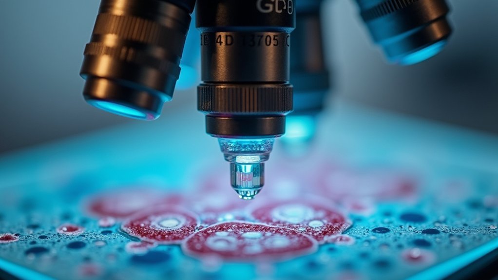
When scientists need to visualize the intricate details of bacterial cells or subcellular structures, they turn to oil immersion objectives, which typically offer magnifications between 100x and 1000x.
These powerful objectives use immersion oil that matches glass’s refractive index, dramatically increasing numerical aperture and resolving power.
You’ll find oil immersion objectives essential for bacterial visualization and fluorescence microscopy, where they reveal tagged subcellular components with exceptional clarity.
However, be aware of their extremely short working distance—usually around 0.2 mm—which requires careful sample preparation.
Regular maintenance is vital; any oil residue left on the lens will compromise image quality.
40X Phase Contrast Objectives: Viewing Transparent Specimens
Unlike standard brightfield objectives, phase contrast objectives transform invisible details in transparent specimens into visible images without requiring stains or dyes. They work by converting phase shifts in light waves into amplitude differences you can see, creating dark objects against a bright background.
When working with phase contrast microscopy, you’ll need:
- Specialized objectives with magnifications from 10x to 100x for examining living cells and microorganisms
- A matching phase contrast condenser to achieve ideal illumination
- Awareness of numerical apertures (NA), typically ranging from 0.25 to 1.4, which affect resolution and image quality
These objectives feature a built-in phase plate that introduces a quarter-wavelength shift, making transparent specimens visible with remarkable clarity while preserving their natural state.
50X Metallurgical Objectives: Surface Structure Analysis
Specially designed for examining opaque materials, metallurgical objectives excel at revealing surface details in metals, alloys, and other solid specimens.
These objectives typically range from 5x to 100x magnification, allowing you to observe grain structures and inclusions at various scales.
What makes these objectives particularly effective is their high numerical aperture (0.9-1.4), which delivers exceptional light-gathering capability and high-resolution images required for metallurgical analysis.
You’ll appreciate their extended working distance when examining thicker specimens, as this prevents lens damage while maintaining image quality.
When you’re analyzing surface features, you can pair these objectives with specialized illumination techniques like brightfield or polarized light to enhance contrast and better visualize structural characteristics of your metallurgical samples.
100X Fluorite Objectives: Advanced Color Correction for Specialized Imaging
The superior color correction capabilities of fluorite objectives make them invaluable for researchers requiring precise chromatic accuracy in microscopy. When you’re conducting high-resolution imaging, these specialized lenses minimize chromatic aberration across red, blue, and violet wavelengths, delivering sharper images than standard achromatic objectives.
- You’ll benefit from higher numerical aperture values, which enhance light-gathering ability and resolution—critical for detailed specimen analysis.
- Your fluorescence microscopy applications will show improved clarity as fluorite objectives maintain focus across the entire field of view.
- You can achieve more accurate results in biological research where distinguishing between differently colored specimen features is essential.
Fluorite objectives represent a significant advancement for materials science and biological research where precise color fidelity under varied lighting conditions directly impacts your analytical outcomes.
Frequently Asked Questions
What Are the Objective Lenses 4x, 10X, 40X, and 100X Used During?
You’ll use the 4x for specimen overview, 10x for larger structures, 40x for finer details without oil, and 100x with immersion oil for examining minute cellular components and organelles.
What Are the Powers of the Objectives on a Microscope?
Microscope objectives typically come in 4x (scanning), 10x (low power), 40x (high power), and 100x (oil immersion). You’ll find these powers let you examine specimens from whole structures to minute cellular details.
What Is the Difference Between Using 40X and 100X Objectives?
40x objectives give you moderate magnification without oil immersion, while 100x objectives provide higher resolution with oil immersion. You’ll get better detail with 100x but you’ll have a shorter working distance and narrower field.
What Are the Most Common Objectives on a Microscope?
You’ll typically find four objectives on standard microscopes: 4x (scanning), 10x (low power), 40x (high power), and 100x (oil immersion). Each offers different magnification levels for various examination needs.
In Summary
When you’re selecting microscope objectives, you’ll find that each magnification level serves a specific purpose in your scientific work. From the overview capabilities of 1X-2X lenses to the subcellular detail revealed by 100X oil immersion objectives, you’re equipped for diverse applications. Match your objective power to your specimen type and research needs, and you’ll achieve ideal clarity and detail in every observation.


Leave a Reply