The best light filters for scientific image enhancement include fluorescence narrowband, adaptive contrast, dual-bandpass, deconvolution, and anti-reflective coated options. You’ll also benefit from neural network-based filters, multi-layer interference filters, phase contrast, polarizing, and optical density filters. Each serves specific purposes from maximizing signal-to-noise ratios to revealing transparent structures. These specialized tools dramatically improve image clarity, contrast, and detail capture in microscopy and other research applications. Discover how these powerful filters can transform your scientific imaging results.
10 Best Light Filters For Scientific Image Enhancement

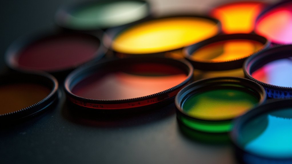
While ordinary photographers may struggle with challenging lighting conditions, scientists and researchers require specialized tools for their imaging needs.
The Optolong L-eNhance and L-eXtreme filters stand out for celestial imaging by blocking unwanted urban light pollution while maximizing transmission of essential wavelengths like H-Alpha and O-III.
Celestial imaging thrives with Optolong filters that reject light pollution while enhancing critical H-Alpha and O-III wavelengths.
For protective applications, Kase Wolverine Neutral filters offer scratch-resistant durability without compromising image clarity.
They’re particularly valuable when working in variable lighting environments.
When you’re capturing fluorescence images, specialized filters can greatly improve edge profile visibility while maintaining quality after compression.
The Haida NanoPro MC Clear-Night Filter excels in nighttime applications by preserving brightness and eliminating unwanted color casts.
For microscopy applications, adaptive filters notably enhance contrast in fluorescence imaging, making biological structures more detectable and easier to analyze.
Fluorescence Narrowband Filters: Maximizing Signal-to-Noise Ratio
You’ll achieve ideal fluorescence imaging results by carefully selecting precise bandpass filters that match your fluorophore’s emission spectra.
When dealing with challenging samples, you can eliminate unwanted autofluorescence by combining narrowband excitation filters with specialized blocking filters that remove background signals.
These high-performance filter combinations prove especially valuable in multi-label imaging applications, where you’ll need to clearly distinguish between multiple fluorescent markers with potentially overlapping emission spectra.
Precision Bandpass Selection
When capturing fluorescence signals in scientific imaging, the precise selection of wavelength ranges can make the difference between meaningful data and unusable noise. High quality narrowband filters enable you to isolate specific emission wavelengths with remarkable accuracy, ensuring ideal visibility of your fluorescent markers.
By tailoring your precision bandpass selection to match the exact excitation and emission profiles of your fluorophores—including specialized options like OIII wavelengths—you’ll dramatically improve your signal-to-noise ratio.
With transmission rates reaching 90% for target wavelengths, you’re capturing the maximum useful signal while effectively blocking background interference.
This customized approach allows you to fine-tune your imaging system to the unique fluorescence characteristics of your specific markers, resulting in clearer differentiation between structures and more reliable experimental outcomes.
Autofluorescence Elimination Techniques
Despite its critical importance in fluorescence microscopy, natural autofluorescence from biological specimens often drowns out your signals of interest.
This is where fluorescence narrowband filters become essential to your imaging toolkit. These specialized filters transmit only specific wavelengths associated with your target fluorophores while blocking unwanted background emissions.
Multi-Label Imaging Applications
Building on autofluorescence elimination principles, the power of fluorescence narrowband filters truly shines in multi-label imaging applications.
When you’re working with multiple fluorophores simultaneously, these specialized filters become essential for distinguishing between overlapping emission spectra.
You’ll achieve superior signal-to-noise ratio by selecting narrowband filters precisely matched to your target fluorophores’ emission wavelengths. With transmission rates exceeding 90% for key wavelengths, you’re capturing maximum signal while effectively blocking unwanted spectral components.
The impact on your fluorescence images is dramatic—enhanced contrast reveals structural details previously lost in background noise, especially in challenging low-light conditions.
Adaptive Contrast Enhancement Filters for Low-Light Microscopy
Adaptive contrast enhancement filters employ sophisticated mathematical models that transform low-light fluorescence images into visually interpretable data, revealing previously obscured biological structures.
You’ll find these filters particularly effective at mitigating Gaussian noise through specialized algorithms that preserve edge definition while suppressing random signal variations.
When implementing these techniques in your microscopy workflow, you’re not only enhancing image quality but also facilitating efficient compression with minimal data loss, maintaining critical visual information for scientific analysis.
Mathematical Filter Models
When examining specimens under low-light conditions, researchers often struggle to discern essential details hidden in the darkest regions of microscopy images. This is where adaptive contrast enhancement filters become invaluable. These sophisticated tools combine mathematical functions to reveal vital information that would otherwise remain invisible.
You’ll find these filters particularly effective when working with images affected by Gaussian noise—they maintain clear edge profiles even in degraded samples. The mathematical models powering these filters strike a best balance between visual clarity and data integrity, ensuring your image processing workflow doesn’t compromise scientific accuracy.
With a compression ratio of 0.9688, these filters also contribute to efficient data storage—a significant advantage when handling large datasets in biomedical imaging research.
The mathematical precision behind these filters transforms obscured details into valuable scientific insights.
Noise Reduction Techniques
Three significant challenges plague low-light microscopy: signal degradation, random noise artifacts, and loss of edge definition. When examining images without proper filtering, you’ll struggle to identify essential details—similar to observing the night sky without a telescope.
Adaptive contrast enhancement filters help reduce these issues by dynamically adjusting pixel values in darker regions while preserving data integrity. You’ll achieve clearer edge profiles and enhanced visibility of faint structures, even in poor lighting conditions.
For best results, combine these adaptive filters with median filtering and Gaussian blurring techniques. This integrated approach not only reveals previously obscured details but also supports efficient data compression (ratio of 0.9688).
Your quantitative accuracy remains intact while random noise artifacts are minimized, ensuring reliable scientific image analysis.
Dual-Bandpass Filters for Urban Laboratory Environments

Scientists working in urban laboratory environments face unique challenges from ambient light pollution that can compromise image quality and data integrity. You’ll find dual-bandpass filters invaluable for fluorescence microscopy in these settings, especially when working with Deep Sky imaging techniques.
| Filter Type | Transmission Rate | Best Application | Light Pollution Reduction | Image Quality Improvement |
|---|---|---|---|---|
| H-Alpha/OIII | >90% | Cellular structures | High | Sharp edge detection |
| Blue/Green | 85-95% | Protein localization | Medium | Enhanced contrast |
| UV/Infrared | 80-92% | DNA visualization | Very high | Reduced background |
| Red/Far-Red | 88-94% | Tissue imaging | High | Improved depth |
| Narrow-band OIII | >95% | Subcellular details | Excellent | Superior clarity |
When ambient light threatens your results, the Oxygen III filter is best for extracting clear biological patterns from otherwise compromised images.
Deconvolution Filters: Eliminating Light Diffraction Artifacts
Although light diffraction artifacts represent a considerable challenge in fluorescence microscopy, deconvolution filters offer a powerful solution for restoring image clarity and precision.
These specialized filters employ algorithms that reverse diffraction-induced blurring, considerably enhancing resolution in your scientific imaging applications.
When you’re working with low-light fluorescence microscopy, you’ll find these filters particularly valuable for:
- Reducing noise and revealing fine structural details that would otherwise remain obscured
- Extracting sharp edge profiles from images degraded by Gaussian noise
- Improving downstream analysis through more accurate segmentation of biological structures
As a preprocessing step, deconvolution techniques dramatically improve your ability to visualize subtle features within complex biological samples, ensuring more reliable quantitative analysis and interpretation of your research data.
Optical Density Filters for Precise Light Transmission Control
Controlling light intensity with precision stands as a fundamental requirement for quality scientific imaging, which is precisely where optical density filters excel.
These specialized filters operate on a logarithmic scale, where each optical density unit increase reduces light transmission tenfold, giving you remarkable control over illumination levels.
In fluorescence microscopy, you’ll find these filters particularly valuable for enhancing contrast and visibility when working with specimens in low-light conditions.
High-quality optical density filters minimize unwanted reflections while preserving the spectral integrity of transmitted light—critical for capturing accurate data in your research.
For ideal results, you can combine these filters with adaptive filtering and deconvolution techniques to further improve image quality.
This integrated approach guarantees you’ll achieve the clarity and precision necessary for advanced scientific imaging applications.
Anti-Reflective Coated Filters for Improved Image Clarity
When capturing precision scientific images, anti-reflective coated filters represent a breakthrough technology that greatly enhances clarity and detail.
You’ll notice immediate improvements in contrast and color accuracy as these multi-layered coatings target specific wavelengths, markedly reducing unwanted reflections and glare.
These high-performance filters deliver:
- Exceptional light transmission efficiency (over 99%), making them ideal for challenging low-light conditions in fluorescence imaging
- Enhanced durability through scratch-resistant materials that guarantee consistent performance across diverse environments
- Versatile compatibility with various imaging systems including fluorescence microscopes and astrophotography equipment
Whether you’re conducting detailed research or capturing celestial objects, these filters allow more light to reach your sensor while maintaining image integrity, giving you clearer results with greater precision.
Neural Network-Based Filters for Real-Time Image Processing

Neural network-based filters transform scientific imaging by using deep learning algorithms to enhance details in challenging, low-light conditions.
You’ll find these advanced filters particularly valuable when working with fluorescence microscopy, where they can extract clear edge profiles from noisy images in real-time.
Their adaptive learning capabilities allow your imaging systems to make immediate adjustments during experiments, dramatically improving visualization without the processing delays common in traditional filter methods.
Deep Learning Applications
As scientists increasingly work with complex microscopy data, deep learning has revolutionized how we enhance low-light fluorescence images in real time.
These advanced neural networks automatically extract features from degraded images, retrieving clear edge profiles even when Gaussian noise obscures important details.
You’ll find deep learning particularly valuable for three key applications:
- Specialized segmentation tools – Cellpose and Stardist excel at identifying faint structures and separating overlapping objects with unprecedented accuracy.
- AI denoising preprocessing – Implementing denoising before analysis dramatically improves downstream segmentation results.
- Customizable filtering solutions – Adapt neural network architectures to address your specific imaging challenges, from low-light conditions to motion artifacts.
With these deep learning approaches, you’ll transform barely visible specimens into clearly defined structures while maintaining scientific integrity in your image processing workflow.
Real-Time Enhancement Techniques
Building on the deep learning foundations we’ve explored, today’s most advanced image processing workflows now operate at unprecedented speeds. Neural network-based filters now enhance images in real-time, dramatically improving visibility in low-light conditions where traditional methods fail.
You’ll find these systems particularly valuable for fluorescence imaging, where they adaptively adjust to varying noise levels and Gaussian interference. The compression efficiency is remarkable—achieving ratios of 0.9688 while preserving visual quality, which helps manage your storage requirements.
When you’re examining blurred specimens, these filters leverage sophisticated mathematical functions to optimize contrast and reveal marginal profiles previously lost in noise.
For medical applications, you’ll notice considerably improved diagnostic accuracy as these technologies transform degraded images into clear, actionable visual data.
Multi-Layer Interference Filters for Spectral Precision
Three fundamental properties make multi-layer interference filters indispensable for scientific imaging: precision, efficiency, and customization.
These sophisticated filters use multiple dielectric layers to control which wavelengths pass through and which are blocked, giving you unprecedented spectral control for your experiments.
You’ll benefit from:
- High transmission rates – up to 90% of desired wavelengths pass through while unwanted light is effectively blocked
- Customizable specifications – tailor the bandwidth and central wavelength to your specific experimental requirements
- Enhanced image quality – especially in fluorescence microscopy where isolating specific emission wavelengths drastically improves contrast
The anti-reflective coatings on these filters guarantee stability and durability, maintaining performance even in demanding laboratory environments.
When precise spectral control matters, multi-layer interference filters deliver exceptional results.
Phase Contrast Filters: Revealing Transparent Structures
Two major challenges have historically plagued biological imaging: observing transparent structures and maintaining specimen viability.
Phase contrast filters offer an elegant solution by transforming invisible phase shifts into visible amplitude changes. You’ll see transparent specimens with enhanced contrast against bright backgrounds without staining procedures that might compromise cell integrity.
When you’re studying live cells or delicate tissue samples, these filters allow you to detect subtle morphological changes that would otherwise remain invisible.
They’re particularly valuable for monitoring cellular processes in real-time, as they preserve the natural characteristics of your specimens while considerably improving image quality.
You can integrate phase contrast filters into various microscopy systems, making them versatile tools for researchers who need to visualize fine structural details in transparent biological materials.
Frequently Asked Questions
How Often Should Light Filters Be Calibrated?
You should calibrate light filters monthly for standard usage, but increase frequency with heavy use. If you’re conducting critical research, you’ll need weekly calibrations. Always recalibrate after environmental changes or contamination incidents.
Are There Portable Filter Solutions for Fieldwork?
Yes, you’ll find several portable filter options for fieldwork. Clip-on filters, lightweight filter wheels, and battery-powered filter systems all work well. Look for weatherproof cases and filters designed specifically for field conditions.
Can Filters Be Stacked Without Degrading Image Quality?
Yes, you can stack filters, but limit it to 2-3 high-quality ones. Beyond that, you’ll face light reduction, vignetting, and potential reflections. Use multi-coated filters to minimize quality degradation when stacking.
What Maintenance Extends the Lifespan of Scientific Filters?
You’ll extend your scientific filters’ lifespan by cleaning them with lens tissue, storing in dust-free cases, avoiding fingerprints, minimizing light exposure when unused, and conducting regular optical performance checks for degradation signs.
How Do Temperature Fluctuations Affect Filter Performance?
Temperature fluctuations can degrade your filters by altering bandpass wavelengths, reducing transmittance, and causing physical warping. You’ll notice decreased contrast, color shifts, and shorter lifespans when you expose filters to inconsistent temperatures.
In Summary
You’ll find that light filters aren’t just accessories but essential tools in scientific imaging. Whether you’re working with fluorescence microscopy or analyzing transparent specimens, the right filter dramatically enhances your results. By choosing filters that match your specific research needs, you’re not just capturing images—you’re revealing details invisible to the naked eye, elevating your research to new levels of precision and discovery.


Leave a Reply