To calculate image scale bars, first photograph a reference object of known size alongside your specimen. Use ImageJ or similar software to measure the reference object in pixels, then establish the scale by setting known physical dimensions to that pixel count. Position scale bars in a non-obtrusive corner with high contrast colors and clear unit labels. You’ll need consistent magnification settings for accurate scale representation. These fundamental techniques will help you create precise, scientifically valid measurements for all your research imagery.
Numeric List of Second-Level Headings

Five key sections comprise the process of calculating accurate scale bars for scientific images.
When creating your documentation, you’ll want to organize your workflow into these essential components:
- Obtaining Reference Objects with Known Dimensions
- Measuring Pixel Distances Using ImageJ or Similar Software
- Using the Set Scale Feature to Establish Conversion Factors
- Placing and Formatting Scale Bars for Maximum Visibility
- Validating Scale Accuracy Through Comparison
Each section represents a critical step in ensuring your scientific photos contain accurate size references.
By following this structured approach, you’ll avoid common pitfalls in scale bar creation.
Remember that proper scaling allows other researchers to interpret your visual data correctly, making it a fundamental aspect of scientific image documentation that shouldn’t be overlooked.
Understanding Pixel-to-Physical Measurement Conversion
To convert pixels to physical measurements, you’ll need to understand the fundamental relationship between your camera’s pixel density and real-world dimensions.
You can establish this conversion by measuring a reference object of known size in your image, such as a microscope stage micrometer or calibration slide.
Once calibrated, your measurements will remain accurate across images taken with the same equipment settings, allowing you to create precise scale bars that communicate true physical dimensions.
Pixel Density Fundamentals
At the core of creating accurate scale bars lies the concept of pixel density, which forms the bridge between digital and physical measurements.
Pixel density, measured in DPI or PPI, tells you how many pixels fit into one inch of physical space. The standard density for most screens is 96 DPI, meaning an image 960 pixels wide would measure 10 inches when printed.
To convert between pixels and physical dimensions, simply divide your pixel count by the DPI. For example, a 1200-pixel wide image at 300 DPI translates to a 4-inch print width.
This conversion is essential for scientific imaging where precise scaling communicates the true size of specimens. When you’re calibrating images, understanding this relationship guarantees your scale bars accurately represent real-world dimensions.
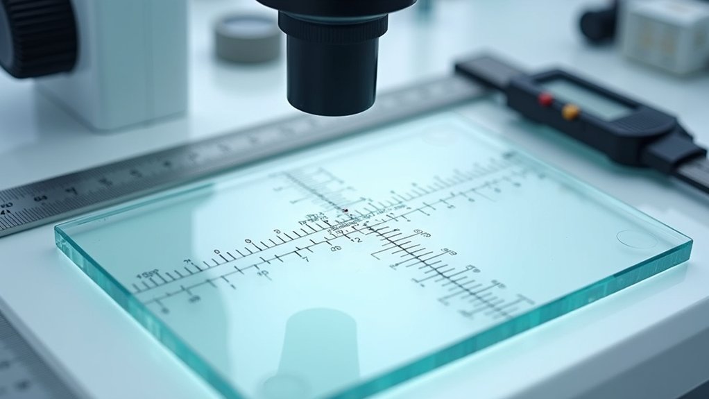
Calibration Using References
Precise calibration forms the foundation of accurate scale bars in scientific imagery. To achieve this, you’ll need a known reference—such as a calibration slide or ruler—that appears in your image.
Using software like ImageJ or Fiji, measure this reference in pixels and input both this value and the actual physical distance to establish the vital pixel-to-measurement conversion ratio.
Always select appropriate units (micrometers, millimeters) during calibration to maintain consistency throughout your research. You can determine the pixel size either from your image’s metadata or by consulting pixel size tables specific to your imaging equipment’s magnification settings.
To guarantee accuracy, validate your calibration by cross-checking measurements against objects of known dimensions, accounting for potential distortions from lens curvature or camera settings that might affect your scale.
Essential Tools for Scale Bar Calibration
While accurate scale bars may seem like minor details in scientific imaging, they require specific tools to affirm proper calibration and representation. You’ll need software like ImageJ with its “Set Scale” feature to input pixel distances and their corresponding real-world measurements. Always verify that you’ve selected the appropriate unit (μm, mm) to maintain scale consistency across experiments.
| Tool | Why It Matters |
|---|---|
| Calibration slide | Provides known reference measurements |
| ImageJ/Fiji | Offers precise scaling functions |
| “Set Scale” feature | Converts pixels to actual dimensions |
| “Show Info” function | Verifies metadata for accurate calibration |
| High-contrast colors | Affirms scale bar visibility |
Don’t forget to position your scale bars where they won’t obscure important image details, using contrasting colors for maximum visibility.
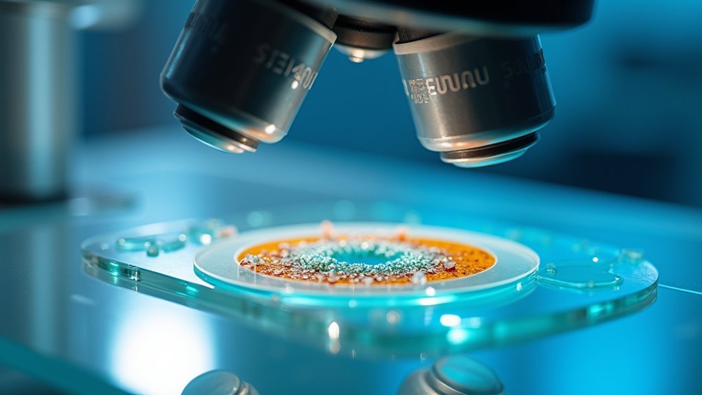
Determining Microscope Magnification Factors
To calculate accurate scale bars, you’ll need to understand how objective lens magnification works in combination with your eyepiece settings.
Your microscope’s total magnification is determined by multiplying the objective lens value (commonly 4x, 10x, 40x, or 100x) by your eyepiece magnification (typically 10x).
This multiplication factor directly impacts your pixel size calculations and must be precisely documented when preparing scientific images for publication or analysis.
Objective Lens Calculations
Understanding objective lens calculations forms the foundation of accurate scale bar creation for microscope images. When you’re working with microscope photography, you’ll need to calculate the width of your field of view based on your objective lens specifications.
Each objective lens (10x, 40x, 100x) has a distinct numerical aperture (NA) that affects resolution and clarity. As you increase magnification, your field of view narrows, requiring more precise specimen positioning.
To determine effective pixel size, divide your field of view by your camera’s sensor resolution. This conversion is essential when creating scale bars, as you’ll need to translate pixels to micrometers.
Use this formula for accurate scale bar length:
Scale Bar (µm) = (Field of View Diameter (µm) / Image Width (pixels)) × Desired Bar Length (pixels)
Eyepiece Multiplier Effects
When calculating accurate scale bars, you’ll need to account for eyepiece multiplier effects, as they greatly impact your total magnification power. The eyepiece multiplier, typically labeled directly on the eyepiece, serves as one of two essential factors in determining your microscope’s total magnification.
To calculate total magnification, simply multiply your objective lens value by the eyepiece multiplier. For instance, pairing a 10x eyepiece with a 40x objective yields 400x total magnification.
Different eyepieces (5x, 10x, 15x) will dramatically alter your field of view and the apparent size of your specimen.
Be vigilant about verifying your eyepiece multiplier against known standards, as even small calibration errors can lead to significant measurement inaccuracies in your scientific images and resulting scale bars.
Capturing Images With Reference Objects
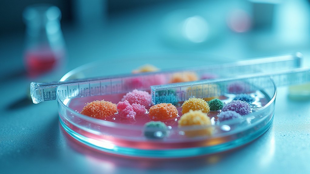
Scientific photography requires accurate scale representation, which you’ll achieve by placing reference objects of known dimensions in your images. Position your reference object at the same focal distance as your subject to prevent distortion. Common items like rulers or coins work best, as they’re easily recognized by viewers unfamiliar with your specimen’s size.
| Reference Object | Best For | Positioning Tip |
|---|---|---|
| Ruler/Scale Bar | Precise measurements | Align parallel to subject |
| Coin | Small specimens | Place at same height as subject |
| Color card | Microscopy | Include in corner of frame |
Always document your distance and magnification settings for consistency in future analyses. Be mindful of your photography angle—perspective shifts can misrepresent the size relationship between your reference object and the subject.
Methods for Measuring Known Distances in Pixels
To measure known distances in pixels, you’ll need to select a reliable reference object in your image and use the straight line selection tool in ImageJ or Fiji to draw across its known dimension.
Once you’ve drawn the line, navigate to “Analyze > Set Scale” and enter the actual length value corresponding to your pixel measurement to establish proper calibration.
You can verify your calibration’s accuracy by measuring other known features in your image or by checking that the scale bar appears proportionally correct for your specimen size.
Pixel-Distance Measurement Methods
Three essential methods exist for accurately measuring known distances in pixels when creating scale bars for scientific images.
The first approach involves using the straight line selection tool to trace a known feature, such as a ruler or calibration slide, directly in your image. You’ll immediately see the distance in pixels displayed in the software’s measurement panel.
The second method requires drawing a line across a standardized reference object within your image. This technique works well when you’ve included calibration scales in your original photography setup.
For the third approach, use multiple reference points to verify your measurements. After obtaining your pixel count, enter this value alongside the known physical distance into the Set Scale dialog, ensuring you’ve selected the appropriate measurement units (mm, μm) for your scientific context.
Reference Object Selection
When selecting reference objects for pixel-distance measurements, you’ll need carefully chosen calibration tools that provide known physical dimensions. Calibration slides and rulers are commonly used reference objects because they offer precise, verifiable measurements that can be accurately captured in your images.
Position your reference object in the same focal plane as your specimen to prevent depth-of-field distortions that could compromise measurement accuracy. Maintain consistent lighting and camera perspective to guarantee both the reference object and specimen are comparably represented in the final image.
Always record the reference object’s known physical dimensions in your image metadata or in separate documentation.
After photographing, measure the object’s pixel dimensions in your image software to establish the pixel-to-unit scale ratio, which you’ll apply to all measurements in that image.
Calibration Scale Techniques
In the dialog box, the “Distance in pixels” field will automatically populate based on your drawn line. Enter the known distance value and specify the unit of measurement.
For consistent measurements across multiple images, enable the “Global” option. When adding scale bars later, they’ll reflect these calibrated measurements.
Always verify your calibration by measuring known dimensions within your image, adjusting as needed to guarantee precision that matches your microscope’s magnification specifications.
Calculating the Scale Ratio for Microscopy Images
Determining the precise scale ratio for microscopic images requires careful measurement and calibration.
You’ll need to use ImageJ or Fiji software to establish accurate measurements for scientific documentation and analysis.
- Measure a known distance in your image using the straight line selection tool, ensuring you’ve identified a reference structure with a confirmed actual size.
- Open the “Set Scale” dialog box and input your known distance value—the software will automatically fill in the pixel distance from your line measurement.
- Validate your calculated scale by comparing it with your microscope’s specifications and expected magnification factor.
- Cross-reference your measurements against known dimensions from calibration slides or other reliable sources to confirm accuracy.
Always recalibrate when changing magnification settings, as the pixel-to-micrometer ratio will vary.
Creating Visually Effective Scale Bars
Creating visually effective scale bars requires attention to both aesthetics and scientific accuracy. Choose high-contrast colors that won’t blend with your image content—avoid red, green, or blue which might disappear against similar colored backgrounds.
Position your scale bar in the lower left corner of images where it remains visible without obscuring critical details. This placement has become standard practice in scientific publications and helps maintain visual clarity.
Use simple units like 100μm or 50μm that readers can quickly understand. Always verify your scale bar accurately reflects the specimen’s actual dimensions by calibrating against known distances using ImageJ or FIJI.
Add the scale bar as your final step in image preparation. This timing prevents complications if you need to resize figures during manuscript revisions, keeping your scale bar proportionally accurate.
Proper Placement and Formatting of Scale Indicators
Now that we’ve established why scale bars matter, let’s focus on their perfect placement and formatting.
Position your scale bar in the lower left corner of your image to maintain visual clarity without obscuring critical elements. Choose high-contrast colors like black or white to guarantee your scale bar remains visible against any background.
Position scale bars in the lower left corner with black or white coloring for maximum visibility without disrupting critical image elements.
- Select best positioning – Lower left corner placement prevents obstruction of important image content
- Use high-contrast colors – Black or white scale bars offer the best visibility across varied backgrounds
- Label with simple units – Mark lengths clearly with straightforward measurements (100 μm, 50 μm)
- Add scale bars last – Insert scale indicators after finalizing all image adjustments to accommodate any resizing
Always avoid red, green, or blue for your scale bars as these colors may interfere with image interpretation.
Batch Processing Scale Bars for Multiple Images
When you’re dealing with dozens or even hundreds of scientific images, manually adding scale bars to each one becomes tedious and error-prone. Photoshop’s “File Automate Batch” feature offers a solution for efficiently applying scale bars across your entire dataset.
First, establish an accurate mm/pixel ratio using a calibration slide with known dimensions. Then create a Photoshop action that automatically adds your scale bar with consistent size and text formatting.
Before processing, organize all images in a designated folder for systematic application. Launch the batch process to apply your recorded scale bar action to all images without manual intervention.
Periodically verify the accuracy of your automated scale bars by comparing them against known measurements, making adjustments as needed to maintain precision across different image dimensions.
Software Options for Automated Scale Bar Generation
Several specialized software tools exist to eliminate the tedium of manual scale bar creation while ensuring scientific accuracy.
These applications offer calibration-based automation that considerably reduces human error in your scientific imagery.
- ImageJ/FIJI provides built-in tools that automatically generate scale bars based on your image calibration, ensuring precise size representation.
- Photoshop streamlines your workflow through actions and batch processing, allowing you to apply consistent scale bars across multiple images.
- Piximetre specializes in measurement and scale bar addition, offering targeted functionality for scientific analysis.
- Custom macros and scripts can further automate scale bar generation in most imaging software.
For specialized research needs, community-developed plugins extend these capabilities, giving you tailored scale bar solutions for particular scientific applications.
Troubleshooting Common Scaling Errors
Despite your best efforts, scaling errors can creep into scientific imagery, compromising the accuracy of your measurements and conclusions. When troubleshooting scale bar issues, first verify that your calibration distance isn’t affected by optical distortions or lens artifacts. This fundamental step guarantees your reference measurement truly represents what’s displayed.
Check that pixel dimensions in your image metadata match the expected values based on your microscope settings and objective. If you spot discrepancies, remeasure using an alternative known distance to cross-validate your initial calibration.
For non-uniform pixel dimensions, access “Image Properties” in ImageJ to confirm consistent width and height values.
Remember to recalibrate regularly with a calibration slide, especially after changing lenses, to prevent cumulative measurement errors that could invalidate your research.
Frequently Asked Questions
How to Calculate the Scale Bar of an Image?
To calculate an image’s scale bar, you’ll measure a known distance in pixels, convert it to actual units, and set the scale in software like ImageJ before adding a visual scale representation.
How Do You Calculate Scale Bar for Biological Drawings?
You’ll calculate a scale bar by measuring a known reference object in your drawing, dividing its actual size by pixel length, then using this scale factor to create an appropriately sized, clearly visible bar.
How to Make a Scale Bar for a Microscope Image?
To make a scale bar for a microscope image, you’ll need to draw a line on your image in ImageJ, set the scale by defining its known length, then add a scale bar through Analyze/Tools.
How Is a Scale Bar Used in Scientific Images?
Scale bars in scientific images let you accurately judge the size of specimens. They’re essential when viewing microscopic objects, providing a standardized reference measurement that helps you interpret dimensions consistently across different images.
In Summary
You’ve now mastered the essentials of calculating accurate scale bars for your scientific images. By understanding pixel-to-measurement conversions and utilizing proper calibration techniques, you’ll guarantee your research photos maintain scientific integrity. Remember to document your magnification methods consistently and verify your scale bars before publication. With practice, adding these critical reference indicators will become second nature in your scientific imaging workflow.


Leave a Reply