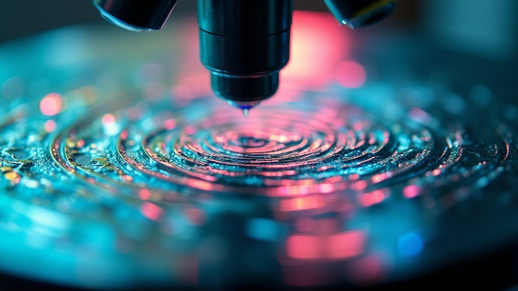
Strange patterns in your microscope light often result from condenser misalignment, dust on optical components, or incorrect diaphragm settings. You’ll typically see diffraction rings, halos, or uneven illumination when components aren’t properly aligned. Check for proper Köhler illumination, clean all lenses with appropriate solutions, and verify your condenser is centered. Adjusting the aperture diaphragm to 65-80% of the objective aperture can dramatically improve your image quality. Discover more specific solutions below.
Understanding Köhler Illumination Problems and Solutions
While Köhler illumination stands as the gold standard for microscopy, achieving perfect illumination requires careful attention to several essential factors.
If you’re seeing strange light patterns, check your substage condenser alignment first. Misalignment between your light source and condenser creates uneven illumination and unwanted aberrations across your specimen.
Examine your diaphragm settings—they should be between 65-80% of your objective aperture for ideal contrast and resolution. An improperly adjusted aperture diaphragm frequently causes vignetting or brightness inconsistencies that compromise image quality.
Aperture diaphragm calibration at 65-80% of objective aperture maximizes contrast while preventing vignetting issues in microscopic imaging.
Don’t overlook your lamp filament position relative to the condenser lens—this relationship is vital for preventing light artifacts.
Finally, regularly clean all optical components to prevent dust and contaminants from creating interference patterns. Proper maintenance eliminates many common illumination problems before they affect your observations.
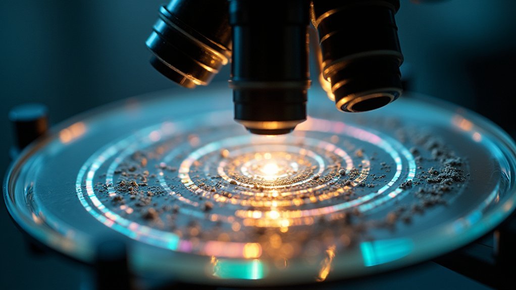
Common Causes of Diffraction Patterns in Microscope Lighting
Beyond basic illumination problems, you’ll sometimes encounter distinctive diffraction patterns that can obscure your specimen’s details. Misalignment between your condenser and light source is often the primary culprit. Check that your optical components are properly centered.
Using incorrect cover glass thickness can cause light refraction—always opt for No. 1½ (0.17 mm) coverslips.
Spherical aberration may result from mismatched objectives and eyepieces or incorrect adjustment of correction collars.
Don’t overlook your diaphragm settings; they should be at 65-80% of the objective aperture to prevent excessive light intensity that creates unwanted diffraction effects.
Finally, contamination on optics scatters light and produces diffraction artifacts. Regular cleaning and maintenance of lenses, slides, and all optical surfaces will greatly reduce these issues.
Condenser Misalignment: Identifying and Correcting Light Artifacts
Condenser misalignment produces telltale artifacts like uneven illumination and strange light patterns that compromise your microscope’s imaging quality.
You’ll need to center your condenser properly under the objective lens and verify that the aperture diaphragm is set between 65-80% of the objective aperture for ideal contrast.
Implementing Köhler illumination will help you eliminate these alignment issues while providing even illumination across your specimen field.
Identifying Condenser Artifacts
When examining specimens under your microscope, you might notice unexpected shadows, uneven lighting, or strange patterns that weren’t present in your sample. These visual disturbances often result from condenser misalignment, which disrupts the optical axis and creates diffraction artifacts that compromise image quality.
You can identify these issues by looking for uneven illumination across your field of view or strange geometric patterns that move when you adjust the focus.
To correct these problems, try adjusting height and centering of your condenser using a centering telescope. For phase contrast microscopy, verify proper alignment between the condenser’s annulus and the objective’s phase ring.
Proper Köhler illumination depends on a well-aligned condenser. Implement regular maintenance to check that your condenser is clean, properly attached, and correctly positioned for best viewing results.
Proper Centration Techniques
Three key techniques will help you achieve proper condenser centration and eliminate frustrating light artifacts from your microscope images. First, adjust the condenser’s height to position the light cone directly on your objective lens’s optical axis. Second, check for uniform illumination by observing a blank field; any shadows or intensity variations indicate condenser misalignment requiring lateral adjustment.
| Technique | Signs of Misalignment | Correction |
|---|---|---|
| Height Adjustment | Halos, dim periphery | Raise/lower condenser until field is evenly lit |
| Lateral Alignment | Shadowing on one side | Use centering screws to position light centrally |
| Phase Contrast | Ring misalignment | Align condenser annulus with phase ring |
Finally, incorporate the phase contrast technique to enhance image quality. Remember that routine microscope maintenance should include checking condenser alignment to guarantee peak performance during all imaging sessions.
Köhler Illumination Fixes
Proper Köhler illumination serves as the foundation for high-quality microscopy, yet many researchers struggle with condenser misalignment issues that create frustrating artifacts.
When you notice strange patterns or uneven brightness across your field of view, your condenser likely needs adjustment.
To fix this, start by focusing your specimen with the low power objective, then adjust the condenser until your field diaphragm appears sharp and fully illuminated.
Verify your condenser aperture diaphragm is set between 65-80% of the objective aperture for ideal contrast and resolution.
Don’t overlook regular cleaning of your condenser and checking that it’s properly seated in its mount.
For precision alignment, use a focusing telescope to align the condenser’s light cone with the microscope’s optical axis.
These steps will eliminate vignetting and restore uniform illumination across your specimens.
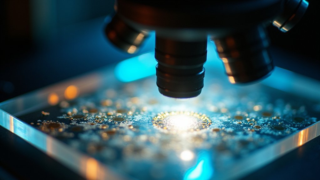
Dust and Contamination Effects on Microscope Illumination
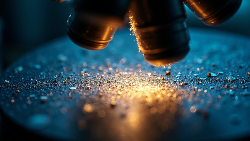
Microscopic invaders in the form of dust particles and contaminants pose a considerable threat to the quality of your microscope illumination. When dust accumulates on optical components, it scatters light and creates strange patterns that diminish image clarity.
You’ll notice halos, glare, or uneven illumination when fingerprints, oil, or debris contaminate your objective lens or condenser.
Don’t overlook the specimen slide itself—dust here creates shadows and obscured areas that reduce contrast in your observations.
To maintain ideal light transmission and prevent distortion, regularly clean all optical surfaces using appropriate lens cleaning solutions and lint-free cloths.
This simple maintenance routine will greatly enhance illumination uniformity and eliminate those puzzling light patterns that interfere with your microscopy work.
Phase Contrast Issues: When Annuli and Phase Plates Misalign
Phase contrast microscopy becomes a puzzle when the annuli and phase plates don’t perfectly align.
You’ll notice unusual image patterns and distortions that obscure your specimen’s details. Each objective requires a specific annulus size; using the wrong one compromises contrast and creates unexpected artifacts.
Mismatched annuli create deceptive artifacts, turning your specimen into an optical illusion rather than a clear scientific observation.
The circular annulus must align precisely with the phase ring on your objective for ideal imaging. If you’re seeing strange patterns, check this alignment first.
Proper Köhler illumination is equally essential—any unevenness in lighting will intensify these issues.
Don’t neglect regular alignment checks of your condenser and objectives. This maintenance preserves the integrity of the phase contrast effect.
How Improper Objective Selection Affects Light Patterns
When you select the wrong objective for your microscopic work, distinctive light patterns immediately reveal the mismatch. High numerical aperture objectives are particularly sensitive—using them with incorrect coverslip thickness creates spherical aberration, producing distorted light patterns and reduced image sharpness.
Watch for vignetting and optical path misalignment when your coverslip doesn’t match the objective’s specifications. Parfocal errors between objectives can create strange light artifacts as focal points fail to converge properly.
Using a dry objective where oil immersion is required introduces aberrations that manifest as unusual patterns.
Don’t overlook the alignment of light source and condenser with your objective. Improper positioning leads to uneven illumination across your field of view.
These issues compound each other, so proper objective selection and setup are critical for artifact-free imaging.
Diaphragm Adjustment Techniques for Eliminating Strange Patterns
The proper adjustment of your microscope’s diaphragm stands as the cornerstone technique for eliminating unwanted light patterns in microscopic imaging. Set your substage condenser aperture to 65-80% of the objective aperture to improve image contrast and reduce diffusion artifacts.
| Technique | Benefit |
|---|---|
| Köhler illumination | Eliminates shadow patterns |
| Light source alignment | Prevents unwanted artifacts |
| Regular diaphragm cleaning | Reduces irregular light patterns |
| Experimental adjustment | Identifies ideal configuration |
Always verify your diaphragm aligns perfectly with the light source to avoid distortion. You’ll notice immediate improvement when proper alignment is achieved. Don’t forget to clean your diaphragm regularly, as contaminants create unusual patterns that compromise image quality. Try different settings while observing your specimen to find the perfect balance for your specific application.
Light Source Problems: Bulb Aging and Uneven Illumination
Light output gradually deteriorates as microscope bulbs age, creating frustrating patterns and color inconsistencies across your field of view. To prevent these issues, replace your bulbs according to the manufacturer’s recommended usage hours rather than waiting for complete failure.
Check for lamp filament misalignment, which commonly causes uneven illumination. Proper adjustment guarantees ideal light distribution for clear imaging.
Misaligned lamp filaments create frustrating illumination patterns. A quick adjustment ensures optimal light distribution for superior microscopic clarity.
Consider upgrading from halogen to LED light sources, as LEDs deliver more consistent illumination over their longer lifespan.
Don’t overlook regular maintenance of your lamp housing and optical components. Dust accumulation greatly contributes to strange lighting patterns and diminished image quality.
A simple cleaning routine for all exposed surfaces will help maintain uniform illumination and prevent the mysterious shadows and patterns that can compromise your microscopy work.
Advanced Troubleshooting for Persistent Illumination Anomalies
When you’re facing persistent illumination artifacts, first check your Köhler illumination setup by verifying proper alignment of the light source, condenser position, and aperture diaphragm settings.
You’ll need to systematically examine the entire optical pathway for contamination, including cleaning objectives, eyepieces, and slides with appropriate solutions for each component type.
Don’t overlook specialized diagnostic techniques such as testing phase ring alignment, filter compatibility, and performing regular microscope calibration to resolve stubborn light pattern anomalies.
Light Artifact Diagnostic Techniques
Five systematic approaches exist for troubleshooting persistent illumination anomalies that confound even experienced microscopists. Start by checking your light source and condenser alignment, as misalignment frequently causes uneven illumination patterns. Next, verify proper Köhler illumination by aligning the lamp filament correctly. Don’t overlook optical components—dirty lenses or filters introduce unwanted light artifacts that compromise image quality.
| Diagnostic Approach | Common Problem | Solution |
|---|---|---|
| Light Source Check | Uneven illumination | Realign source with optical axis |
| Condenser Inspection | Poor contrast | Adjust aperture to 65-80% of objective |
| Component Cleanliness | Scattered artifacts | Clean all optical surfaces |
| Phase Contrast Setup | Interference patterns | Align phase annulus with phase plate |
For phase contrast techniques, verify proper alignment between the phase annulus and phase plate to eliminate interference patterns.
Köhler Illumination Troubleshooting
Despite meticulous setup procedures, persistent illumination anomalies often plague microscopy work requiring advanced troubleshooting techniques.
When your microscope shows irregular light patterns, first verify your light source alignment with the condenser and check that the filament is properly centered.
Adjust your condenser aperture diaphragm to 65-80% of the objective aperture to achieve ideal Köhler illumination and eliminate vignetting.
Don’t overlook cleanliness—dust or oil on optical components can scatter light creating unwanted patterns.
Set your condenser height to match your current objective, as mismatches cause aberrations and uneven illumination.
Finally, inspect the complete optical path for misalignments, particularly in phase contrast setups where phase annulus and plate positioning are critical.
These subtle adjustments often resolve those frustrating illumination issues that compromise your microscopy results.
Optical Pathway Contamination Solutions
Stubborn illumination problems often stem from overlooked contamination along the microscope’s optical pathway. Inspect your objectives, condensers, and filters for dust, oil, or fingerprints that disrupt light transmission. Clean affected components with appropriate lens tissue and solutions to restore peak illumination.
If strange patterns persist after cleaning, check for misalignment among optical components. Improper positioning can create interference patterns or vignetting that mimic contamination effects. Use a focusing telescope to thoroughly examine the optical pathway and identify hidden obstructions.
For particularly resilient issues, consider recalibrating your microscope’s illumination system. Verify your light source functions correctly, as lamp irregularities can distribute light unevenly.
Remember that even minor residues can cause significant aberrations, so maintain a regular cleaning schedule to prevent future contamination issues.
Frequently Asked Questions
What Might Be the Reasons for Blurry or Distorted Images When Using a Microscope?
Your microscope’s blurry images may result from misaligned light sources, incorrect coverslip thickness, vibrations, parfocal errors, or dirty optics. Regular maintenance and proper setup will improve your imaging quality greatly.
What Are the Sources of Error When Using a Light Microscope?
You’ll encounter microscopy errors from improper focusing, incorrect illumination, dirty lenses, poor sample preparation, and wrong condenser settings. Vibration, lens aberrations, and misalignment also compromise your observations during light microscopy.
How to Get a Clear Image on a Microscope?
To get a clear microscope image, you’ll need to properly align your light source, adjust the condenser aperture, focus using low magnification first, clean all optical components, and implement Köhler illumination for uniform lighting.
Why Is My Microscope Not Focusing?
Your microscope isn’t focusing properly due to possible vibrations, incorrect focal adjustment, parfocal errors, thick specimens, or dirty optical components. Try starting with a low-power objective and make certain your slides are properly positioned.
In Summary
You’ll often find that strange light patterns in your microscope aren’t mysterious at all. They’re typically caused by misaligned condensers, dust, incorrect phase contrast settings, or improper diaphragm adjustments. Don’t ignore these issues—they’ll affect your image quality and data accuracy. With proper maintenance, alignment checks, and understanding of Köhler illumination principles, you’ll quickly resolve these problems and return to clear, artifact-free microscopy.


Leave a Reply