
For live cell imaging, oblique illumination angles of 30-45 degrees work best, offering enhanced contrast while minimizing phototoxicity. You’ll get clearer visibility of cellular structures with Köhler illumination, which provides uniform brightness and reduces damaging hotspots. DIC microscopy’s polarized light configuration reveals three-dimensional details without fluorescent labeling, while properly aligned phase contrast techniques amplify structural differences in transparent specimens. The ideal setup balances optical clarity with minimal light exposure to protect your delicate living samples.
The Science Behind Oblique Illumination for Cell Visibility
While traditional illumination techniques often struggle with contrast issues, oblique illumination revolutionizes live cell imaging by directing light rays at specific angles to the specimen.
You’ll notice immediately how this approach enhances cell visibility by creating shadows that highlight cellular structures while considerably reducing background noise.
By optimizing the angle of illumination, you’re effectively minimizing out-of-focus light—a vital advantage when examining thicker specimens.
Precise angle optimization eliminates background interference—essential when studying thick biological samples under the microscope.
This technique excels at revealing low-contrast features that would otherwise remain invisible, letting you observe rapid cellular processes with remarkable clarity.
What’s particularly valuable is oblique illumination’s compatibility with other methods like DIC and phase contrast microscopy, further improving structural delineation without increasing phototoxicity.
This means you can extend observation times without damaging delicate live cells.
Köhler vs. Critical Illumination for Live Cell Imaging
When setting up your live-cell imaging system, you’ll notice Köhler illumination provides superior brightness uniformity across your specimen, preventing the hotspots that critical illumination often creates.
Your choice between these methods greatly impacts cell viability, as critical illumination’s direct light application increases the risk of photodamage and photobleaching during extended observations.
Köhler illumination requires more careful optical alignment but rewards your effort with enhanced image quality and reduced phototoxicity—essential considerations when your research depends on maintaining cellular health during imaging sessions.
Comparison of Brightness Uniformity
Because illumination quality directly impacts experimental outcomes, the uniformity of brightness serves as an essential factor in live cell imaging.
When you use Köhler illumination, you’ll achieve even light distribution across your entire field of view, eliminating the hotspots and shadows that plague essential illumination techniques. This consistency is important for maintaining accurate contrast and resolution, especially when imaging thicker specimens.
Essential illumination’s direct light pathway often creates uneven brightness patterns that can compromise data quality and increase phototoxicity in sensitive cells.
Photodamage Consideration Differences
Photodamage represents a fundamental challenge when imaging live cells under microscopic illumination. When comparing illumination techniques, Köhler illumination typically offers superior protection against phototoxicity by distributing light more uniformly and reducing intensity at the specimen.
| Feature | Köhler Illumination | Essential Illumination |
|---|---|---|
| Light Distribution | Uniform, diffuse | Direct, concentrated |
| Phototoxicity Risk | Lower | Higher |
| Cell Viability | Better preserved | More compromised |
You’ll find that Köhler’s controlled light paths minimize out-of-focus light that contributes to photodamage. For ideal live-cell imaging results, consider your specific cell types’ sensitivity when choosing between these methods. The trade-off between image quality and photodamage risk is vital—Köhler’s lower light intensity with longer exposure times generally enhances cell viability compared to essential illumination’s higher intensity approach.
Practical Setup Requirements
Three essential components distinguish Köhler illumination setup from vital illumination when preparing for live-cell microscopy.
First, you’ll need proper alignment of the light source, condenser, and objective lenses to achieve even light distribution across your field of view. This precise configuration prevents hotspots that can cause photodamage to delicate specimens.
Second, you must optimize your illumination angle to control light intensity, which directly reduces photobleaching risks during extended imaging sessions. This adjustment is particularly critical for time-lapse experiments where cells are observed over hours.
Finally, your setup should facilitate reduced background fluorescence to enhance signal-to-noise ratios. With Köhler illumination, you’ll capture clearer images of dynamic cellular processes compared to critical illumination, which often introduces artifacts and compromises detail visibility in live-cell imaging.
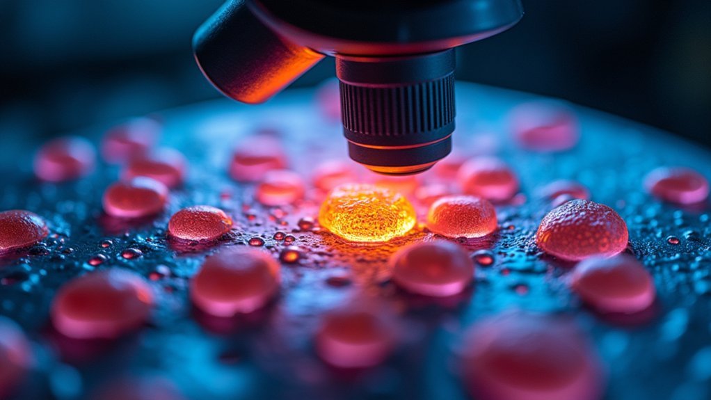
Optimizing Darkfield Illumination Angles for Cellular Detail
When setting up darkfield microscopy for live cell imaging, the angle of illumination serves as the critical factor that determines image quality and detail visibility.
You’ll achieve ideal results when positioning your light source at angles between 30 and 60 degrees relative to the specimen plane. These lighting angles maximize scattered light from cellular structures while minimizing unwanted background glare.
Always use a high numerical aperture condenser to improve collection of scattered light, enhancing resolution of fine details.
Take time to properly align your light source—even minor misalignments can dramatically reduce contrast enhancement. Adjust both angle and intensity until you’ve achieved clear visualization.
This technique’s greatest advantage is preserving cell viability by eliminating the need for staining, allowing you to conduct real-time monitoring of dynamic cellular processes without compromising specimen health.
Phase Contrast Techniques: Manipulating Light Paths for Enhanced Contrast

Phase contrast microscopy transforms how you’ll observe living cells by manipulating light paths through careful positioning of the annular diaphragm.
You’ll achieve ideal contrast when your diaphragm perfectly aligns with the phase plate, ensuring diffracted and direct light interact properly at the image plane.
For challenging specimens, you can combine phase contrast with oblique illumination techniques, which adds a three-dimensional effect and reveals fine cellular details that standard lighting methods might miss.
Oblique Illumination Benefits
Four significant advantages make oblique illumination a powerful technique in phase contrast microscopy for live cell imaging.
When you direct light at an angle to your specimens, you’ll immediately notice enhanced contrast that reveals transparent structures without staining. This preservation of cells in their natural state allows you to observe authentic cell morphology and behavior.
By controlling light paths, you’ll reduce out-of-focus light that creates background noise, resulting in remarkably sharp images of your samples.
- Minimal phototoxicity – Your live cells remain healthier during extended observation periods
- Enhanced visualization – You’ll see delicate organelles and membranes with surprising clarity
- Dynamic observation – You can track cellular events in real-time without disrupting normal functions
Annular Diaphragm Positioning
Building on the advantages of oblique illumination, proper annular diaphragm positioning stands as the foundation of effective phase contrast microscopy. When you align this component correctly, you’ll create an annular cone of light that enhances contrast by amplifying phase differences in transparent specimens without staining.
| Diaphragm Width | Application | Benefits |
|---|---|---|
| Narrow | Thin specimens | Highest contrast, reduced noise |
| Medium | Standard cells | Balanced resolution, good detail |
| Wide | Thick specimens | Better depth penetration |
| Variable | Dynamic processes | Adaptable to changing conditions |
Fine-tuning your annular diaphragm’s position during live-cell imaging helps mitigate out-of-focus light, essential for capturing sharp images of cell membranes and organelles. Ideal light transmission through proper alignment minimizes background noise, allowing you to visualize cellular structures with remarkable clarity during dynamic events.
DIC Lighting Geometry: Maximizing Three-Dimensional Cell Structure
When properly configured, differential interference contrast (DIC) microscopy provides unparalleled visualization of three-dimensional cellular structures.
Witness cellular architecture come alive through perfectly calibrated DIC microscopy, revealing depth and detail beyond conventional imaging capabilities.
By leveraging polarized light through a Nomarski prism, you’ll create an interference pattern that reveals detailed morphology without fluorescent labeling, eliminating concerns about phototoxicity in live cells.
The ideal lighting geometry depends on precise alignment of the polarizer and analyzer angles.
Adjust these components to enhance contrast and reveal fine cellular structures that would otherwise remain invisible in conventional brightfield microscopy.
- Experience the thrill of watching dynamic cellular processes unfold in real-time
- Marvel at the clarity of intricate cytoskeletal elements without staining
- Discover hidden details as three-dimensional imaging brings depth to previously flat cellular landscapes
Balancing Illumination Angles to Minimize Phototoxicity
While DIC microscopy offers exceptional structural visualization, the light exposure necessary for imaging can harm your living specimens over time. You’ll need to carefully balance illumination angles to minimize phototoxicity while maintaining adequate contrast.
| Approach | Benefit |
|---|---|
| 30-45° incident angles | Enhanced contrast with reduced exposure |
| Low-intensity light | Extends viable imaging duration |
| Shorter exposure times | Decreases cumulative cell damage |
| Structured illumination | Improves spatial resolution with less light |
| Ideal excitation wavelengths | Reduces damage in fluorescence microscopy |
Frequently Asked Questions
How to Image Live Cells?
To image live cells, you’ll need a fluorescence microscope, controlled temperature, and correct media. Use low-intensity light, minimize exposure time, and employ spinning disk confocal or TIRF for better resolution with less phototoxicity.
What Microscope Can Be Used to View Live Cells?
You can use inverted microscopes, widefield fluorescence, confocal laser scanning, spinning disk confocal, or light-sheet fluorescence microscopy for live cells. Each offers different advantages depending on your specific imaging needs.
Can Bright Field Microscopy Be Used on Living Cells?
Yes, you can use bright field microscopy on living cells. It’s gentle enough to maintain cell viability, though you’ll often need contrast enhancement techniques to clearly visualize cellular structures and processes.
What Microscopy Techniques Can Be Used in Live-Cell Imaging?
You can use widefield fluorescence, spinning disc confocal, two-photon excitation, and light-sheet microscopy for live-cell imaging. Each technique offers different advantages for visualizing cellular processes with minimal phototoxicity to living specimens.
In Summary
You’ll achieve the best live cell imaging by tailoring your illumination to your specific research needs. Oblique angles reveal surface details, while phase contrast and DIC offer superior internal structure visualization with minimal phototoxicity. Remember to adjust your Köhler illumination carefully and consider darkfield techniques for transparent specimens. When you balance contrast requirements against cell viability concerns, you’ll capture meaningful data while keeping your specimens healthy.


Leave a Reply