Focus stacking on your Mac transforms microscopy by overcoming depth of field limitations, especially at high magnifications where only a thin slice (less than 1 μm at 100x) remains sharp. You’ll capture entire 3D structures in one detailed composite image, enhancing visual clarity and analytical precision. Mac-specific options like Focus Stacker and Helicon Focus make this accessible with user-friendly interfaces. The right hardware-software combination will revolutionize how you document and analyze microscopic specimens.
Why Focus Stack Microscope Images on Your Mac?

When you’re examining specimens under high magnification, the extremely shallow depth of field—often less than 1 μm at 100x magnification—can limit what you see in a single image.
Focus stacking solves this problem by combining multiple images captured at different focal planes into one thorough, sharp composite.
Focus stacking transforms limited depth-of-field limitations into stunning visual clarity by merging multiple focal planes into a single, comprehensive image.
Your Mac offers powerful tools specifically designed for this process. Applications like Focus Stacker and Helicon Focus leverage macOS’s native support for Apple M1 chips and Metal rendering to process these image stacks efficiently.
This approach, borrowed from macro photography techniques, lets you reconstruct complex 3D structures into detailed 2D images that reveal your specimen’s intricate features with remarkable clarity.
You’ll achieve greater analytical precision while maintaining the visual context that single-plane images simply can’t provide.
Understanding the Limitations of Microscope Depth of Field
Although microscopes reveal fascinating detail, they struggle with a fundamental optical limitation: extremely shallow depth of field. When you’re viewing specimens at high magnifications, only a thin slice—often just micrometers thick—appears in focus at any moment.
| Magnification | DOF Range | Visibility Challenge | Solution |
|---|---|---|---|
| 4x | ~50μm | Minor layering issues | Single capture may suffice |
| 10x | ~15μm | Partial specimen blur | Basic focus stacking helpful |
| 40x | ~2.5μm | Severe focus limitations | Multiple stacked images required |
| 100x | <1μm | Extreme shallow focus | Advanced focus stacking essential |
This inherent constraint prevents you from seeing your entire specimen clearly at once. Focus stacking overcomes this limitation by combining multiple images taken at different focal planes, creating a single composite with everything in sharp focus.
How Focus Stacking Transforms Microscope Photography
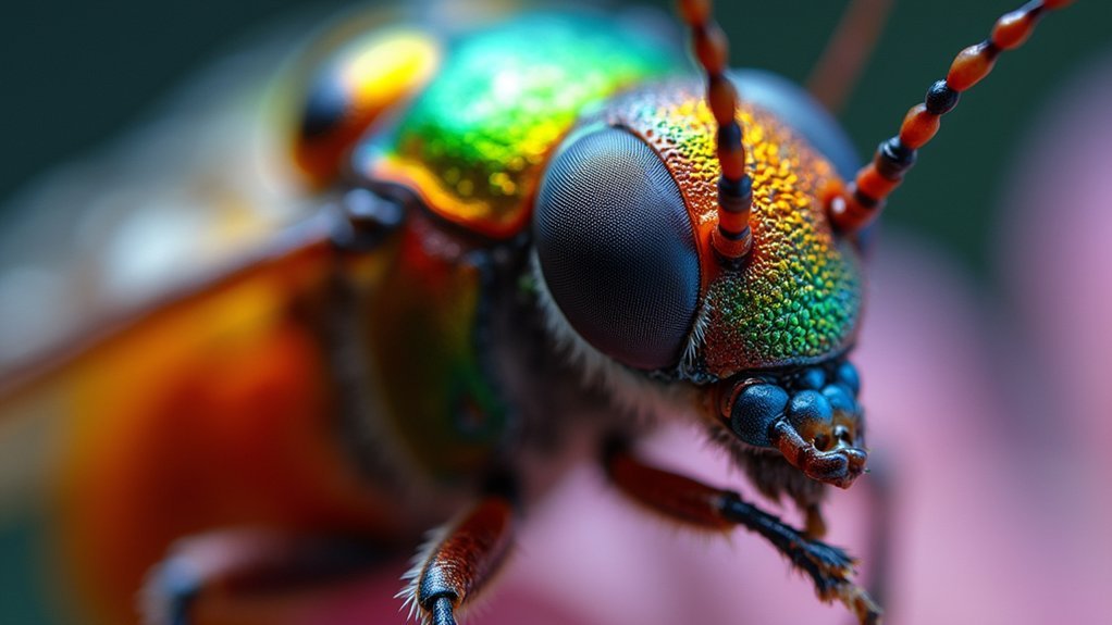
Focus stacking transforms your microscopy by merging multiple images at different focal planes to create clarity through multiple layers that would be impossible with a single shot.
You’ll notice greatly enhanced detail visibility as intricate structures snap into focus, revealing features that would otherwise remain blurred or hidden.
This technique expands your depth field well beyond the 1 μm limitation at high magnifications, allowing you to capture and present three-dimensional specimens with remarkable precision.
Clarity Through Multiple Layers
While peering through a microscope reveals fascinating miniature worlds, the limited depth of field often frustrates photographers and researchers alike. At high magnifications, your focal plane may be just a few micrometers deep, leaving vital details blurred and hidden.
Focus stacking overcomes this limitation by combining multiple images captured at different focal planes. You’ll see intricate structures with unprecedented clarity as the technique merges only the sharpest areas from each image into one composite. The result is an enhanced depth of field that reveals your specimen’s complete three-dimensional structure.
Using specialized software like Helicon Focus or Focus Stacker on your Mac, you can easily align and blend these images automatically.
This process transforms your microscopy work, allowing you to document and analyze complex biological specimens and materials with remarkable precision and detail.
Enhanced Detail Visibility
When viewing microscopic subjects, you’ll quickly discover that traditional single-shot photography simply can’t reveal the complete picture.
With depth of field measuring mere micrometers at high magnifications, vital details often remain blurred or completely invisible.
Focus stacking transforms your microscopy work by selectively blending the sharpest parts from multiple images taken at different focal depths.
This technique dramatically enhances detail visibility across your entire specimen, revealing intricate structures that would otherwise remain hidden.
You’ll capture fine features with exceptional clarity, allowing for more accurate scientific documentation and analysis.
Expanded Depth Field
The remarkable limitation of traditional microscope photography lies in its restricted depth of field, which becomes increasingly problematic at higher magnifications. When you’re working with high numerical aperture objectives, only a thin slice of your specimen appears sharp while everything else blurs into obscurity.
Focus stacking elegantly solves this challenge by combining multiple images captured at different focal planes. The result is an expanded depth of field that reveals your specimen’s complete three-dimensional structure in a single composite image.
You’ll capture intricate cell structures and surface textures that would otherwise remain partially obscured in conventional microscopy. This technique transforms your microscopic imaging capabilities, allowing you to create high-resolution composites that display complex specimens with unprecedented clarity—essential for both scientific analysis and creating compelling educational materials from your microscopic explorations.
Essential Hardware Requirements for Microscope Focus Stacking
The hardware you’ll select for microscope focus stacking directly impacts your final image quality, with full-frame cameras offering superior detail capture compared to smartphone alternatives.
You’ll need a reliable camera-to-microscope connection through either a dedicated microscope camera adapter or a T-mount that guarantees proper alignment and stability.
Your connection method must eliminate any potential vibration that could compromise image sharpness between successive focus layers.
Equipment Choice Matters
Selecting appropriate hardware dramatically impacts your focus stacking results, especially for microscope imaging on Mac systems.
For high-quality microscope images, you’ll need a full-frame camera like the Nikon D800 or Canon EOS 5DS that captures fine specimen details.
Don’t overlook the importance of a motorized focusing rail—the StackShot system from Cognisys provides the precise incremental adjustments crucial for professional stacking.
Pair this with remote control software such as Helicon Remote to minimize manual errors during capture.
Choose a camera with live view capabilities for real-time monitoring and adjustment.
For ideal results, verify compatibility with modern microscope cameras like the Nikon DS-Ri2 or Sony Alpha 6600.
These components work together seamlessly with your Mac to produce exceptionally detailed composite images.
Camera-to-Microscope Connection
Moving from equipment selection to implementation, proper connection between your camera and microscope forms the backbone of a successful focus stacking system.
You’ll need specialized adapters like the LM Digital Adapter to create a secure, precise camera-to-microscope connection that eliminates any potential movement that could ruin your stacked image.
When working with high-magnification objectives (like 100x), even microscopic vibrations can compromise image quality.
These objectives have extremely shallow depth of field—often just micrometers—making proper mounting critical. Your adapter should be compatible with both your camera model and microscope make.
The entire system works in concert: your full-frame camera captures detail-rich frames while the motorized rail makes precise movements, all controlled through software like Helicon Remote to produce seamlessly stacked microscope images with exceptional depth.
Comparing Top Focus Stacking Software Options for Macos
When diving into focus stacking on your Mac, you’ll find several powerful software options that cater to different needs and budgets.
Focus Stacker stands out as a popular choice, offering both automatic stacking algorithms and manual retouch modes for just €20 on the Mac App Store. While effective at alignment, be aware it may produce some artifacts.
Helicon Focus excels with misaligned images and offers a free trial with full features before purchase.
If you’re budget-conscious, consider GIMP—a free alternative supported by numerous online tutorials—or ChimpStackr, an open-source utility designed specifically for automatic alignment and stacking.
Each option balances different strengths in cost, user-friendliness, and advanced features, allowing you to choose based on your specific microscopy needs.
Step-by-Step Guide to Capturing Image Sequences for Stacking
Before you can use any of those powerful Mac stacking applications, you’ll need proper source material to work with.
Start by setting up your microscope on a stable surface to reduce vibrations during capture. A motorized focusing rail is crucial for precise control when capturing your 10-20 image sequence at different focal depths.
For ideal microscope image capture:
- Use your camera’s Live View to monitor exposure settings before starting your sequence
- Move through focal planes methodically, guaranteeing each depth is properly represented
- Save your images in TIFF or RAW format for maximum quality when processing on your Mac
These high-quality source images will guarantee your stacking software has everything needed to create incredibly detailed composite microscope images with complete depth of field.
Mastering Focus Stacker: A Mac-Native Solution
Focus Stacker gives you unparalleled depth control for your microscope images with its advanced stacking algorithm that precisely combines sharp areas from multiple captures.
You’ll appreciate the native macOS integration that leverages Metal technology and M1 optimization, creating a seamless and efficient workflow specifically designed for Mac users.
The intuitive interface lets you import various image formats and export in high-quality 16-bit TIFF or 8-bit JPEG/PNG, allowing for maximum flexibility in your microscopy post-processing.
Perfect Microscopy Depth Control
Although microscopy provides incredible detail, the limited depth of field often forces compromises—until now.
Focus Stacker gives you perfect depth control by intelligently combining multiple images from different focal planes into one stunning composite that’s fully sharp throughout.
The advanced algorithm handles the complex work for you:
- Automatic detection identifies sharp areas across your image set, eliminating guesswork
- Seamless integration merges only the clearest parts of each frame, preserving maximum detail
- Planar stitching capability aligns and combines multiple frames for wider field coverage
You’ll appreciate how this Mac-native application simplifies what was once a technical challenge.
With support for various formats including Apple Raw and high-quality export options, you’ll never sacrifice detail when creating perfectly focused microscopy images.
Mac-Optimized Stacking Workflow
When working with microscope imagery on your Mac, you’ll appreciate how Focus Stacker delivers a truly native experience designed specifically for macOS.
The application runs flawlessly on Apple M1 processors with performance optimized for recent macOS versions.
You can import various image formats, including Apple Raw, making it perfect for handling high-resolution microscope images.
The advanced automatic stacking algorithm intelligently combines sharp areas from multiple focus points into one composite image with enhanced depth.
Export your finished work in 16-bit TIFF or 8-bit JPEG/PNG formats, preserving quality for further analysis or sharing.
The built-in planar stitching feature is particularly valuable for microscope work, allowing you to create detailed composites of complex sample structures without switching between applications.
Advanced Techniques for Processing Complex Microscope Specimens

Despite their incredible resolution capabilities, high-magnification microscope objectives struggle with limited depth of field, creating a significant challenge for three-dimensional specimens.
When you’re working with intricate biological structures or materials, focus stacking becomes an essential image processing technique to reveal complete detail.
For complex specimens, you’ll need to:
- Employ automated focus rails like StackShot that can capture precise incremental frames while minimizing vibration and movement
- Utilize Mac-compatible software such as Helicon Focus or Focus Stacker to blend only the sharpest portions of each image
- Consider 3D visualization options in your stacking software to extract additional structural information from your dataset
This approach overcomes the limitations of high numerical aperture objectives, ensuring your scientific observations retain both exceptional resolution and thorough depth.
Integrating Focus Stacked Images Into Scientific Research
The technical mastery of focus stacking opens new possibilities for scientific communication and analysis. When you incorporate focus stacked images into your research, you’re providing colleagues with detailed visualizations that reveal intricate structures normally impossible to capture in a single frame.
By integrating focus stacked images into publications and presentations, you’ll enhance the quality of your scientific outputs, making complex 3D structures more accessible in 2D format. This technique is particularly valuable when documenting specimens with varied topography in biology or materials science.
Your Mac’s Focus Stacker software streamlines this integration process, allowing you to efficiently compile image sets for analysis and presentation.
The result: more compelling evidence, clearer documentation, and ultimately more impactful research that fellow scientists can better understand and verify.
Troubleshooting Common Issues in Microscope Image Stacking
Even the most advanced focus stacking setups can encounter frustrating technical hurdles. When your image stacking process isn’t producing the crisp, detailed results you expect, several common issues might be at play. Proper alignment is critical—even minimal shifts between frames can create artifacts that ruin your final composite.
Perfect focus stacking requires pixel-perfect alignment—even microscopic shifts can transform masterful composites into frustrating artifacts.
If you’re experiencing problems with focus stacking on your Mac, try these approaches:
- Check for macOS path randomization issues that might prevent plugin detection—you may need to relocate ImageJ or adjust settings manually.
- Experiment with alternative plugins in Fiji ImageJ if Stack Focuser fails to process your images.
- Verify consistent focus distances between captured images to prevent processing loops.
When troubleshooting persists, don’t hesitate to consult community forums where fellow microscopists share valuable solutions.
Frequently Asked Questions
What Is the Point of Focus Stacking?
Focus stacking helps you create images with enhanced depth of field by combining multiple photos taken at different focus points. You’ll capture sharp details across the entire subject that would normally be impossible in a single shot.
What Does Focus Do on a Mac?
On your Mac, focus allows you to combine multiple images with different focal points into one sharp composite. You’ll achieve enhanced depth of field, especially useful for microscopy or macro photography projects.
What Is the Difference Between Focus Bracketing and Focus Stacking?
Focus bracketing is when you take multiple photos at different focus points, while focus stacking is the process of combining those images into one final photo with extended depth of field. You’ll need both for peak sharpness.
What Is Macro Focus Stacking?
Macro focus stacking is a technique where you combine multiple images taken at different focus points to create one perfectly sharp photo with extended depth of field, especially useful when photographing tiny subjects up close.
In Summary
You’ve now discovered how focus stacking can dramatically transform your microscope images on a Mac. With the right hardware and software, you’ll reveal details previously hidden by depth of field limitations. Whether you’re using Focus Stacker or another solution, you’re equipped to create stunning composite images that showcase your specimens in remarkable clarity. Don’t let flat, partially-focused images limit your microscopy work—embrace focus stacking for truly professional results.



Leave a Reply