For successful fluorescence photography, you’ll need high-CRI LEDs (90+) that precisely match your fluorophores’ excitation wavelengths. Use fast-switching capabilities (300μs) to reduce photobleaching while balancing intensity levels—low (20-40%) for live imaging, medium (40-70%) for general use, and high (70-100%) for maximum clarity. Incorporate proper filters to minimize spectral overlap and implement adequate cooling systems to maintain LED performance. The right setup will transform your fluorescence imaging results from ordinary to extraordinary.
11 Second-Level Headings for “Best LED Lighting Setup For Fluorescence Photo Success”
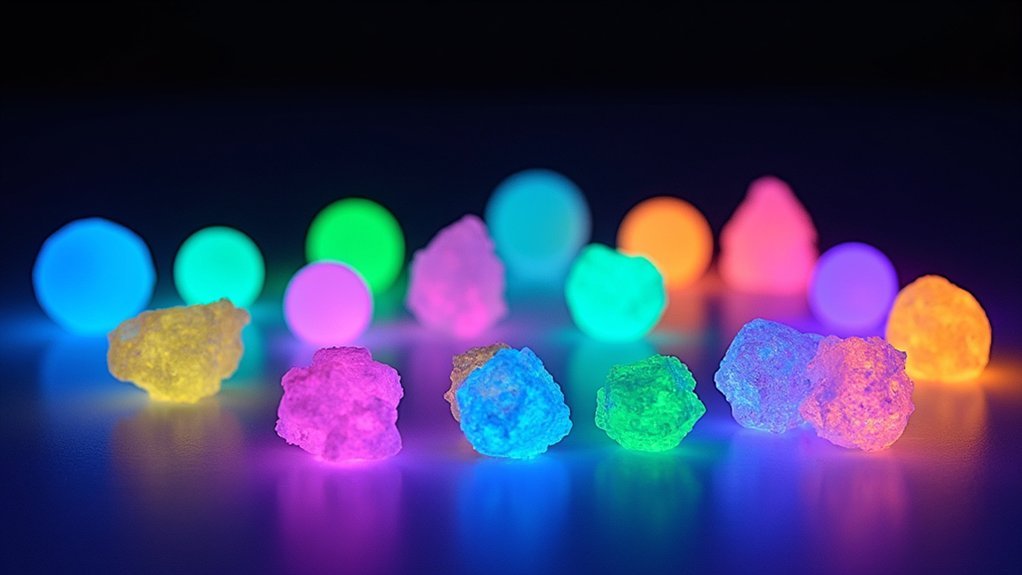
Include sections addressing “Fast-Switching Capabilities for Multi-Fluorochrome Imaging” to highlight the operational efficiency advantages.
LED technology enables instantaneous switching between fluorochromes, dramatically reducing image acquisition time and photobleaching effects.
Don’t forget headings about “Heat Management and Energy Efficiency” that emphasize LED’s practical benefits in laboratory settings.
A technical section on “Incorporating Filters to Reduce Bleedthrough” will help readers understand how to enhance image clarity.
Conclude with “Setting Up Your Complete LED System” to provide actionable setup advice.
These headings create a logical flow that covers both the scientific principles and practical applications of LED lighting for fluorescence microscopy.
Understanding Key Fluorescence Microscopy Lighting Requirements
To achieve ideal fluorescence microscopy results, you’ll need light sources that precisely match your fluorophores’ excitation wavelengths with minimal spectral overlap.
Your LED system must deliver consistent illumination intensity throughout imaging sessions, as fluctuations can compromise data reproducibility and quantitative analysis.
Beyond spectral precision, the stability of your light source directly impacts experimental consistency, allowing for reliable comparisons between imaging sessions and reduced photobleaching of your specimens.
Lighting Spectral Quality
When selecting an LED lighting setup for fluorescence microscopy, spectral quality stands as the paramount consideration for successful imaging. Your excitation light must precisely match the absorption spectra of your fluorochromes for peak results. Modern high-performance monochromatic LEDs offer narrow bandwidth illumination that efficiently triggers fluorescence while minimizing background noise.
| LED Feature | Benefit | Application |
|---|---|---|
| Specific wavelengths | Targeted excitation | Multi-fluorochrome imaging |
| Adjustable intensity | Reduced photobleaching | Live cell observation |
| Fast switching (300μs) | Precise timing control | Time-lapse experiments |
Your illumination system’s spectral purity directly impacts contrast and signal-to-noise ratio. Systems like Colibri demonstrate how individually controlled LEDs allow you to simultaneously observe multiple fluorochromes with minimal crosstalk, enhancing your imaging capabilities while reducing phototoxicity risks.
Power Stability Matters
Four essential stability factors determine the quality of your fluorescence imaging results.
First, consistent illumination intensity prevents variations that can compromise quantitative analysis accuracy. LED technology excels here with superior emission stability compared to traditional light sources.
Second, precise intensity control in percentage steps helps you minimize phototoxicity when imaging live specimens. You’ll appreciate this feature during extended imaging sessions.
Third, LEDs’ rapid switching capability (approximately 300 microseconds) enables efficient sequential imaging without light intensity degradation between captures.
Finally, uniform light output guarantees reproducible results across multiple experiments.
LED technology’s minimal thermal output and reliable power delivery maintain this consistency throughout the light source’s extended lifespan, eliminating frequent bulb replacements and recalibrations that plague other illumination systems.
Selecting High-CRI LEDs for Accurate Fluorophore Excitation
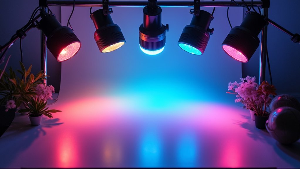
Why do high-quality LEDs matter so critically in fluorescence applications? When you’re capturing fluorescence imagery, the accuracy of your light source directly impacts your results.
High-CRI LEDs (90+) replicate natural sunlight properties, ensuring precise fluorophore excitation and true color representation in your microscopy work.
Natural light fidelity is essential—high-CRI LEDs deliver the spectral precision your fluorophores demand for accurate imaging results.
For ideal performance, select LEDs with narrow wavelength emissions (20-30nm bandwidth). These specialized lights reduce background noise and dramatically enhance image clarity.
Look for LEDs designed specifically for fluorescence applications, as they often feature customizable output wavelengths tailored to your specific fluorochromes.
Consider pairing your high-CRI LEDs with low-pass filters to block unwanted longer wavelengths that might interfere with excitation.
This combination, along with the inherent stability of quality LEDs, provides the consistent illumination necessary for reproducible, professional-grade fluorescence imaging.
Optimal Wavelength Selection to Match Fluorescent Dyes
Now that you’ve chosen high-quality LED sources, you’ll need to precisely match their wavelengths to your specific fluorescent dyes. Ideal wavelength selection directly impacts your fluorescence intensity and image quality.
- Select monochromatic LED lights that align with your dye’s absorption spectrum (e.g., 488nm for GFP)
- Consider wavelength conversion technology for challenging excitation ranges like the “green gap”
- Match your LED’s peak emission to your fluorescent dyes’ excitation bandwidth for maximum efficiency
- Use proper filters to isolate excitation wavelengths and minimize spectral overlap
- Remember that different fluorophores require different excitation wavelengths, so you’ll need specific LED lights for each
Designing a Modular LED Configuration for Multiple Fluorophores
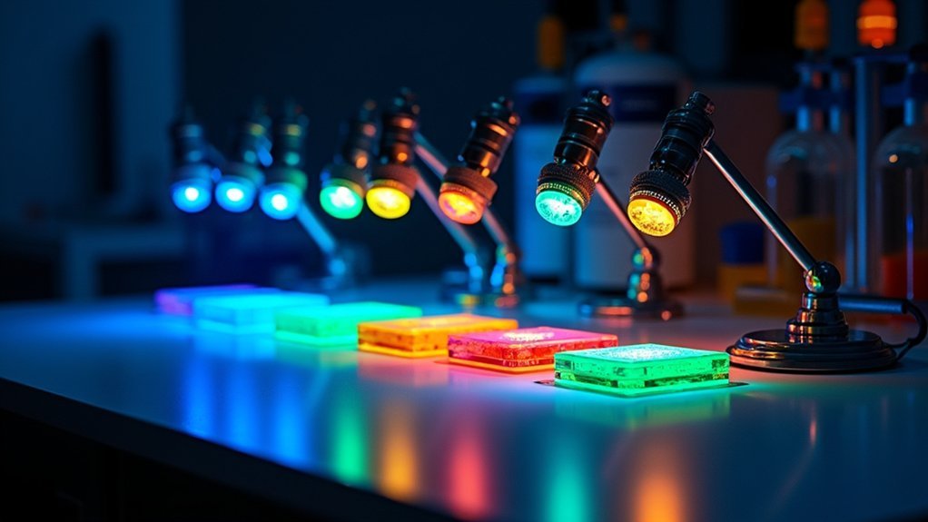
You’ll maximize your fluorescence imaging capabilities by implementing interchangeable wavelength filters that precisely match your target fluorophores’ excitation spectra.
Quick-swap excitation arrays allow you to shift between different fluorescent dyes without time-consuming system reconfiguration, essential for capturing dynamic cellular processes.
Multi-channel light control systems let you independently adjust intensity levels for each LED, minimizing phototoxicity while ensuring ideal excitation of multiple fluorophores during sequential or simultaneous imaging.
Interchangeable Wavelength Filters
When working with multiple fluorophores in a single experimental setup, implementing interchangeable wavelength filters becomes essential for achieving precise excitation and clear imaging results.
You’ll need to match specific wavelengths to your fluorescent proteins’ absorption spectra for ideal visualization without cross-excitation.
- Select high-performance monochromatic LEDs tailored to your specific fluorophores
- Incorporate neutral density filters to control light intensity and minimize phototoxicity
- Look for systems with fast switching times (around 300 μsec) for sequential imaging
- Verify your setup allows for reproducible intensity settings across different experiments
- Consider modular systems like Colibri that facilitate rapid wavelength changes
Quick-Swap Excitation Arrays
Designing an efficient quick-swap excitation array transforms how researchers approach multi-fluorophore experiments by eliminating the bottlenecks of traditional filter wheels.
You’ll achieve seamless shifts between up to four individually controlled LED light sources, allowing you to observe multiple fluorochromes simultaneously without mechanical switching.
These modular configurations match excitation wavelengths precisely to your fluorescent dyes’ absorption spectra, optimizing signal intensity and experimental outcomes.
With lightning-fast switching speeds of approximately 300 μsec, you’ll gain precise control over illumination timing—crucial for minimizing phototoxicity in live cell applications.
Multi-Channel Light Control
Unlike traditional single-source illumination systems, modular LED configurations empower you to simultaneously control multiple excitation channels with unprecedented precision.
With multi-channel light control, you’ll maximize your fluorescence microscopy capabilities while eliminating mechanical filter switches that slow down imaging.
- Select high-performance monochromatic LEDs matched to your specific fluorophores
- Control up to four individual channels in systems like Colibri
- Adjust intensity in precise percentage steps for each LED independently
- Reduce phototoxicity in live cell applications through enhanced illumination
- Achieve stable, reproducible results with long-lasting LED light sources
This modular approach enables rapid sequential imaging and simultaneous observation of multiple fluorochromes.
You’ll experience enhanced imaging efficiency while maintaining superior contrast—critical for both routine applications and advanced research requiring multiple fluorescence channels.
Managing Light Intensity for Maximum Signal-to-Noise Ratio
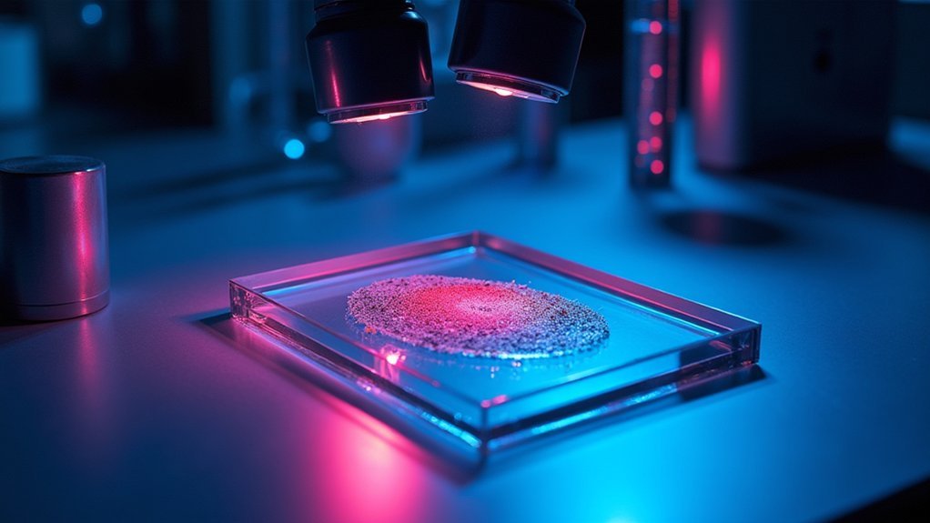
Although many factors contribute to successful fluorescence imaging, optimizing light intensity stands as perhaps the most critical element for achieving excellent signal-to-noise ratio (S/N). You’ll need to balance your LED’s output carefully—too dim and your signal disappears, too bright and you’ll damage live specimens.
| Intensity Level | S/N Impact | Sample Considerations |
|---|---|---|
| Low (20-40%) | Weak signals, poor contrast | Safe for extended live imaging |
| Medium (40-70%) | Good detail, moderate clarity | Limited exposure recommended |
| High (70-100%) | Maximum clarity, best S/N | Risk of phototoxicity, bleaching |
LEDs offer the precision you need, with microsecond switching speeds (≈300 μsec) allowing you to adjust intensity on-the-fly. Match your LED’s wavelength closely to your fluorochrome’s excitation peak for even greater S/N improvements with less intensity.
Heat Management Solutions for Extended Imaging Sessions
Implement strategic thermal dissipation techniques to protect your live specimens during long fluorescence imaging sessions.
You’ll need to integrate cooling systems like small fans or heat sinks near your LED light sources to maintain ideal temperatures throughout extended experiments.
These heat management solutions not only preserve sample integrity but also enhance LED performance and longevity, ensuring consistent illumination quality for your most critical imaging applications.
Thermal Dissipation Strategies
Three critical components define successful thermal management in LED lighting setups for fluorescence imaging. Effective heat dissipation prevents light output degradation while extending your equipment’s lifespan.
You’ll need a thorough approach to maintain ideal thermal conditions during those marathon imaging sessions.
- Install quality heat sinks and fans to efficiently disperse heat from LED components
- Implement pulsed illumination instead of continuous operation to minimize heat buildup
- Monitor LED temperature regularly with thermal sensors to prevent overheating
- Use thermal insulation techniques to protect both equipment and sensitive samples
- Choose LED arrays specifically designed with advanced heat dissipation capabilities
Cooling System Integration
While your LED setup generates exceptional fluorescence excitation, it also produces heat that can compromise image quality over time.
Implementing thorough heat management solutions is essential for maintaining performance during extended imaging sessions.
You’ll benefit from incorporating active cooling systems such as fans or liquid cooling mechanisms that prevent LED temperatures from reaching critical thresholds.
Mount heat sinks directly to your LED modules to efficiently dissipate thermal energy and preserve stable light output.
For best results, consider housing your setup in a temperature-regulated imaging chamber that protects both your equipment and biological samples.
Install thermal sensors to monitor LED temperatures in real-time, allowing you to make immediate adjustments before overheating occurs.
These proactive cooling strategies will extend your system’s lifespan while ensuring consistent illumination quality throughout your fluorescence microscopy work.
Pulsed vs. Continuous Illumination Techniques
When setting up your fluorescence imaging system, choosing between pulsed and continuous illumination can greatly impact both image quality and sample viability.
Pulsed illumination with LEDs offers significant advantages for live cell imaging by reducing phototoxicity while maintaining signal clarity.
- LEDs provide instant ON/OFF capabilities, eliminating mechanical shutters and enabling precise control over exposure times.
- Pulsed techniques promote healthier cell proliferation during extended imaging sessions.
- Adjustable intensity levels during pulses enhance your signal-to-noise ratio.
- LED pulsing systems stabilize optical power more effectively than continuous sources.
- Continuous illumination offers steady capture conditions but increases phototoxicity risk.
For most live sample applications, you’ll find pulsed illumination delivers the best balance between image quality and sample preservation, particularly when your experiment requires extended observation periods.
Minimizing Phototoxicity and Photobleaching With LED Controls
The precise control offered by pulsed LED systems directly addresses one of the most significant challenges in fluorescence microscopy: phototoxicity and photobleaching.
You’ll benefit from LED technology’s remarkably fast switching capabilities (approximately 300 μsec) and adjustable intensity settings that reduce harmful exposure during live cell imaging.
By implementing pulsed rather than continuous illumination, you’re allowing cells to recover between light exposures, promoting healthier proliferation throughout your experiments.
Lower intensity settings further minimize phototoxicity while still providing sufficient excitation for effective fluorescence imaging.
Choose specific wavelengths, particularly green LEDs, to mitigate unwanted fluorescence and improve image quality.
The exceptional stability and long lifespan of LEDs also guarantee consistent performance over time, eliminating disruptions from frequent replacements that could compromise your experimental conditions.
Essential Light Modifiers for Fluorescence Photography
Although LED systems provide excellent illumination for fluorescence imaging, incorporating specialized light modifiers greatly enhances your results by controlling beam characteristics and reducing unwanted artifacts.
When working with LED light sources, proper modification is essential for preserving specimen integrity while maximizing signal detection.
- Diffusers and scrims soften harsh LED illumination, minimizing phototoxicity during live cell imaging
- Softboxes provide contained diffusion with adjustable positioning to optimize the benefits of LEDs for your samples
- Neutral density (ND) filters used with LED systems help control intensity and prevent photobleaching
- Strategic placement of reflectors improves light distribution while maintaining required excitation wavelengths
- Combining multiple modifiers creates balanced illumination that preserves sample integrity while enhancing fluorochrome performance
Integrating LED Systems With Microscope Optics
Successfully integrating LED systems with microscope optics represents the next frontier in fluorescence imaging after mastering light modifiers.
You’ll find that modern high-performance LEDs perfectly match the excitation bandwidth of common fluorescent dyes, delivering precisely targeted illumination for your specimens.
Systems like Colibri exemplify effective integration, allowing you to control up to four different LEDs without mechanical switching—significantly improving your workflow efficiency.
Seamless LED integration eliminates mechanical delays, enhancing experimental throughput and precision in multi-fluorophore imaging workflows.
These LED light sources provide fast switching capabilities (around 300 μsec), enabling rapid sequential imaging while minimizing phototoxicity in your live cell samples.
The compact size and minimal heat output of LED systems make them ideal for direct integration with microscope optics.
You’ll benefit from their stability and longevity, ensuring consistent illumination and reproducible results throughout your fluorescence microscopy experiments.
Frequently Asked Questions
What Is the Best LED Color for Visibility?
For visibility in general, you’ll find blue (450-490 nm) and green (500-550 nm) LEDs most effective. They’re brightest to human eyes, provide excellent contrast, and require less power while offering greater visibility in various environments.
What Is the Best Lighting for Professional Pictures?
For professional pictures, you’ll want high CRI LED lights (94+) with adjustable color temperature. Use softboxes or diffusion panels to soften light and reduce shadows. Continuous lighting helps you perfect exposure before shooting.
Is Fluorescence More Efficient Than LED?
No, fluorescence isn’t more efficient than LED. Fluorescence is a process that emits light, while LEDs are actually devices that can efficiently produce light. You’ll find LEDs are used to trigger fluorescence in microscopy.
How Do You Take Good Pictures With LED Lights?
You’ll get great LED-lit photos by positioning lights at 45° angles, using diffusers to soften harsh shadows, adjusting white balance on your camera, and controlling brightness to prevent overexposure or dark spots.
In Summary
You’ll achieve outstanding fluorescence photography results when you’ve optimized every aspect of your LED setup. Remember to match wavelengths precisely to your fluorophores, control illumination intensity to prevent photobleaching, and utilize proper filters. With the right modular configuration, high-CRI LEDs, and appropriate light modifiers, you’re ready to capture stunning fluorescence images while maintaining sample integrity. Keep experimenting to find your perfect balance of clarity and preservation.


Leave a Reply