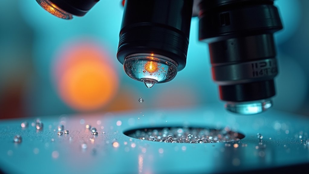
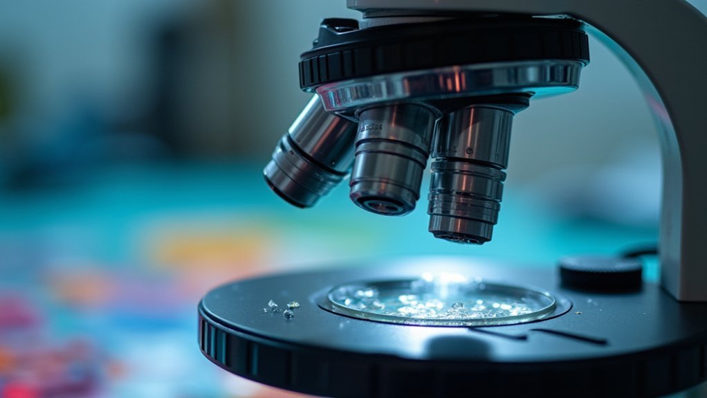
A microscope’s depth of field is primarily limited by its numerical aperture (NA). When you increase the NA to improve resolution, you’ll sacrifice depth of field. Higher magnification objectives further narrow this focus range. The wavelength of light also matters—shorter wavelengths reduce depth of field while improving resolution. Your aperture settings play a vital role too, with smaller apertures extending focus depth but potentially reducing overall image clarity. Discover how focus stacking techniques can help you overcome these inherent limitations.
The Science Behind Numerical Aperture and Its Impact on Depth of Field

When you’re examining specimens under a microscope, numerical aperture (NA) fundamentally determines what you can see and how clearly you can see it. This critical value measures your lens’s ability to gather light and resolve fine details.
There’s an inverse relationship between NA and depth of field—as NA increases, your focal plane becomes thinner.
Low-power objectives (4x) with NA values of 0.05-0.1 provide greater depth of field, allowing you to see more layers of your specimen simultaneously. In contrast, high-power objectives (100x) with NA values approaching 0.95 deliver exceptional resolution but at the cost of a remarkably shallow focus range.
The fundamental trade-off in microscopy: low-power objectives show depth, high-power objectives reveal detail—never both simultaneously.
This trade-off is unavoidable in microscopy: you’ll always balance resolution against depth of field when selecting objectives for your specific imaging needs.
How Magnification Affects Focus Range in Microscopy
As you increase magnification on your microscope, you’ll notice the focus range becoming progressively narrower, creating a significant practical challenge for specimen examination.
This relationship between magnification and depth of field is inversely proportional—higher magnification drastically reduces the thickness of your specimen that appears in focus.
For instance, at 20X magnification, your depth of field may reach about ±500 nanometers, allowing more of your sample to be visible simultaneously.
However, when you switch to 100X, this focus range shrinks dramatically to just ±0.1-0.2 microns.
This reduction occurs because higher magnifications typically employ objectives with larger numerical aperture values, which inherently decrease depth of field.
Understanding this relationship helps you develop effective focusing strategies when examining specimens at various magnifications.
Fixed Apertures vs. Variable Apertures: Understanding the Trade-offs

Microscope aperture systems fundamentally determine how much control you’ll have over your depth of field during specimen examination.
Fixed apertures offer simplicity but limit your ability to adapt to different samples, locking you into a predetermined focus range. In contrast, variable apertures provide flexibility to adjust your depth of field as needed.
When working with complex specimens, consider these key trade-offs:
- Reducing aperture size (higher f/number) increases depth of field but may introduce diffraction that reduces overall resolution.
- Larger apertures (lower f/number) enhance detail visibility but create shallower focus ranges that can’t capture all sample components simultaneously.
- Higher numerical aperture settings improve resolution of fine details but sacrifice the depth of field needed for samples with varying heights.
Wavelength of Light and Its Relationship to Depth of Field
The fundamental relationship between wavelength and depth of field follows a direct mathematical correlation, where longer wavelengths produce greater depths of field as governed by the diffraction limit.
You’ll notice significant variations in focus depth when switching between different colored light sources, with red light maintaining more of your sample in focus simultaneously compared to blue light.
While blue light offers superior resolution due to its shorter wavelength, you’re trading that enhanced detail for a shallower depth of field—a critical consideration when imaging thick or uneven specimens.
Wavelength-DOF Mathematical Relationship
When exploring the fundamental constraints of optical microscopy, you’ll discover that light’s wavelength plays a crucial role in determining depth of field (DOF). The mathematical relationship between wavelength and DOF is governed by the diffraction-limited resolution formula: 1.22λ/NA.
As wavelength (λ) increases, diffraction patterns enlarge, directly reducing your ability to resolve fine details and consequently limiting DOF.
This inverse relationship means you’ll experience:
- Shorter wavelengths (blue light) produce shallower DOF but higher resolution
- Higher numerical aperture (NA) objectives magnify the wavelength’s influence on DOF
- Red light (longer wavelength) creates deeper DOF but sacrifices some resolution
For high-magnification work, the wavelength effect becomes especially critical, impacting your imaging clarity when examining intricate structures where precise focus is essential.
Color Impact Variations
While examining specimens under a microscope, you’ll notice that different colored light produces distinctly variable imaging results. This variation stems directly from the relationship between wavelength and depth of field (DOF).
| Color Light | Wavelength Effect | Impact on DOF |
|---|---|---|
| Blue | Shorter wavelength | Shallower DOF, higher resolution |
| Green | Medium wavelength | Balanced DOF and resolution |
| Red | Longer wavelength | Greater DOF, reduced resolution |
Blue light allows you to resolve finer details due to less diffraction but creates a narrower plane of focus. Red light, conversely, provides a more forgiving DOF at the expense of image quality. When working with high numerical aperture (NA) objectives, these color effects become even more pronounced. Understanding these wavelength interactions helps you optimize your microscope settings for either maximum detail or extended focus depth.
Blue Light Advantage
Despite what the previous section suggests, blue light offers significant advantages for microscopy applications requiring enhanced resolution. When you’re working with blue light (approximately 450 nm), you’ll notice improved depth of field compared to longer wavelengths like red light (650 nm). This occurs because blue light creates smaller diffraction patterns, maintaining focus across broader distances.
- Blue light’s shorter wavelength minimizes light ray spread, creating a more defined focus area for superior image clarity.
- Combining blue light with higher numerical aperture (NA) enhances image sharpness while decreasing depth of field.
- For examining fine structural details, blue light’s properties help overcome diffraction limitations.
The quantifiable relationship between wavelength and depth of field makes blue light particularly valuable when you need to resolve minute features in your specimens while maintaining ideal focus.
Practical Solutions for Overcoming Limited Depth of Field
Because microscope users often struggle with limited depth of field when examining three-dimensional specimens, several practical solutions have emerged to address this challenge.
You can decrease the numerical aperture (NA) by using lower magnification objectives, which naturally increases depth of field along the optical axis. For a simple yet effective approach, try a 0.5x Barlow lens, which allows your objective to work at a more favorable distance without significant quality loss.
Focus stacking techniques, where you capture multiple images at different focal planes and combine them digitally, offer remarkable results for stationary specimens.
Focus stacking transforms depth limitations into opportunities, revealing complete specimen details through digital fusion of multiple focal planes.
Modern microscopes with Extended Depth of Field (EDOF) features can also save you considerable time. Additionally, strategic use of field stops can improve depth of field, though careful placement is essential to avoid vignetting.
Focus Stacking: Extending the Effective Depth of Field Digitally

When traditional microscopy fails to capture every detail of your three-dimensional specimens, focus stacking offers a powerful digital solution. This technique involves capturing multiple images at different focal planes, then combining them to create a single image with extended depth of field (DOF). You’ll achieve remarkable image detail that would be impossible with conventional microscopy.
- By layering several focal planes, you’re fundamentally bypassing the physical limitations of your microscope’s optics.
- The quality of your final image directly correlates with the number of layers you capture—more layers mean greater detail.
- While effective for stationary specimens, focus stacking requires specialized software and additional time investment.
This technique particularly shines with three-dimensional samples where traditional microscopy would force you to compromise between magnification and depth of field.
Frequently Asked Questions
What Limits the Range of Microscopy?
Your microscope’s range is limited by numerical aperture, magnification, wavelength of light, sample preparation quality, and optical aberrations. These factors constrain both resolution capabilities and the depth you can effectively visualize.
What Three Things Affect Depth of Field?
Three key factors affecting depth of field are numerical aperture (higher NA creates shallower DOF), aperture size (smaller aperture increases DOF), and magnification (higher magnification decreases DOF). You’ll notice these relationships when adjusting microscope settings.
What Factors Might Limit Your Ability to Control the Depth of Field?
You’re limited by objective design constraints, minimum aperture sizes, diffraction effects at small apertures, sensor resolution limitations, and mechanical constraints of your microscope that prevent precise adjustments to numerical aperture and focus position.
What Is the Depth of Field of a Microscope?
Your microscope’s depth of field is the range above and below the focal plane where specimens remain sharp. It’s typically ±500nm at 20X magnification, decreasing to ±100-200nm with high-power oil immersion lenses.
In Summary
You’ve seen how depth of field in microscopy is primarily limited by numerical aperture, magnification, aperture design, and light wavelength. As you increase magnification or numerical aperture for better resolution, you’ll sacrifice depth of field. Don’t worry—techniques like focus stacking can help you overcome these physical limitations, allowing you to create composite images with extended depth while maintaining the high resolution you need for detailed specimen analysis.


Leave a Reply