
Critical illumination creates superior scientific images by focusing your light source directly onto specimens, delivering exceptional brightness that reveals minute details. You’ll capture sharper images with enhanced contrast, especially when using homogeneous light sources like sunlight. While it may show filament structures, you can minimize this by carefully adjusting your condenser’s aperture diaphragm. Though modern microscopy often favors Köhler illumination, critical illumination offers simplicity and remarkable illumination power for specific applications. The perfect balance awaits below.
The Fundamentals of Critical Illumination in Microscopy
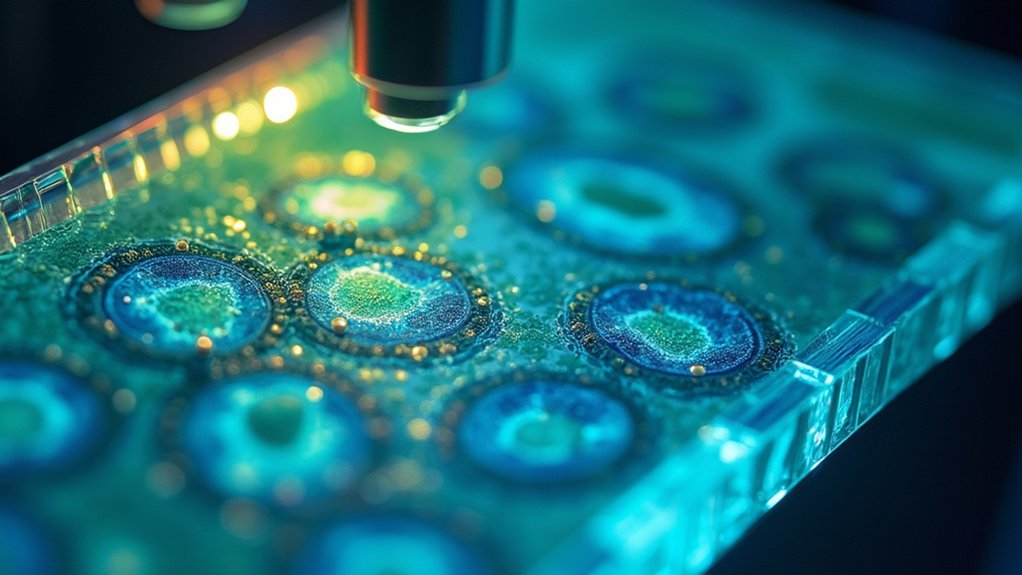
When examining the intricacies of microscopic worlds, critical illumination serves as one of the foundational lighting techniques available to researchers. Also known as Nelsonian illumination, this approach focuses an image of the light source directly onto your specimen through the condenser.
You’ll notice the illumination focuses with remarkable brightness, though the evenness of illumination can be compromised by revealing structures like bulb filaments in your final image.
To improve image resolution, you can adjust the aperture diaphragm and incorporate homogeneous light sources such as sunlight or ground glass diffusers.
Optimal microscopic resolution demands strategic aperture adjustments and uniform light sources like diffused sunlight.
While scientific light microscopy has largely shifted toward Köhler illumination for superior uniformity, critical illumination remains valuable for basic applications due to its simplicity.
Optimizing Light Source Placement for Maximum Detail Capture
Although critical illumination offers exceptional brightness, your light source’s precise positioning can make or break image quality. Position your light directly above the specimen to achieve evenness of illumination and minimize shadows that might obscure fine details.
For ideal uniformity, incorporate homogeneous light sources like sunlight or use ground glass diffusers. You’ll need to carefully adjust both angle and distance of your illumination to reduce glare and unwanted reflections. Proper alignment with the microscope’s optical axis ensures maximum light reaches your specimen, greatly enhancing detail resolution.
Don’t settle for your initial setup—experiment with different placements during your imaging session. By systematically varying your light source position, you’ll discover the perfect configuration that maximizes contrast and reveals subtle features in your specimen that might otherwise remain hidden.
Balancing Contrast and Brightness for Scientific Accuracy
Proper light source positioning reveals detail visibility, but mastering the interplay between contrast and brightness takes your scientific imaging to new heights.
Critical illumination excels by focusing the light source directly onto your specimen, creating illumination that’s remarkably bright while enhancing contrast in key areas.
You’ll need to make careful adjustment to achieve ideal image quality. While critical illumination can produce uneven lighting, you can fine-tune it to highlight specific features during scientific analysis.
Consider using homogeneous illumination sources like flames or sunlight to improve evenness across your specimen.
Comparing Critical and Köhler Illumination Techniques
Scientists must understand the fundamental differences between critical and Köhler illumination to select the best technique for their research needs.
When you’re using critical illumination, the light source is focused directly onto your specimen, creating bright but potentially uneven light distribution. You’ll notice the light source’s structures, like bulb filaments, appearing in your images.
Köhler illumination, conversely, delivers superior image clarity and evenness across your sample. It eliminates visible light source artifacts but requires additional optical components, including an adjustable condenser and centerable diaphragm. This makes the setup more complex but markedly improves specimen illumination quality.
While homogeneous light sources can enhance critical illumination’s performance, they can’t match Köhler’s consistent brightness.
Modern scientific microscopy mainly favors Köhler illumination for its ability to produce uniform, high-quality images essential for detailed research applications.
Practical Steps for Achieving Perfect Microscope Light Adjustment

Understanding the differences between illumination techniques is only half the battle; you’ll need to apply this knowledge through precise adjustment procedures.
Begin by setting eyepiece diopters to zero and adjusting the interpupillary distance for comfortable viewing. Select your objective lens (start with 10X) and center your sample for ideal illumination.
The key to perfect light adjustment involves three critical steps:
- Focus on your sample, then adjust the field diaphragm until its edge is clearly visible.
- Center the condenser and gradually open the field diaphragm just beyond your field of view.
- Set the condenser aperture diaphragm to match your objective’s numerical aperture (about 90% area illumination).
These adjustments guarantee that light properly illuminates your specimen, maximizing image clarity and revealing fine structural details that might otherwise remain invisible.
Frequently Asked Questions
What Is the Purpose of Critical Illumination?
You use critical illumination to focus the light source directly onto your specimen, enhancing visibility of fine details and surface characteristics. It’s simpler than Köhler illumination while still providing bright, high-contrast images.
Why Are Different Degrees of Illumination Required When Using a Microscope and Why?
You need different illumination levels in microscopy because specimens vary in thickness and transparency, and your objective’s NA demands specific light intensity to reveal ideal detail and contrast in your samples.
How Does Köhler Illumination Work?
Köhler illumination works by focusing your light source’s image onto the specimen plane. You’ll adjust the condenser and field diaphragm to fill the objective’s back focal plane, ensuring even illumination without visible light source artifacts.
What Is the Illumination System of a Light Microscope?
In your microscope, the illumination system consists of a light source, condenser lens, and objective lens. They work together to direct light through your specimen, allowing you to view it with peak clarity.
In Summary
You’ve now seen how critical illumination can transform your scientific imaging. By precisely positioning your light source and mastering contrast adjustments, you’ll capture considerably more detail than with standard techniques. Don’t settle for mediocre microscopy when these straightforward adjustments can dramatically improve your results. Apply these principles consistently, and you’ll produce images that reveal subtle structures others might miss entirely.


Leave a Reply