For scientific photography, you’ll get best results using Aperture Priority for depth control, Shutter Priority for motion capture, and Manual mode for complete precision. Program mode offers quick adaptability while Spot and Center-weighted metering guarantee accurate exposure of specimens. Pair these with focus stacking techniques for complex structures and histogram monitoring for perfect exposure. Adding exposure compensation and ISO adjustments helps balance detail and lighting challenges in challenging laboratory conditions. Discover how these modes transform your scientific imaging workflow.
10 Best Camera Exposure Modes For Scientific Photography
When capturing images for scientific purposes, choosing the right exposure mode can greatly impact your results.
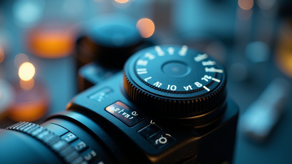
Aperture Priority Mode (A/Av) is often ideal as it lets you control depth of field while the camera handles shutter speed—perfect for detailed specimen documentation.
Control depth of field while leaving shutter speed to the camera—ideal for capturing precise specimen details in scientific photography.
If you’re photographing fast-moving subjects like chemical reactions, Shutter Priority (S/Tv) helps freeze motion by letting you set specific shutter speeds while the camera adjusts aperture accordingly.
For specialized scientific imaging such as astrophotography or macro work, Manual Mode offers complete control over all exposure parameters.
This precision is essential when documenting critical research.
In unpredictable lighting situations, Program Mode can be valuable, allowing quick adjustments while maintaining control over ISO and other important settings.
Pair these modes with Matrix metering for optimal scientific documentation.
Aperture Priority (A/Av) Mode for Depth Control in Specimen Photography
When photographing specimens, you’ll find Aperture Priority mode gives you precise control over background blur by allowing direct adjustment of f-stops while the camera handles exposure.
You can create clean, distraction-free backgrounds with larger apertures (f/2.8-f/4) or capture detailed structural features with smaller apertures (f/8-f/16) depending on your documentation needs.
For specimens with complex three-dimensional structures, Aperture Priority also facilitates focus stacking techniques where you’ll shoot multiple images at different focus points to combine later for extended depth of field.
Subheading Discussion Points
Why do scientific photographers prioritize depth of field control when documenting specimens? When capturing detailed images of scientific subjects, you need every vital feature in sharp focus.
Aperture Priority (A/Av) mode gives you direct control over this essential element of the exposure triangle while automating shutter speed adjustments.
By selecting smaller apertures (f/11-f/16), you’ll achieve greater depth of field, ensuring complete specimen documentation with maximum detail. This mode shines in changing light conditions, letting you maintain consistent image quality without constantly adjusting exposure settings.
You’ll also discover creative possibilities beyond mere documentation. Larger apertures can artistically isolate specific features, drawing attention to particular details while still maintaining scientific accuracy.
This balance of technical control and artistic flexibility makes Aperture Priority indispensable for scientific photography work.
Controlling Background Blur
Controlling specimen photography involves more than just capturing details—it’s about making those details stand out. Aperture Priority (A/Av) mode gives you precise depth of field control while automatically adjusting shutter speed for proper exposure.
| Aperture Setting | Effect | Best For |
|---|---|---|
| Wide (f/1.4-f/4) | Shallow depth of field, pronounced blur | Isolating specimens from distractions |
| Medium (f/5.6-f/8) | Moderate focus range | Balancing subject detail with context |
| Small (f/11-f/16) | Extended depth of field | Scientific documentation requiring full clarity |
You’ll find exposure compensation particularly useful when working with highly reflective or dark specimens. Adjust it up or down to maintain accurate detail representation while preserving your chosen aperture setting. This approach lets you work quickly in changing environments or with live subjects while maintaining creative control.
Focus Stacking Applications
Many scientific specimens exhibit complex three-dimensional structures that exceed the depth of field limitations of even the smallest apertures. For these challenging subjects, focus stacking in Aperture Priority mode provides a powerful solution.
You’ll achieve consistently sharp images by maintaining control over your depth of field while the camera handles exposure.
When employing focus stacking techniques:
- Set your camera to Aperture Priority mode and select smaller apertures (f/8-f/16) to maximize depth of field for each shot.
- Mount your camera on a tripod to guarantee perfect alignment between frames as you adjust focus points.
- Capture multiple images at different focus distances, then use specialized software to blend only the sharpest portions into a single, detailed composite.
This approach guarantees your scientific specimens are rendered with exceptional clarity and detail throughout the entire subject.
Manual Mode: The Gold Standard for Consistent Microscopy Results
When capturing microscopic specimens with scientific precision, manual mode stands as the unrivaled choice for serious researchers and technicians. This approach gives you complete control over exposure parameters, allowing you to maintain consistent aperture settings that deliver your desired depth of field for intricate structural details.
You’ll benefit from manually setting your ISO to minimize noise—critical when documenting minute cellular structures where every detail matters. The ability to lock in specific exposure settings guarantees that any variations between images reflect actual specimen differences rather than camera adjustments.
Manual mode also enables precise white balance control, enhancing color fidelity that helps distinguish between different cellular structures. For sequential imaging of similar specimens, this consistency becomes invaluable, making manual mode truly indispensable for professional microscopy work.
Spot Metering Mode for Precise Sample Illumination
Spot metering mode represents the next level of precision for scientific photographers who need exacting control beyond what manual mode offers.
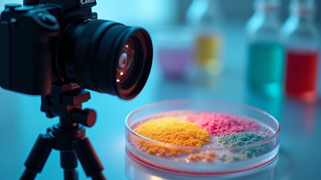
When you’re working with laboratory samples that contrast sharply with their backgrounds, spot metering guarantees your exposure calculations focus solely on the critical areas—typically just 1-5% of your frame. This precision proves invaluable when documenting microscopy specimens or chemical reactions where accurate detail representation determines analytical quality.
For ideal scientific photography using spot metering:
- Position your metering point directly on the most important element of your sample.
- Take test shots to confirm precise illumination of your subject without background interference.
- Consider combining spot metering with exposure compensation for samples with unusual reflectivity or translucency.
Center-Weighted Metering for Balanced Microscope Field Exposure
You’ll find center-weighted metering particularly valuable for maintaining specimen illumination balance across your microscope field, as it prioritizes exposure readings from the central area where most specimens are positioned.
By focusing exposure calculations on the central portion, you’ll achieve greater focal point precision for detailed scientific subjects while still accounting for surrounding areas.
This metering technique effectively reduces edge falloff in microscope photography, preventing the darkening that often occurs at image peripheries while keeping your main specimen properly exposed.
Specimen Illumination Balance
Three key challenges in scientific specimen photography revolve around achieving proper exposure across an unevenly lit field of view. Center-weighted metering offers an effective solution by prioritizing the central area where your specimen typically resides while still accounting for surrounding areas.
When balancing specimen illumination, you’ll need to:
- Set your camera to center-weighted metering mode to guarantee the microscope’s focused specimen receives proper exposure priority.
- Apply exposure compensation (+/-) based on specimen characteristics – increase for dark specimens and decrease for bright or reflective samples.
- Take test shots and review histograms to verify detail preservation in both highlight and shadow areas.
This approach provides consistent results even when your microscope creates dramatic lighting differences between the specimen and background, preserving the critical details needed for scientific analysis.
Focal Point Precision
Precision at the focal point represents the cornerstone of effective scientific photography, particularly when working with microscope imagery.
When capturing detailed specimens, center-weighted metering should be your go-to exposure mode. This technique prioritizes the center of your frame—where your specimen typically resides—ensuring accurate exposure readings despite varying background light conditions.
For ideal results, always position your subject centrally and pair this metering mode with appropriate shutter speed adjustments.
You’ll achieve better detail preservation by fine-tuning ISO and aperture manually while letting center-weighted metering handle the exposure balance. This approach proves especially valuable when working with microscope fields that contain high contrast elements, as it prevents bright backgrounds from underexposing your specimen.
The result: precisely exposed scientific images with exceptional focal point clarity.
Reducing Edge Falloff
While documenting specimens through microscopes, edge falloff often compromises the quality of scientific images by creating uneven illumination across the field of view.
You’ll find center-weighted metering particularly effective in countering this issue by prioritizing exposure calculation for the central portion of your frame where critical specimen details typically reside.
When selecting ideal exposure modes for microscope photography, consider these advantages:
- Center-weighted metering maintains consistent lighting across specimens, preserving the dynamic range necessary for accurately documenting subtle features.
- By focusing exposure calculations on the central area, you’ll reduce the influence of peripheral lighting variations that can skew readings.
- This metering technique guarantees your primary subject receives ideal exposure even when background brightness fluctuates.
This approach proves especially valuable in controlled laboratory environments where precision and clarity are paramount for scientific documentation.
Matrix/Evaluative Metering for Complex Biological Samples
When photographing intricate biological specimens, matrix or evaluative metering serves as an invaluable tool for achieving balanced exposures across varied lighting conditions. This advanced metering system analyzes your entire frame, employing sophisticated algorithms to assess brightness, contrast, and composition throughout the scene.
You’ll find matrix metering particularly useful when working with biological samples that contain both light and dark areas. The system prevents overexposure in high-contrast scenarios, such as when capturing transparent or reflective specimens. By evaluating multiple zones within the frame, it delivers a thorough exposure that preserves detail in both highlights and shadows.
For best results, combine matrix metering with aperture priority mode. This pairing maintains your desired depth of field while the camera’s evaluative system handles the exposure calculations, ensuring every important detail of your specimen remains visible.
Program Mode With Exposure Compensation for Rapid Documentation
Program Mode offers two significant advantages for scientific photographers requiring quick documentation: speed and adaptability. When you’re working in conditions with unpredictable lighting, this mode helps you maintain good exposure without needing complex manual adjustments. You’ll save valuable time while ensuring accurate subject representation.
To maximize Program Mode’s effectiveness:
- Use exposure compensation to fine-tune brightness levels when documenting specimens in challenging lighting conditions.
- Make rapid adjustments knowing your camera retains new settings until you manually change them.
- Preserve image detail by quickly adapting to lighting changes while maintaining critical scientific information.
This approach balances efficiency with quality, letting you focus on capturing data rather than camera settings, particularly valuable when documenting time-sensitive experiments or specimens.
Shutter Priority (S/Tv) Mode for Moving Microorganisms
Capturing rapidly moving microorganisms requires precise control over motion blur, which is why Shutter Priority (S/Tv) mode proves invaluable for scientific photographers.
When you’re documenting fast-moving bacteria or protozoa, selecting shutter speeds of 1/1000s or faster will freeze their motion, preserving the fine details essential for accurate analysis.
What makes S/Tv mode particularly useful is that your camera automatically adjusts the aperture to maintain proper exposure while you focus on controlling motion.
This becomes essential when working in variable laboratory lighting conditions.
Don’t forget to monitor your ISO settings, especially in low light situations.
You’ll need to find the balance between increasing ISO for proper exposure and avoiding excessive noise that could compromise the scientific integrity of your images.
Bracketing Mode for High Dynamic Range Specimen Capture

Photographing specimens with extreme light and dark areas presents a significant challenge that goes beyond the capabilities of standard exposure modes.
Bracketing mode offers a powerful solution by capturing multiple shots at different exposure settings, preserving details in both shadows and highlights that are critical for scientific analysis.
When working with high-contrast specimens in fields like biology or geology, you’ll benefit from:
- Adjusting your camera’s bracketing settings (typically 1-3 stops) to capture the full dynamic range of challenging specimens
- Taking a sequence of images (underexposed, correctly exposed, and overexposed) of the same subject without moving the camera
- Combining these images later using HDR software to reveal fine details that would otherwise be lost in a single exposure
This technique effectively overcomes your camera’s inherent dynamic range limitations, ensuring no critical specimen details are lost.
Live View Histogram-Based Exposure for Scientific Accuracy
You’ll find mastering histogram-based exposure essential for maintaining scientific accuracy as it provides immediate visual feedback on your image’s tonal distribution.
By monitoring the live view histogram, you can quickly identify and correct exposure problems before they compromise your scientific documentation.
This real-time approach lets you adjust settings on the fly, ensuring each image captures the precise color values and details required for reliable research results.
Histogram-Based Exposure Mastery
Why rely on guesswork when your camera’s live view histogram offers precise exposure guidance for scientific photography? This powerful tool displays your image’s tonal distribution in real-time, allowing you to make immediate adjustments to shutter speed and ISO while ensuring no critical details are lost in shadows or highlights.
When mastering histogram-based exposure, focus on:
- Maintaining a balanced histogram that fills the graph without touching either edge—this preserves both shadow and highlight details essential for scientific documentation.
- Using exposure compensation in conjunction with histogram readings to fine-tune exposure for specific scientific details.
- Checking your histogram regularly during shooting sessions, especially when lighting conditions change, rather than trusting the potentially misleading camera preview.
The histogram is your scientific accuracy ally, providing objective feedback beyond what your eyes perceive.
Real-Time Exposure Adjustment
While traditional exposure methods rely on after-the-fact analysis, modern digital cameras equipped with Live View offer a transformative approach through real-time histogram monitoring. You’ll see immediate feedback on your exposure levels, allowing you to make precise adjustments before capturing the image.
| Adjustment | Effect | Scientific Benefit |
|---|---|---|
| Aperture | Controls light volume | Enhances depth of field for specimen clarity |
| Shutter Speed | Affects exposure time | Prevents motion blur in dynamic processes |
| Change your ISO | Adjusts sensitivity | Balances noise levels for analytical accuracy |
Frequently Asked Questions
What Is the Best Camera for Botanical Photography?
You’ll want a camera with macro capabilities, high resolution (20+ MP), manual controls, wide aperture (f/2.8+), and good ISO performance. The Sony Alpha or Nikon Z series offer excellent options for botanical photography.
What Mode Do Professional Photographers Shoot In?
Professionals don’t use just one mode. You’ll find them switching between Aperture Priority for depth control, Shutter Priority for action, and Manual for precise settings, depending on their specific shooting needs.
What Is the Benefit of Using P Mode for Learning Photography?
P mode helps you learn photography by automating initial exposure settings while letting you adjust them. You’ll understand how aperture and shutter speed affect images without being overwhelmed by technical details right away.
What Camera Setting Should You Experiment With to Get Better Exposure When Shooting an Event Like a Prom or Wedding Reception That’s Dimly Lit?
You should experiment with wider apertures (f/1.8-f/4), higher ISO settings, and exposure compensation (+1 or +2 EV). Don’t forget to contemplate using bounce flash to illuminate subjects without harsh shadows.
In Summary
You’ve now explored the essential exposure modes that’ll transform your scientific photography. Whether you’re capturing microscopic specimens or documenting experiments, these techniques will help you achieve consistent, accurate results. Remember to choose the appropriate mode based on your specific research needs. With practice, you’ll develop an intuitive understanding of which exposure settings work best for different scientific applications.


Leave a Reply