
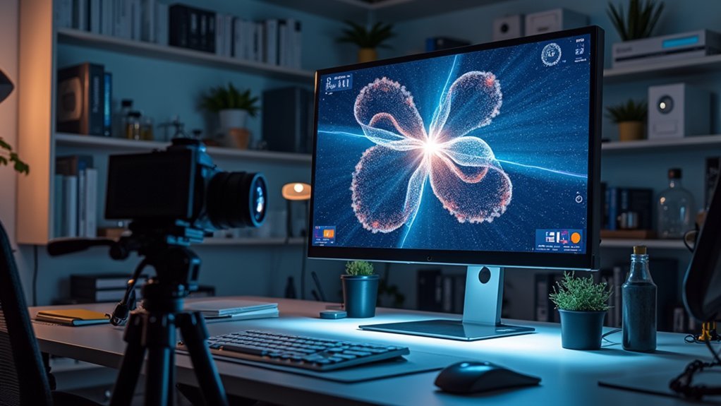
Scientific imaging automation requires specialized time-lapse software to capture cellular processes effectively. Your best options include LR Timelapse for professional de-flickering, DaVinci Resolve 17 for high-resolution editing, and the EVOS FL Auto system for lab work. Configure custom protocols with specific time intervals, enable Z-stack imaging for structural detail, and implement metadata extraction for efficient data management. The right software combination will transform your research workflow and elevate your scientific findings.
Numeric List of Second-Level Headings
Five key software options dominate the time-lapse editing landscape.
When implementing Automated imaging systems for your research or creative projects, you’ll want to evaluate these essential tools:
- Timelapse Lite (TLDF Lite) – Free solution with unlimited image editing and built-in color grading
- LR Timelapse – Professional tool with free trial for up to 400 images and superior de-flickering
- DaVinci Resolve 17 – Powerful free video editor supporting high-resolution Time-Lapse Imaging
- Five Day Deal promotions – Educational bundles including time-lapse presets at significant savings
- EVOS FL Auto Imaging System – Scientific-grade automated system with customizable scanning options
Each option offers unique advantages, whether you’re conducting laboratory research requiring precise image collection or creating stunning visual sequences for creative projects.
Understanding Time-Lapse Software for Scientific Research
While creative professionals often use time-lapse techniques for aesthetic purposes, scientific researchers rely on specialized time-lapse software to document and analyze cellular processes that occur over extended timeframes.
These tools enable you to capture critical cellular events like division and migration through automated imaging and real-time monitoring.
When selecting time-lapse software for your laboratory, consider these essential capabilities:
- Customizable imaging protocols that integrate with fluorescence microscopy to visualize specific cellular structures
- Z-stack creation options for three-dimensional tissue analysis
- Image enhancement tools to adjust brightness and contrast for improved clarity
Advanced software options range from extensive platforms like DaVinci Resolve to specialized solutions like LR Timelapse, with free alternatives available for projects with limited funding.
Key Features of Effective Microscope Automation Tools
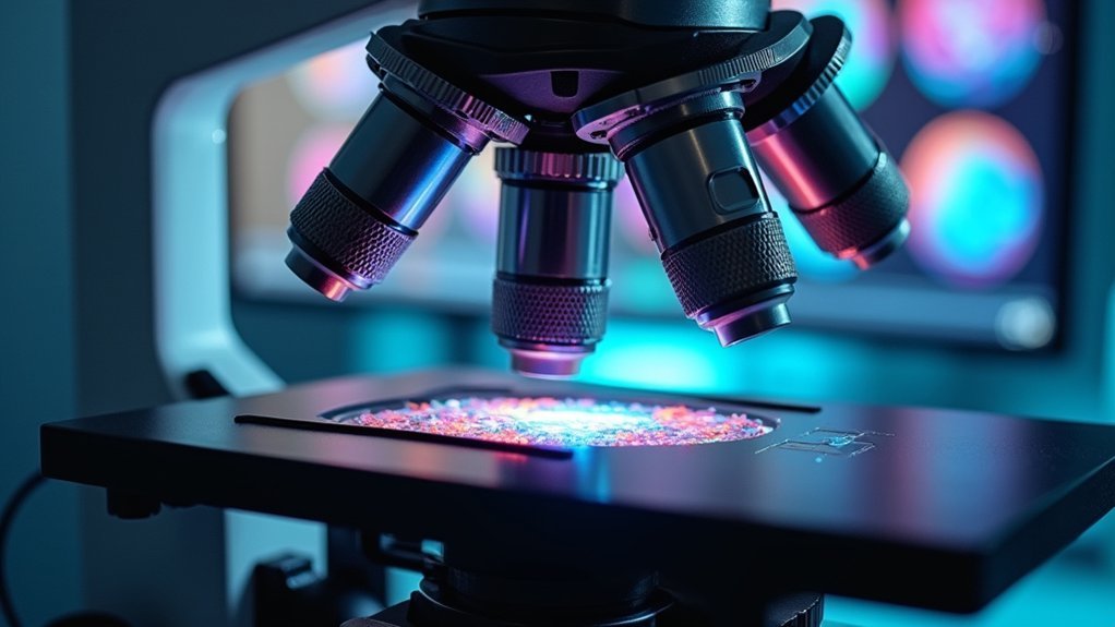
Because cellular processes often occur over extended periods, effective microscope automation tools must provide seamless time-lapse capabilities to capture critical moments.
The EVOS FL Auto Imaging System exemplifies this functionality, allowing you to monitor cell division and migration at precisely timed intervals.
Look for systems offering Z-stack imaging to visualize structures at multiple focal planes, revealing details invisible with standard techniques.
Custom vessel mapping and automated scan routines greatly reduce your experimental imaging time while maintaining accuracy.
Quality software should include essential image processing features—scale bars, contrast adjustment, and grid overlays—to enhance data presentation.
For maximum versatility, choose systems that integrate with fluorescent reagents like CellLight and NucBlue Live, enabling real-time visualization of specific cellular components during your time-lapse imaging experiments.
Setting Up Your First Automated Imaging Workflow
Now that you’re familiar with the key features to look for, let’s establish a practical workflow for your automated imaging experiments. The EVOS FL Auto Imaging System offers an excellent platform for beginners, allowing you to capture biological processes over extended periods.
Begin by creating a custom vessel map for your specific sample type, which will greatly streamline your setup process.
Then configure your time-lapse imaging parameters:
- Set appropriate time intervals (12-minute intervals work well for 24-hour cell division studies)
- Enable Z-stack imaging to capture multiple focal planes for thorough structural detail
- Define the experiment duration based on your biological process of interest
After data collection, transfer your image sequences to editing software like DaVinci Resolve 17 to compile a high-resolution video that effectively communicates your scientific findings.
Time-Lapse Software Comparison: Free vs. Premium Options

Choosing the right time-lapse software depends largely on your project requirements and budget constraints. Free options like Timelapse Lite provide basic editing features including color grading and denoising, but limit you to 720p resolution—ideal for social media but insufficient for professional work.
For more demanding projects, premium options like LR Timelapse offer advanced capabilities such as flicker reduction and Holy Grail time-lapse support. While costly, these specialized tools deliver professional results that free alternatives can’t match.
DaVinci Resolve 17’s free version offers a middle ground with unlimited resolution support, though you’ll need to convert raw files to DNG format first.
If you’re looking to enhance your skills, consider educational bundles like Five Day Deal, which provides time-lapse presets and guides at significant discounts.
Optimizing Image Quality in Extended Capture Sessions
When conducting extended time-lapse captures, maintaining consistent image quality becomes increasingly challenging as environmental variables fluctuate. Your EVOS FL Auto Imaging system requires proper adjustment to deliver reliable results during long timelapse experiments.
For ideal cellular imaging quality:
- Set your Z-stack step size at 0.366 μm for HeLa cells following the Nyquist formula, ensuring you capture detailed subcellular structures without unnecessary data volume.
- Utilize de-flickering and denoising algorithms in software like Timelapse Lite to eliminate lighting inconsistencies that commonly develop during extended captures.
- Maintain cells in an onstage incubator with controlled temperature and humidity to minimize stress responses that can alter morphology and compromise image integrity.
After capture, incorporate scale bars during post-processing and adjust brightness/contrast to enhance visualization of critical cellular events.
Cell Division Visualization: Software Requirements
With proper image quality established for your time-lapse captures, the focus shifts to software capabilities needed specifically for visualizing cell division events.
High-quality imaging sets the foundation; now it’s time to identify software that excels at capturing dynamic cell division processes.
When studying mitosis, you’ll need platforms like the EVOS FL Auto Imaging System that can program precise imaging intervals—typically every 12 minutes—across multiple fluorescence channels.
Your software should support Z-stack imaging to capture different cellular planes, providing thorough morphological details during cell division.
Look for analysis tools like Timelapse that enable you to query image recognition data to identify specific cellular events efficiently.
For peak visualization, choose software that integrates with fluorescent markers such as CellLight Histone 2B-GFP for chromatin and CellLight Mitochondria-RFP for organelle tracking.
Advanced features including color grading and de-flickering capabilities guarantee your cell division time-lapse sequences render with scientific precision.
Data Management Strategies for Large Image Collections
Managing your extensive time-lapse image collections requires thoughtful database architecture that accommodates growth while maintaining quick access.
You’ll benefit from Timelapse software’s automated metadata extraction capabilities, which pull dates, times, and locations directly from your images, dramatically reducing manual data entry.
Implement search optimization techniques by categorizing unusual images and utilizing the software’s ability to query specific subsets based on your defined criteria or imported image recognition data.
Database Architecture Considerations
As time-lapse projects generate massive volumes of images, establishing a robust database architecture becomes essential for long-term success. When designing your database structure, implement metadata standards such as Dublin Core to guarantee consistent organization and seamless retrieval of your visual assets.
An RDBMS solution offers powerful querying capabilities and maintains relationships between images and their associated data points.
For peak performance, you’ll want to:
- Leverage cloud storage or distributed file systems to accommodate growing collections while maintaining accessibility
- Implement image compression techniques to reduce storage requirements without sacrificing quality
- Create efficient indexing methods to accelerate search functions across your dataset
Don’t overlook the importance of regular backups and integrity checks to protect your valuable research data from corruption or loss.
Automated Metadata Extraction
Beyond database architecture, your time-lapse projects can reach new efficiency levels through automated metadata extraction tools.
Timelapse software automatically retrieves dates, times, and locations from your images, eliminating tedious manual entry when managing large collections.
You’ll benefit from the software’s ability to extract user-defined metadata, preserving project-specific information without additional input.
Timelapse also intelligently categorizes problematic images like dark or corrupted files, maintaining data integrity across your dataset.
The extracted information is saved in Excel-compatible formats, simplifying database imports and downstream analysis.
You can further enhance your metadata management by importing and querying image recognition data from systems like Microsoft Megadetector, seamlessly integrating automated detection results with your existing metadata for thorough dataset management.
Search Optimization Techniques
The sheer volume of images in time-lapse projects demands robust search capabilities to prevent workflow bottlenecks. When using Timelapse, implementing visual search tools like magnifying glass and pan/zoom functions allows you to efficiently examine cell counting results across thousands of frames.
To maximize search efficiency:
- Leverage metadata filters to quickly isolate images by date, time, or location—particularly valuable when tracking cell division patterns over extended periods.
- Create custom data field templates specific to your cell counting parameters, enabling precise searching across experimental variables.
- Utilize Microsoft Megadetector integration to automatically categorize images, letting you focus only on frames containing relevant cellular structures.
These techniques transform overwhelming image collections into manageable, searchable datasets that support rigorous scientific analysis while dramatically reducing time spent on manual sorting.
Custom Protocols for Different Experimental Designs
Creating experiment-specific templates with your time-lapse software will save you hours of setup time and guarantee consistency across related studies.
You’ll maximize research efficiency by designing workflows that automatically apply your preferred imaging intervals, Z-stack parameters, and fluorescent channel settings to new experiments.
These customized protocols can be shared with lab members, allowing your entire team to implement standardized imaging procedures while still accommodating variations in vessel types and experimental conditions.
Experiment-Specific Templates
When designing complex time-lapse experiments, you’ll need data structures that perfectly match your research parameters—which is exactly what experiment-specific templates provide.
Timelapse’s template functionality allows you to create custom data field formats tailored to your specific experimental design, ensuring all relevant metadata is captured consistently.
The Timelapse Template Guide walks you through creating templates that incorporate your unique project requirements:
- Create custom categories and parameters aligned with your research goals for more efficient data organization
- Streamline analysis by designing templates that allow for targeted searching and filtering of results
- Maintain consistency across multiple experiments, making comparative analysis more reliable
Efficient Workflow Design
Because research needs vary dramatically across different experimental designs, customized protocols become essential for maximizing efficiency in time-lapse imaging workflows. Your Auto Cell Imaging System can accommodate these variations through tailored settings that optimize data collection.
| Workflow Element | Efficiency Enhancement |
|---|---|
| Vessel Maps | Simplifies setup for different vessel types, ensuring consistent imaging |
| Z-stack Integration | Captures detailed cellular structures across multiple focal planes |
| Automated Scan Routines | Reduces variability and saves time in data collection |
| Custom Data Fields | Streamlines project-specific information organization |
Enhancing Scientific Analysis Through Visual Documentation
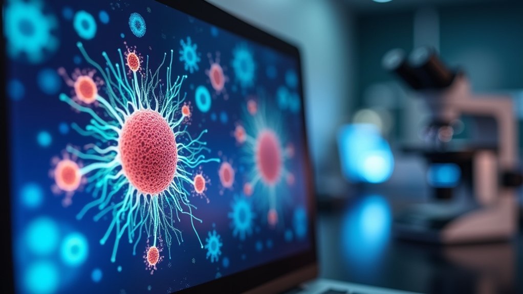
As researchers explore complex biological phenomena, time-lapse imaging emerges as a powerful tool for visualizing dynamic cellular processes that would otherwise remain hidden.
By combining fluorescent proteins like Histone 2B-GFP with automated systems, you’ll transform raw data into compelling visual narratives that reveal cellular behaviors over extended timeframes.
Transform cellular mysteries into visual stories through automated fluorescence imaging that reveals life’s hidden dynamics across time.
Your time-lapse documentation offers three critical advantages:
- Captures dynamic events such as cell division and migration with precision, revealing temporal relationships invisible in static images
- Enables multi-channel visualization when using different fluorescent markers (like mitochondria-RFP) to track multiple cellular components simultaneously
- Provides quantifiable data for statistical analysis while maintaining visual context that helps communicate findings to broader audiences
Z-stack integration further enhances your analysis by adding depth to time-series, creating thorough 4D datasets of cellular activity.
Integrating Multiple Fluorescence Channels in Time-Lapse Studies
You’ll need to master multi-channel acquisition techniques to simultaneously capture distinct cellular structures, such as nuclei with CellLight Histone 2B-GFP and mitochondria with CellLight Mitochondria-RFP.
When setting up your EVOS FL Auto Imaging System, make certain proper calibration of exposure time and gain for each channel to prevent spectral overlap and obtain high-quality images.
These optimized settings will enable you to visualize protein interactions in real time, revealing dynamic cellular events and relationships between different components that wouldn’t be apparent with single-channel imaging.
Multi-channel Acquisition Techniques
While traditional microscopy captures a single view of cellular activity, multi-channel acquisition opens up an entirely new dimension to time-lapse imaging.
Using the EVOS FL Auto Imaging System, you’ll capture multiple fluorescence channels simultaneously, revealing complex cellular processes in remarkable detail.
Multi-channel acquisition techniques enable you to:
- Track different cellular components like histone-labeled nuclei and mitochondria using specific fluorescent markers (GFP/RFP)
- Observe dynamic interactions between proteins and structures in real-time during cell division or migration
- Combine Z-stack imaging with multi-channel acquisition for thorough 3D visualization of cellular structures
For ideal results, pair your acquisition with advanced editing software like DaVinci Resolve to integrate your channels into cohesive presentations that showcase the full complexity of your cellular studies.
Visualizing Protein Interactions
When cellular proteins interact in living systems, they create dynamic patterns that tell vital stories about biological function. By integrating multiple fluorescence channels like CellLight Histone 2B-GFP (green) and Mitochondria-RFP (red), you’ll capture these interactions in real time.
Using time-lapse imaging at 12-minute intervals, you can monitor how proteins localize during significant cell processes such as division and migration. This approach reveals otherwise hidden dynamics of multipolar mitosis and normal cellular functions.
For deeper analysis, incorporate Z-stack imaging techniques to visualize three-dimensional protein relationships within the cell. The right software tools enable quantification across multiple channels, transforming your visual data into measurable interactions.
This thorough approach provides vital insights into cellular mechanisms that static imaging simply can’t reveal.
Troubleshooting Common Time-Lapse Imaging Challenges
Even the most carefully planned time-lapse projects can encounter technical hurdles that affect the final output quality. Proper calibration of your imaging system is essential to prevent flickering effects caused by inconsistent brightness across frames. When you notice variations in light intensity, utilize de-flickering algorithms available in most time-lapse software packages to smooth out these discrepancies.
Your lighting setup requires special attention to maintain consistent conditions:
- Regularly check and replace light sources to avoid fluctuations that impact image quality
- Monitor temperature changes that might affect your equipment or biological samples
- Consider adjusting ISO settings or applying denoising algorithms when facing image noise issues
For long-duration experiments, temperature-controlled incubators will help maintain stable conditions, ensuring your time-lapse data remains scientifically valid and visually cohesive.
Advanced Processing Techniques for Scientific Publication
To elevate your time-lapse imaging for scientific publication, mastering advanced processing techniques becomes essential as journal standards continue to rise.
Start by implementing Z-stack imaging with Nyquist-calculated step sizes to reveal cellular structures invisible to standard methods.
Enhance your work by incorporating CellLight fluorescent reagents for visualizing specific cellular components during real-time division and migration studies.
You’ll achieve publication-quality results using specialized software like LR Timelapse, which offers superior de-flickering capabilities critical for Scientific and its subsidiaries’ acceptance standards.
Apply professional color grading and denoising workflows to considerably improve clarity in your time-lapse sequences.
Don’t overlook educational webinars focusing on proper antibody selection and sample preparation—these resources will help you master the advanced processing techniques required for high-impact publications.
Frequently Asked Questions
Can Time-Lapse Software Compensate for Laboratory Temperature Fluctuations?
Yes, advanced time-lapse software can compensate for temperature fluctuations by using algorithmic corrections. You’ll find many programs offer temperature normalization features that adjust imaging parameters automatically when your lab’s temperature changes.
How Do Different File Formats Affect Long-Term Archival of Imaging Data?
File formats impact data longevity greatly. You’ll want to use lossless formats like TIFF for preservation, while JPEG or MP4 saves space. Consider open standards for future accessibility and include metadata for context.
What Security Measures Protect Proprietary Research Captured Through Automated Imaging?
You’ll need encryption, access controls, and secure backups to protect your proprietary research. Use two-factor authentication, watermarking, audit trails, and data use agreements when sharing images with collaborators.
Are Cloud-Based Time-Lapse Solutions Reliable for Sensitive Scientific Applications?
Cloud-based time-lapse solutions can be reliable for sensitive research, but you’ll need to verify their encryption standards, data sovereignty compliance, and backup protocols. Consider hybrid solutions that maintain local copies of critical data.
How Does Automated Imaging Software Handle Unexpected Power Outages?
You’ll find most robust software includes automatic data backup and recovery mechanisms. If power fails, they’ll save your current state, resume imaging when power returns, and alert you about the interruption.
In Summary
You’ll find that investing time in the right time-lapse software pays significant dividends for your scientific imaging. Whether you’re choosing free options or premium solutions, automation will transform your research efficiency and data quality. Don’t hesitate to experiment with advanced features as you become comfortable. With proper setup and troubleshooting knowledge, you’ll capture compelling visual documentation that strengthens your scientific publications.


Leave a Reply