To measure microscope resolution like an expert, calculate the theoretical limit using the formula d = λ/(2NA), where λ is wavelength and NA is numerical aperture. Capture images of fluorescent beads (100-200nm) and analyze the Point Spread Function’s Full Width at Half Maximum (FWHM). Control environmental factors like vibration, temperature, and humidity for accurate results. Regular calibration with standardized samples guarantees reliable measurements. The following techniques will transform your microscopy precision from amateur to professional.
Measure Microscope Resolution Like A Lab Expert
When determining your microscope’s true capabilities, you’ll need to adopt the full width at half maximum (FWHM) approach for precise resolution measurement. This technique analyzes the intensity profile of the point spread function (PSF), calculating resolution as FWHM ≈ λ/(2NA).
Start by calibrating your system with reference samples like standardized beads. This establishes a reliable baseline that minimizes measurement variability.
For practical resolution calculations, apply the Abbe diffraction limit formula: d = λ/(2NA). For example, using a 514 nm light source with a numerical aperture of 1.45 yields a 177 nm lateral resolution.
Control environmental factors including light quality, temperature, and humidity during your measurements. Implement statistical analysis across multiple readings to verify precision, ensuring your PSF size and point brightness are optimized for accurate resolution determination.
Understanding Resolution and Diffraction Limits
Resolution in microscopy extends beyond mere measurement—it represents the fundamental boundary of what your instrument can reveal.
When you’re evaluating your microscope’s capabilities, understand that the diffraction limit, expressed as d = λ/(2NA), establishes the physical constraints of light-based imaging.
Your microscope’s resolution depends on four key factors:
Understanding microscope resolution requires mastering wavelength, numerical aperture, point spread function, and Airy disc patterns—the gatekeepers of optical clarity.
- Wavelength of light – shorter wavelengths (blue/violet) reveal finer details
- Numerical aperture of your objective – higher NA translates to better resolution
- Point spread function width – the FWHM measurement indicates actual resolution
- Airy disc pattern – two points are resolvable when their Airy discs are sufficiently separated
When you measure resolution practically, you’re fundamentally determining how successfully your microscope overcomes these diffraction-imposed limitations.
Calculating Theoretical Resolution Using Numerical Aperture
To calculate your microscope’s theoretical resolution limit, you’ll need to apply specific formulas that incorporate both the wavelength of light and numerical aperture of your system.
The lateral resolution depends on the equation d = λ/(2NA), where shorter wavelengths and higher NA values yield better results.
For example, using green light (514 nm) with a numerical aperture of 1.45 produces a lateral resolution of approximately 177 nm.
The axial resolution can be determined using d = 2λ/(NA)². With the same parameters, you’d achieve about 488 nm axial resolution.
For more precise measurements, apply the Rayleigh Criterion: R = 1.22λ/(NA_obj + NA_cond).
Remember, your theoretical limit can only be reached when all optical components are perfectly aligned.
To maximize resolution, always use the highest possible numerical aperture and shorter wavelengths.
Point Spread Function and Full Width at Half Maximum Measurements
Understanding how your microscope depicts a single point of light reveals its true resolution capabilities. The Point Spread Function (PSF) shows how your microscope distributes light from a point source, while the Full Width at Half Maximum (FWHM) gives you a practical measurement of your microscope’s resolution.
The microscope’s true power lies in how it portrays a single light point, revealing resolution through PSF and FWHM measurements.
To determine FWHM and assess resolution accurately:
- Calculate your theoretical resolution using FWHM ≈ λ/(2NA) (for example, green light at 514 nm with NA 1.45 yields 177 nm resolution)
- Capture images of calibration samples like fluorescent beads
- Measure the intensity profile across your point source image
- Determine the width at half-maximum intensity for your actual PSF
Remember that your numerical aperture considerably impacts your PSF shape. Regular calibration guarantees you’re achieving ideal resolution for your experimental needs.
Practical Methods for Measuring Microscope Resolution
When you’re ready to evaluate your microscope’s actual performance, several practical methods will help you quantify its true resolution capabilities.
Begin by imaging fluorescent beads of known diameter and measure the FWHM of their intensity profiles, which directly relates to your microscope resolution through the formula FWHM ≈ λ/(2NA).
For standardized measurements, apply the Rayleigh Criterion (R = 1.22λ/(NA_obj + NA_cond)) which accounts for both objective and condenser numerical aperture values.
Control your experimental variables carefully—maintain consistent distances between points, optimize fluorophore brightness, and minimize environmental noise.
If you need to push beyond diffraction limits, implement superresolution techniques like STED or SMLM.
These methods can greatly enhance resolution by manipulating emission areas or precisely localizing sparse fluorescent probes.
Calibration Standards and Reference Materials
When selecting calibration beads for microscopy, you’ll want to choose fluorescent beads with precise diameters that match your expected resolution range.
DNA origami standards offer nanometer-scale precision for super-resolution techniques, providing fixed-distance reference points that reveal your microscope’s true resolving power.
You should calibrate under your typical imaging conditions to guarantee measurements reflect real-world performance rather than idealized scenarios.
Calibration Beads Selection
Why do calibration beads serve as the gold standard for microscope resolution testing? These fluorescent microspheres provide standardized reference points with consistent size and shape, allowing precise measurement of your microscope’s resolving power.
When selecting calibration beads for determining the resolution of a microscope, focus on these essential criteria:
- Size range – Choose beads between 100-200 nm to effectively test high NA objectives
- Brightness level – Select beads with strong fluorescence signals that stand out clearly from background
- Photostability – Verify beads resist photobleaching during extended imaging sessions
- Compatibility – Match bead fluorescence to your microscope’s filter sets
Remember to perform FWHM calculations on bead intensity profiles to quantitatively assess your system’s resolution capabilities and complement these measurements with standard resolution test strips.
DNA Origami Standards
Beyond traditional calibration beads, DNA origami offers unprecedented precision for microscope resolution assessment.
These nanoscale structures provide fixed distances between fluorescent probes, serving as exact reference points for your optical microscope’s performance evaluation.
You’ll benefit from DNA origami’s customizable dimensions, which allow you to create tailored calibration standards for resolution assessment and point spread function (PSF) calibration.
Their nanometer-level precision helps you overcome measurement variability issues that plague other methods.
Be prepared to address signal-to-noise ratio challenges when working with these standards.
Despite handling difficulties, implementing DNA origami represents a significant advancement in your resolution measurements toolkit.
For superresolution microscopy especially, these standards enable the reliable, reproducible measurements necessary for cutting-edge research.
Optimizing Environmental Conditions for Accurate Measurements
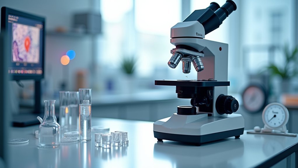
Although microscope resolution depends heavily on optical components, the environment in which you conduct measurements can dramatically impact your results.
Creating ideal environmental conditions requires attention to several key factors that directly affect imaging quality.
- Minimize vibrations – Establish your microscope on a stable platform away from foot traffic, equipment with moving parts, or building vibrations that can blur images even at the nanoscale.
- Maintain consistent temperature control – Even slight thermal fluctuations can alter refractive indices and distort optical performance.
- Monitor humidity levels – Keep humidity within manufacturer-recommended ranges to prevent condensation on lenses and specimens.
- Enhance your light source – Guarantee adequate illumination strength with proper adjustments to avoid over/underexposure.
Calibrate your microscope regularly under your specific working conditions to establish baseline performance and guarantee measurement reliability.
Frequently Asked Questions
How to Measure Microscope Resolution?
You’ll measure microscope resolution by imaging calibration beads, calculating the PSF’s FWHM (approximately λ/2NA), and conducting statistical analysis of multiple measurements while maintaining stable environmental conditions for reliable results.
How Do You Calculate the Limit of Resolution of a Microscope?
You’ll calculate a microscope’s resolution limit using Abbe’s formula: d = λ/(2NA), where λ is the light wavelength and NA is numerical aperture. For axial resolution, use d = 2λ/(NA)². Verify with calibration standards.
What Determines Resolution of a Microscope?
Resolution of your microscope is determined by the wavelength of light, numerical aperture (NA) of your lenses, and quality of your optical system. Higher NA and shorter wavelengths will give you better resolution.
What Must One Be Aware of to Determine the Resolution of a Microscope System?
You must understand the relationship between numerical aperture and wavelength, know both objective and condenser NA values, calibrate using reference samples, and consider environmental factors. The PSF’s FWHM provides practical resolution measurement.
In Summary
You’ve now mastered both theoretical and practical methods for measuring microscope resolution. By understanding diffraction limits, calculating numerical aperture values, and using calibration standards properly, you’ll consistently achieve accurate measurements. Remember to optimize your environmental conditions and regularly check your point spread function. With these techniques, you’re equipped to evaluate any microscope’s true performance like a seasoned laboratory professional.



Leave a Reply