
For successful live specimen imaging, first maintain ideal conditions including 37°C temperature, proper pH, and appropriate gas levels for your specific cell type. Second, use phenol red-free media with minimal fluorophore concentrations to reduce background and phototoxicity. Third, pre-configure your microscope with the lowest possible light intensity and shortest exposure times that still yield clear images. These fundamental preparations will greatly enhance your imaging quality while preserving specimen viability throughout your experiment.
Optimizing Environmental Conditions for Live Cell Viability
When preparing specimens for live-cell imaging, maintaining physiological conditions is critical for achieving both reliable results and prolonged cell survival. Your cell culture must be kept at approximately 37°C to prevent focus drift and maintain cellular processes during imaging experiments.
Monitor pH levels using a synthetic buffer like HEPES, which provides stability even when samples are temporarily removed from your incubator. Control humidity within your imaging chamber to prevent evaporation that can disrupt osmotic balance.
Maintain carbon dioxide levels at 5-10% to support proper cellular metabolism and pH stability. Different cell types require specific oxygen levels, so adjust accordingly.
Remember that your light source can generate heat, potentially disrupting the delicate environmental balance you’ve established for successful live-cell imaging.
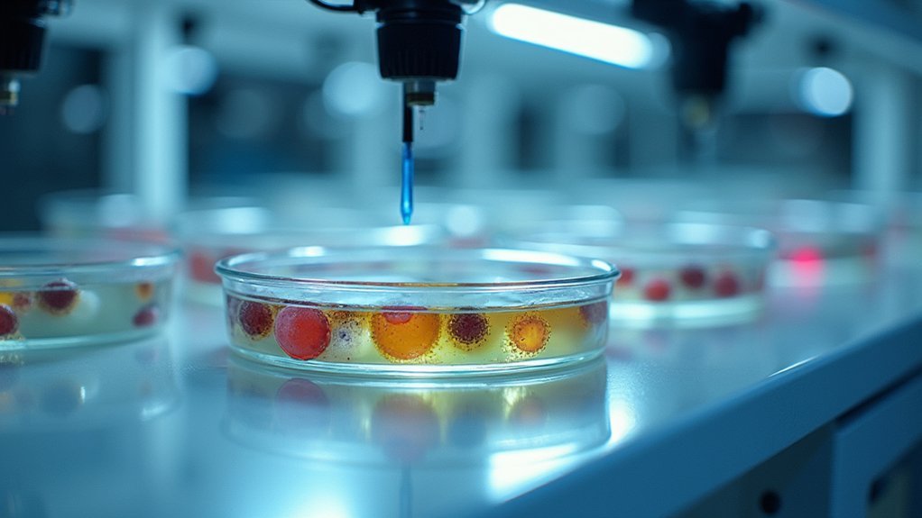
Selecting Appropriate Imaging Media and Labeling Techniques
Choosing the right imaging media forms the foundation of successful live-cell microscopy. Your media must contain essential nutrients while maintaining proper pH, osmolarity, and humidity to support cell viability.
For fluorescence imaging, opt for phenol red-free media to reduce autofluorescence and improve clarity.
When labeling your specimens, select bright, specific fluorophores that work at lower concentrations to minimize phototoxicity during live imaging sessions.
Consider using fluorescent proteins that emit in the red spectrum to tag cellular structures, as these typically cause less damage to cells.
Always use the lowest possible light intensity and shortest exposure times that still yield adequate signals for your specific specimen types.
These careful choices will greatly extend your imaging window while maintaining ideal cell viability.
Minimizing Phototoxicity Through Proper Illumination Control

Phototoxicity represents one of the greatest challenges for live-cell imaging, as even moderate light exposure can damage your specimens and compromise experimental results. To minimize phototoxicity, reduce illumination intensity by selecting lower power settings and using photostable fluorophores that require shorter exposure times.
| Strategy | Benefit |
|---|---|
| Widefield microscopy with deconvolution | Enhanced resolution with decreased exposure |
| Red-shifted fluorescent probes | Lower phototoxicity for extended imaging |
| Autofocus systems | Maintains focus without unnecessary illumination |
Implement gradual illumination techniques to prevent abrupt exposure changes that induce cellular stress. During your imaging experiment, these approaches will help maintain healthier live specimens while still capturing high-quality data, especially for time-lapse studies where cumulative light damage can greatly impact results.
Frequently Asked Questions
How Do You Prepare Cells for Live Cell Imaging?
You’ll maintain ideal growth conditions, handle cells gently, use bright red spectrum fluorescent labels, set up environmental controls for temperature and humidity, and employ noninvasive techniques with minimal illumination to reduce phototoxicity during imaging.
What Are the Disadvantages of Live Cell Imaging?
Live cell imaging disadvantages include phototoxicity from light exposure, complex environmental control requirements, potential artifacts from fluorescent labels, challenges capturing dynamic events, and signal degradation during long-term imaging due to photobleaching. You’ll need specialized equipment and expertise.
What Is a Live Cell Imaging Solution?
A live cell imaging solution is a specialized medium you’ll use to keep your cells alive during microscopy. It maintains proper pH, nutrients, and environmental conditions so you can observe cellular processes in real-time.
What Microscopy Techniques Can Be Used in Live Cell Imaging?
You can use several microscopy techniques for live cell imaging: fluorescence, confocal, phase contrast, and brightfield. Time-lapse imaging is also valuable for capturing dynamic cellular processes over extended periods.
In Summary
You’ll achieve superior live specimen imaging by optimizing environmental conditions, selecting appropriate media and labeling methods, and controlling illumination to minimize phototoxicity. These three essential steps aren’t just technical procedures—they’re critical for maintaining cellular integrity and capturing authentic biological processes. Master these fundamentals and you’ll transform your imaging results from merely adequate to truly exceptional and scientifically valuable.


Leave a Reply