Successful live cell microscopy requires precise environmental control (37°C, 5% CO₂, 90% humidity) to maintain cell viability. You’ll need phenol red-free media to reduce autofluorescence and gentle handling techniques to minimize stress. Use photostable fluorophores with shorter exposure times to reduce phototoxicity, and implement reliable autofocus systems. Proper equipment integration—from humidity chambers to specialized culture dishes—ensures consistent time-lapse capture. These foundational elements will transform your imaging experiments from frustrating to revealing.
What Makes Live Cell Culture Microscopy Successful?
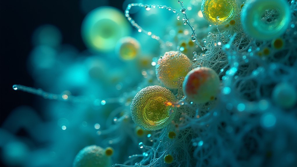
While capturing still images of fixed cells provides valuable snapshots, successful live cell culture microscopy reveals the dynamic nature of cellular processes as they unfold in real time.
For your live cell imaging experiment to succeed, you’ll need precise control of environmental conditions—temperature, CO₂ levels, and humidity—to maintain cell viability.
Specialized equipment with temperature-controlled stages and environmental chambers prevents thermal drift while preserving your cells’ physiological state.
When using fluorescence microscopy, choose photostable probes and low-light imaging techniques to minimize phototoxicity, enabling long-term imaging without compromising cellular health.
Don’t overlook cell culture preparation; proper media and gentle handling techniques produce reliable, reproducible results.
Finally, implement autofocus and tracking features in your imaging software to monitor dynamic events and maintain focus throughout extended observation periods.
Maintaining Optimal Temperature and Environmental Control
The success of your live cell imaging hinges on recreating physiological conditions that keep your cells happy and behaving naturally.
To maintain sample integrity throughout your imaging applications, you’ll need to control the cellular environment precisely.
- Temperature regulation – Keep cells at the ideal 37°C for mammalian systems to guarantee natural cellular processes continue during observation.
- CO₂ levels – Maintain 5% concentration to stabilize pH in culture media, supporting proper metabolism.
- Humidity control – Target 90% relative humidity to prevent media evaporation and avoid osmotic stress.
- Monitoring systems – Implement stage-top incubators and regular parameter checks to guarantee consistent conditions.
Selecting the Right Culture Media for Imaging Experiments
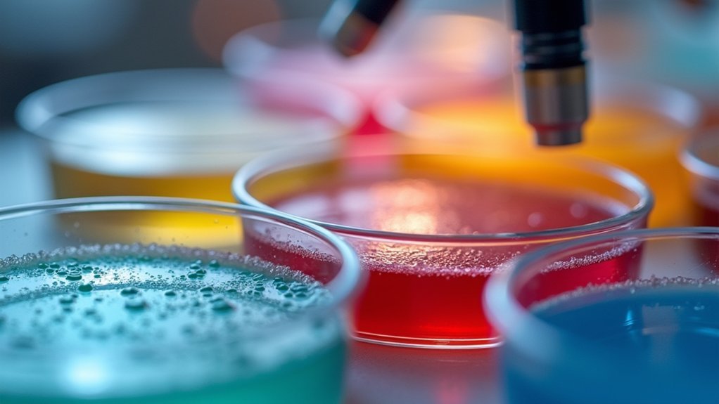
Selecting the right culture media for your imaging experiments requires careful consideration of background fluorescence factors.
You’ll want to use phenol red-free media and reduce serum concentrations to minimize autofluorescence that can interfere with your fluorescence microscopy results.
Proper buffering systems in your media will maintain pH stability throughout your imaging session, ensuring consistent cellular behavior and preventing artifacts.
Minimizing Background Fluorescence
Background fluorescence presents one of the most notable challenges in live cell imaging, often obscuring the specific signals researchers aim to capture.
To maximize imaging quality and preserve cellular integrity during fluorescence imaging, you’ll need to optimize your culture media conditions.
To minimize background fluorescence in live cell imaging experiments:
- Select phenol red-free media to considerably reduce autofluorescence and improve signal clarity.
- Lower serum concentrations in your culture media to minimize non-specific background signals.
- Use specialized media formulations tailored to your specific cell type’s biochemical requirements.
- Monitor and adjust pH, osmolarity, and nutrient levels regularly, as these factors directly impact background fluorescence.
These adjustments will help you achieve clearer fluorescent signals and more precise visualization of labeled cellular structures.
Buffering for Stability
When conducting live cell imaging experiments, proper media buffering becomes essential to maintain cellular homeostasis throughout your observation period. Select media formulations with adequate buffering capacity—typically bicarbonate systems or HEPES—to guarantee stable pH levels during extended imaging sessions.
You’ll need to contemplate how your media supports cell growth while minimizing background fluorescence. Reducing serum concentration can greatly improve image clarity.
Always pre-warm your media to avoid thermal shock when transferring cells to imaging chambers.
Don’t overlook osmolarity monitoring, as changes can dramatically alter cellular behavior and compromise your results. The ideal imaging media strikes a balance between nutritional requirements (amino acids, vitamins, trace elements) and optical properties.
This careful equilibrium assures your cells remain viable and physiologically relevant throughout your microscopy sessions.
Minimizing Phototoxicity During Extended Observation
Although essential for visualization, prolonged light exposure during live cell microscopy presents a significant challenge for researchers.
You’ll need to balance quality imaging with maintaining cell viability during live cell imaging experiments. Phototoxicity occurs when excited fluorophores generate harmful free radicals that damage cells and alter their behavior.
To minimize phototoxicity while preserving image quality:
- Select high signal-to-noise, photostable fluorophores that require shorter exposure time
- Implement hardware or software autofocus systems to optimize image acquisition efficiency
- Adjust time-lapse imaging parameters to reduce illumination intensity and duration
- Monitor cellular responses throughout experiments, making real-time adjustments as needed
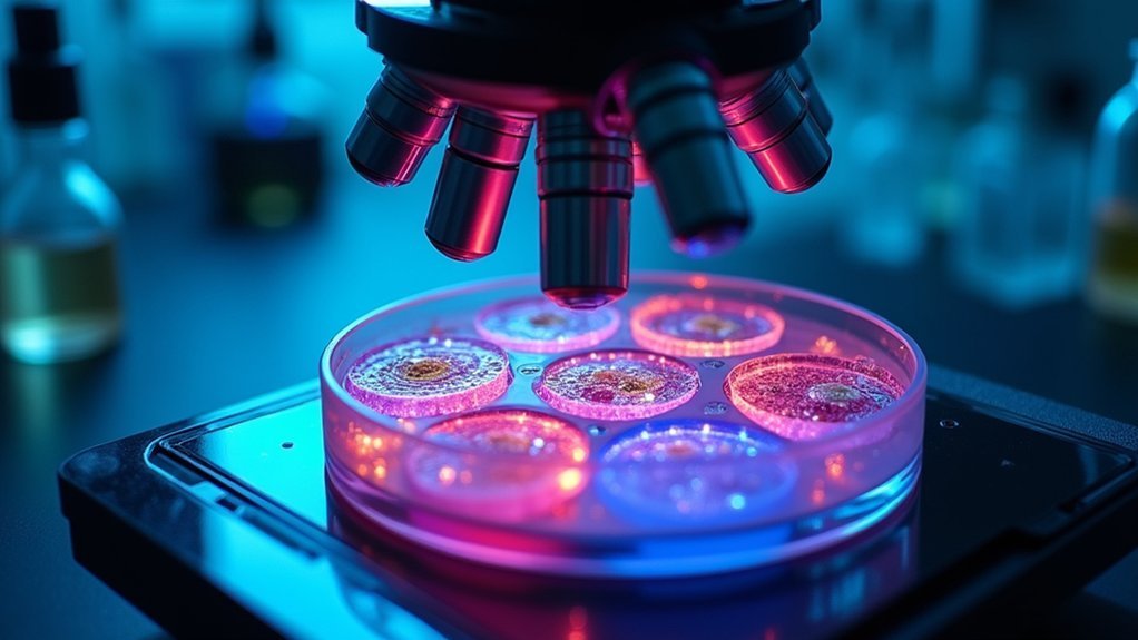
Essential Equipment for High-Quality Time-Lapse Capture
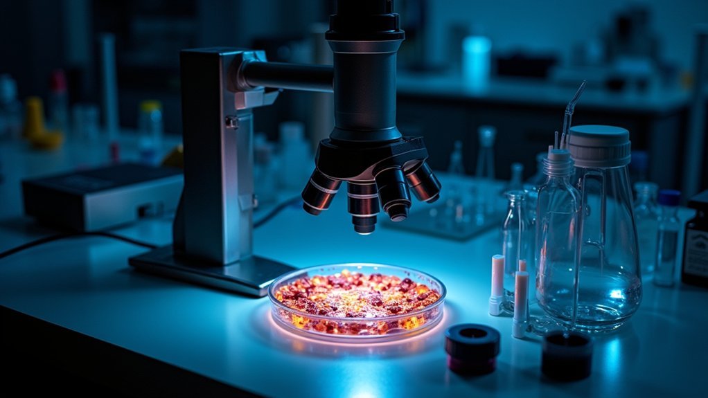
Successful time-lapse microscopy depends on specialized equipment that maintains cellular viability while capturing high-resolution images over extended periods. You’ll need a microscope stage with precise temperature control to guarantee ideal growth conditions for your live cells throughout extended imaging sessions.
| Equipment | Function | Impact on Time-Lapse Experiments |
|---|---|---|
| CO₂ incubator | Regulates gas concentrations | Maintains physiological pH levels |
| Humidity chamber | Prevents media evaporation | Preserves osmotic balance |
| Specialized culture dishes | Facilitates clear observation | Minimizes contamination risk |
Don’t overlook the importance of advanced imaging software with real-time analysis capabilities. These programs allow you to capture and process dynamic cellular events as they unfold. When all these elements work together, you’ll achieve consistent, high-quality time-lapse capture while maintaining cellular health.
Proper Cell Preparation Techniques for Improved Viability
Before initiating any live cell imaging experiment, you’ll need to master proper cell preparation techniques that greatly impact cellular health and experimental outcomes.
Unlike imaging fixed cells, live-cell imaging experiments require meticulous attention to maintaining cell viability throughout observation.
To optimize your preparation process:
- Select appropriate culture media with nutrients tailored to your specific cell type, and pre-warm it to physiological temperature to prevent thermal shock.
- Handle cells gently during transfer and plating to minimize mechanical stress and maximize survival rates.
- Maintain strict sterility throughout your workflow to prevent contamination that could compromise experimental integrity.
- Label all samples clearly before adding fluorescent tags to guarantee proper identification and tracking during extended imaging sessions.
Balancing Resolution and Cell Health in Real-Time Imaging
When pursuing high-resolution live cell imaging, you’re constantly maneuvering a fundamental tradeoff between image quality and cellular health.
To enhance this balance, select photostable fluorophores that deliver strong signals with minimal light intensity, reducing phototoxicity while maintaining necessary resolution.
Implement advanced techniques like deconvolution and binning to improve image quality without extending exposure times. You’ll preserve cell health while still capturing clear, detailed images.
Don’t underestimate the importance of environmental control—stable temperature and CO₂ levels are essential for maintaining cellular integrity during long-term imaging sessions.
Remember to calibrate your imaging system regularly and adjust parameters based on your specific cell type.
Effective Focus Strategies for Multi-Dimensional Acquisition
Beyond optimizing resolution and cell health, maintaining proper focus throughout your multi-dimensional acquisition forms the backbone of meaningful live cell imaging. Unlike imaging fixed cells, live specimens require strategies that balance acquisition rate with preventing damage to the cells.
Four important factors for maintaining focus position:
- Implement hardware autofocus systems to enhance accuracy and reduce photobleaching that compromises cell viability.
- Utilize software autofocus routines tailored to your specific assay needs for reliable performance across varying sample conditions.
- Regularly calibrate focus drives and integrate tracking features for sustained Z-axis focus during long-term experiments.
- Employ transmitted light for initial focusing to minimize exposure time and phototoxicity while maintaining high-resolution imaging capabilities.
These strategies will notably improve your success when capturing dynamic cellular events over extended periods.
Data Management Solutions for Large Video Datasets
As your live cell imaging experiments generate massive amounts of data, implementing efficient management solutions becomes critical to your research success.
You’ll need robust storage systems capable of handling terabytes of time-lapse imaging files. Invest in high content analysis software with advanced algorithms that automate data sorting and extract meaningful insights.
Consider cloud storage options for scalable management, remote access, and secure backup of your large video datasets.
Don’t overlook data compression techniques that reduce file sizes while preserving image quality, making transfers faster and storage more efficient.
Establish regular organization and archiving practices to maintain an optimized workflow. With these systems in place, you’ll quickly retrieve relevant datasets and streamline your analysis processes, allowing you to focus on the science rather than searching for files.
Advanced Fluorescence Techniques for Living Specimens
While managing your data efficiently opens up new research possibilities, the imaging techniques you select ultimately determine what cellular processes you can observe and analyze.
Today’s advanced fluorescence techniques offer unprecedented insights into living specimens:
- Fluorescence Recovery After Photobleaching (FRAP) – Measure molecular mobility and dynamics within live cells to understand intracellular trafficking.
- Förster Resonance Energy Transfer (FRET) – Visualize nanoscale molecular interactions in real time using energy transfer between proximal fluorophores.
- Fluorescence Lifetime Imaging Microscopy (FLIM) – Assess molecular microenvironments by measuring fluorescence decay times, revealing conformational changes.
- Label-free approaches like Coherent Anti-Stokes Raman Scattering (CARS) – Image biochemical processes without external fluorophores.
Complement these techniques with genetically encoded fluorescent proteins and quantum dots for enhanced brightness and photostability, allowing extended observation of multiple cellular components simultaneously.
Frequently Asked Questions
What Are the Advantages of Looking at Live Cells Under the Microscope?
You’ll see dynamic processes in real-time: migration, division, and responses to stimuli. You can perform functional assays, observe molecular interactions, capture temporal changes, and gather more extensive data for better biological understanding.
What Microscopy Techniques Can Be Used in Live Cell Imaging?
You can use widefield, confocal, spinning-disc confocal, multiphoton, and TIRF microscopy for live cell imaging. Each technique offers unique advantages for visualizing dynamic cellular processes with varying resolution and phototoxicity levels.
What Are the Problems With Live Cell Imaging?
Your live cell imaging challenges include phototoxicity from illumination, high intensity light damaging cells, environmental control difficulties, potential artifacts from fluorescent tags, and resolution limitations when balancing image quality with cell health.
What Is the Difference Between Fixed and Live Cell Imaging?
In live cell imaging, you’re observing cells in their natural state over time, capturing dynamic processes. With fixed cells, you’ve preserved them at a specific moment, sacrificing their natural behavior for better structural detail.
In Summary
You’ll succeed in live cell culture microscopy by balancing environmental controls with minimal phototoxicity. Invest in quality equipment while selecting appropriate media for your experiments. Remember, it’s not just about high resolution—it’s about maintaining cell health throughout observation. Master focus strategies and data management for multi-dimensional acquisitions. By integrating these approaches, you’ll capture meaningful biological dynamics in real-time while preserving specimen integrity.


Leave a Reply