Live cell time-lapse imaging captures dynamic cellular processes in real-time over extended periods. You’ll need an inverted fluorescence microscope with environmental controls, sensitive cameras, and automated focusing systems. Prepare cells at appropriate density, use supplemented imaging media, and minimize phototoxicity with low laser power and short exposure times. Regular monitoring of cell morphology guarantees viability throughout your experiment. Proper data management and consistent environmental parameters will transform your ability to observe cellular behavior otherwise invisible in fixed-cell studies.
12 Second-Level Headings for “What Is Live Cell Time-Lapse Imaging Protocol?”
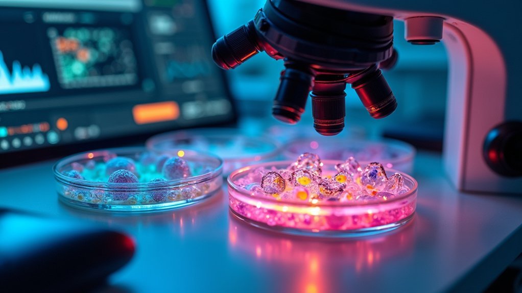
When organizing an extensive guide on live cell time-lapse imaging, you’ll need structured headings that cover all critical aspects of this sophisticated technique.
Consider these essential second-level headings for thorough coverage:
Essential headings create the framework for comprehensive understanding of complex technical procedures in cellular research.
- Principles of Live-Cell Imaging
- Experimental Setup and Equipment Requirements
- Environmental Control Systems
- Cell Preparation and Viability Considerations
- Fluorescent Labeling Strategies
- Phototoxicity Mitigation Techniques
- Image Acquisition Parameters
- Time-Lapse Interval Selection
- Autofocus and Drift Compensation
- Data Management and Analysis
Each section should address specific challenges researchers face when conducting extended cellular observations.
For example, under Environmental Control, you’ll detail temperature, humidity, and pH maintenance systems.
Under Fluorescent Labeling, include protein tag selection and expression levels that balance visibility against cellular stress.
Phototoxicity coverage should emphasize exposure reduction strategies essential for maintaining natural cellular behavior.
The Fundamentals of Live Cell Time-Lapse Imaging
To obtain high-quality live cell time-lapse data, you’ll need specialized equipment including an inverted microscope with incubation capabilities, appropriate cameras, and automated stage controls.
You should optimize your acquisition parameters by carefully balancing exposure time, light intensity, and imaging frequency to minimize phototoxicity while capturing sufficient temporal resolution.
Proper environmental control systems must maintain stable temperature, humidity, and CO₂ levels throughout your experiment to guarantee cell viability and physiologically relevant results.
Equipment and Setup
Successful live cell time-lapse imaging depends fundamentally on having the right equipment and proper setup before any experiment begins.
You’ll need specialized imaging systems like widefield or confocal microscopes equipped with sensitive detectors and automated focus systems to capture high-quality live image cells while minimizing phototoxicity.
Environmental conditions must be precisely controlled using an onstage incubator that maintains stable temperature (within ±0.1°C), CO2 levels, and humidity throughout your imaging session.
Select appropriate imaging media such as LCIS to prevent osmolarity fluctuations and maintain cellular physiological conditions.
Before starting, carefully calibrate your illumination intensity and exposure times to balance image resolution with cell viability.
If using fluorescence microscopy, be especially mindful of light exposure, as excessive illumination can damage cells and compromise your experimental results.
Acquisition Parameters
Setting appropriate acquisition parameters forms the cornerstone of successful live cell time-lapse imaging.
You’ll need to carefully balance illumination intensity and exposure duration to minimize phototoxicity while maintaining image quality. Sensitive detectors are invaluable, allowing you to capture clear images with lower light levels, thereby reducing cellular damage.
When configuring your imaging protocol, prioritize these critical factors:
- Illumination control – Use the minimum light intensity required for adequate signal detection
- Exposure optimization – Keep exposure times as short as possible while capturing necessary details
- Fluorophore selection – Choose bright, specific fluorophores that require less excitation energy
Remember that autofocusing features are essential for maintaining focus throughout long experiments, especially as cells move and environmental conditions fluctuate during extended observation periods.
Essential Equipment and Setup Requirements
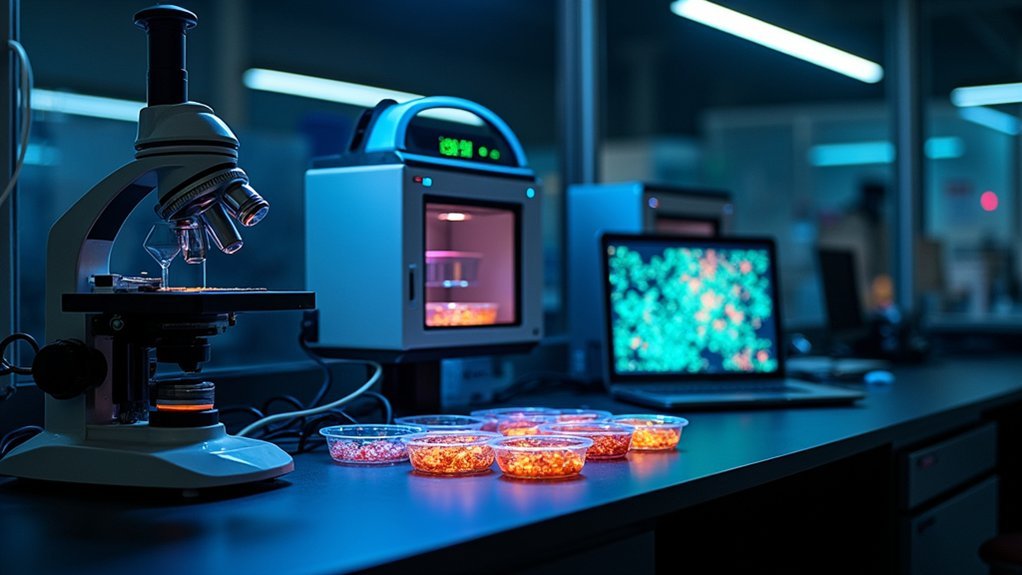
Four significant components form the foundation of any live cell time-lapse imaging system.
First, you’ll need a high-quality fluorescence microscope paired with an environmental control chamber that maintains ideal temperature, humidity, and gas conditions for your cells.
Second, a sensitive camera is essential to capture low-intensity signals without damaging cells during extended imaging sessions.
Third, you’ll require appropriate fluorescent probes that offer brightness and specificity while minimizing phototoxicity.
Automated focusing systems are vital for maintaining image clarity throughout your experiment.
Finally, you’ll need robust imaging software to control your microscope and analyze the resulting data.
Don’t overlook specialized labware requirements—glass-bottom dishes or multi-well plates compatible with microscopy will minimize interference with imaging quality while supporting cell growth.
Cell Preparation and Viability Considerations
Proper cell preparation begins with determining your ideal seeding density, which varies by cell type and should allow for both individual cell tracking and natural cell-cell interactions.
You’ll need to optimize your imaging media by supplementing with appropriate buffers and nutrients that maintain physiological conditions while reducing background fluorescence.
To minimize phototoxicity effects, you should use the lowest possible laser power and exposure times, select less harmful fluorophores, and incorporate antifade reagents in your experimental design.
Optimal Seeding Density
Because cellular density directly impacts experimental outcomes, determining the correct seeding concentration is a critical first step in time-lapse imaging protocols. For 24-well plates, aim for 10,000-50,000 cells per well to maintain ideal seeding density without triggering altered cellular behavior.
When preparing your experiment, consider these essential factors:
- Cell line characteristics – Different cell lines have unique growth rates and doubling times that affect their spatial requirements.
- Post-seeding adhesion time – Allow 4-6 hours for cells to properly attach before beginning live-cell time-lapse analysis.
- Monitoring requirements – Regularly assess cell morphology to confirm your chosen density supports healthy growth.
Remember that overcrowding leads to nutrient depletion and waste accumulation, compromising cell viability and potentially skewing your experimental results.
Media Optimization Techniques
Successful time-lapse imaging hinges on precise media optimization tailored to your specific cell types. When selecting media formulations, consider using specialized commercial options that maintain proper osmolarity, pH, and nutrient levels required for extended imaging periods.
Confirm your cell culture media contains adequate amino acids, vitamins, and trace elements specific to your cell type. Incorporate carbon dioxide and bicarbonate buffer systems to regulate physiological pH throughout the experiment.
Using larger volumes of media helps prevent detrimental shifts in oxygen levels and osmolarity during long-term imaging sessions.
Before beginning your experiment, assess cell viability to determine if adjustments to your optimization strategy are needed. This proactive approach greatly enhances experimental accuracy and confirms cells remain healthy throughout the imaging process, yielding more reliable data for your research.
Minimizing Phototoxicity Effects
Light exposure during time-lapse imaging presents a significant challenge for maintaining cell viability, as even moderate illumination intensities can trigger phototoxic damage.
To minimize phototoxicity during live-cell imaging, you’ll need to implement several strategic approaches that protect your samples while maximizing data quality.
Consider these essential techniques:
- Select red-spectrum fluorescent probes that require lower energy excitation, reducing cellular stress compared to blue or green fluorophores.
- Shorten exposure times and limit imaging channels to decrease the overall light dose your cells receive during experiments.
- Replace traditional UV sources with LED-based illumination systems that offer more precise control over light intensity.
Additionally, using an onstage incubator will maintain ideal temperature and CO2 conditions, further supporting cell viability during extended imaging sessions.
Environmental Control Parameters for Long-Term Imaging
When conducting live cell time-lapse imaging, maintaining ideal environmental conditions is essential for cell survival and experimental accuracy.
You’ll need precise temperature control via heated air or infrared systems to guarantee cellular health throughout long-term imaging sessions.
For pH regulation, implement a bicarbonate buffer system with controlled CO₂ levels that mimic physiological conditions.
Using larger media volumes helps prevent osmolarity shifts and oxygen depletion, particularly critical during shorter experiments.
For extended studies, invest in specialized environmental control chambers that monitor and adjust multiple parameters simultaneously.
Remember that transformed cell lines generally tolerate environmental fluctuations better than primary cells, making them preferable for lengthy time-lapse experiments.
These environmental control parameters require careful optimization based on your specific cell type and experimental duration.
Selection of Appropriate Fluorescent Labels and Probes
Choosing suitable fluorescent labels represents a critical decision point in live cell time-lapse imaging that directly impacts both image quality and cell viability.
Fluorophore selection lies at the intersection of experimental success and sample preservation in microscopy.
You’ll need bright, specific fluorophores at minimal concentrations to reduce cytotoxicity while maintaining visibility. Fluorescent proteins like GFP and its variants offer excellent stability for long-term live-cell imaging of dynamic cellular processes.
When selecting your labeling strategy, consider:
- Using red-spectrum probes to minimize phototoxicity, as they require lower excitation intensities
- Optimizing signal intensity by adjusting exposure times and light intensity to maintain cell health
- Evaluating quantum dots for multicolor applications where photobleaching resistance is essential
Balance your need for visual data with cellular health by choosing labels that provide clear signals while minimizing potential damage to your specimens.
Optimizing Illumination to Minimize Phototoxicity
Since excessive light exposure represents the primary threat to cell viability during time-lapse imaging, implementing strategic illumination protocols becomes essential for successful experiments.
You’ll need to use the lowest possible illumination intensity and limit exposure duration to reduce cellular damage during imaging experiments.
Consider switching to LED-based systems to decrease harmful UV light exposure, greatly improving live cells’ survival rates.
Maintain stable environmental conditions with an onstage incubator to enhance cellular resilience against phototoxicity.
Use autofocus mechanisms sparingly by setting focus beacons for specific areas only.
Optimize your imaging approach by increasing camera gain rather than light intensity, and carefully select only necessary fluorophores.
These adjustments will help you achieve high-quality images while preserving cell viability throughout your time-lapse experiments.
Image Acquisition Settings and Frame Intervals
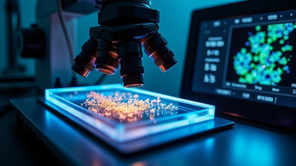
Building upon proper illumination strategies, effective image acquisition settings and frame intervals form the backbone of successful time-lapse experiments.
You’ll need to carefully balance exposure time and light intensity to minimize phototoxicity while maintaining adequate signal-to-noise ratio for your live cell imaging.
When configuring your protocol, consider these critical factors:
- Select frame intervals based on your biological process—ranging from seconds for rapid events to minutes for slower processes—guaranteeing you capture relevant dynamics without excessive illumination.
- Implement reliable autofocus systems to compensate for drift caused by temperature fluctuations and cell movements during extended imaging sessions.
- Monitor environmental conditions consistently, maintaining proper temperature and CO2 levels throughout acquisition to guarantee cell viability and reproducible results.
Focus Stability Techniques for Extended Recordings
Although even the most carefully designed time-lapse experiments can fail due to focus drift, maintaining stable focus throughout extended recordings represents one of the most critical challenges you’ll face in live cell imaging.
To achieve reliable focus stability, implement an autofocus system that continuously adjusts to live cells’ positions and conditions during long-term imaging.
You’ll benefit from using a generous volume of culture medium to prevent osmolarity and oxygen fluctuations that compromise image quality.
Install an onstage incubator for precise temperature control, as thermal variations can greatly impact focus.
Before starting extended recordings, always calibrate and test your autofocus system to identify potential issues.
Consider incorporating brightfield imaging alongside fluorescence techniques to establish reliable reference points for your imaging systems, ensuring consistent focus throughout your experiment.
Data Management Strategies for Large Time-Lapse Datasets
When undertaking time-lapse imaging experiments, you’ll quickly generate massive datasets that require thoughtful management strategies. Efficient data storage becomes critical as file sizes grow exponentially with each captured timepoint.
Time-lapse imaging generates enormous datasets—plan your storage strategy before files overwhelm your research capacity.
Consider implementing cloud-based platforms or high-capacity local servers to handle these large time-lapse datasets.
For ideal data management:
- Establish structured naming conventions for files and folders to easily retrieve specific experiments.
- Utilize data compression software to reduce overall file size without compromising image quality.
- Implement backup protocols across multiple locations to prevent data loss.
Don’t underestimate the value of specialized data management software with tagging and search capabilities. These tools will save you countless hours when analyzing results, allowing you to focus on research insights rather than hunting for files.
Image Processing and Analysis Methods
Once you’ve collected your raw time-lapse data, effective processing and analysis become vital to extract meaningful biological insights.
Start with background subtraction and noise reduction to enhance image clarity, followed by stabilization to correct for sample drift during acquisition.
When analyzing your processed images, you’ll typically track specific cellular structures over time or quantify fluorescence intensity changes that indicate biological activity.
Software tools like ImageJ or Fiji offer powerful features for these analysis methods. For complex time-lapse imaging datasets, consider implementing machine learning approaches to automate detection and quantification.
Always validate your findings against appropriate controls to guarantee your results reflect actual biological processes rather than imaging artifacts.
This validation step is essential for maintaining scientific rigor in your time-lapse experiments and producing reliable, reproducible data.
Troubleshooting Common Issues in Live Cell Imaging
Even with careful preparation and analysis techniques, you’ll inevitably encounter challenges during live cell imaging experiments.
Maintaining cell viability during long imaging sessions requires troubleshooting several common issues:
- Phototoxicity – Reduce illumination intensity and optimize exposure times to prevent damage to live cells and fluorophores. Always use the minimum light necessary for adequate signal detection.
- Environmental instability – Monitor and maintain consistent temperature, humidity, and gas concentrations. Regularly calibrate your autofocus system to prevent image blurriness during extended time-lapse sequences.
- Media and labeling problems – Check imaging media pH and osmolarity regularly as fluctuations affect experimental outcomes.
Optimize your fluorescent labeling concentrations to minimize background fluorescence while maintaining clear cellular visualization without inducing cytotoxicity.
Frequently Asked Questions
What Is the Live Cell Imaging Method?
Live cell imaging allows you to observe cellular processes in real-time using microscopes and fluorescent markers. You’ll maintain proper environmental conditions while capturing images over time to study dynamic cellular behaviors without harming cells.
What Is the Time-Lapse Imaging Technique?
Time-lapse imaging is a technique where you’ll capture sequential images at set intervals over time, allowing you to observe and analyze dynamic cellular processes that might be missed in static imaging approaches.
What Are the Problems With Live Cell Imaging?
You’ll face phototoxicity, environmental instability, fluorophore cytotoxicity, and photobleaching during live cell imaging. You’re constantly balancing resolution with cell viability, as higher illumination improves images but damages cells.
What Is the Difference Between Live Cell Imaging and Fixed Cell Imaging?
You’re capturing dynamic processes in real-time with live cell imaging, while fixed cell imaging gives you a static snapshot. Live imaging requires controlled environments and minimal light, but fixed imaging allows harsher conditions.
In Summary
You’ve now explored the complete workflow for live cell time-lapse imaging. By carefully considering cell preparation, environmental controls, and focus stability while implementing proper data management strategies, you’ll capture meaningful cellular dynamics. Remember, troubleshooting is part of the process. As you refine your protocol, you’ll develop expertise that transforms these technical challenges into routine procedures, ultimately revealing the fascinating secrets of living cells in real-time.


Leave a Reply