Phase contrast microscopy allows you to observe living cells without staining by converting invisible phase shifts into visible contrast. You’ll need a specialized microscope with proper objectives and an annular diaphragm. For best results, guarantee correct alignment, control light intensity to prevent cell damage, and maintain stable environmental conditions for extended imaging sessions. Master these fundamentals, and you’ll capture clear, dynamic cellular behaviors while avoiding common artifacts that can compromise your observations.
Numeric List of Second-Level Headings

When organizing your study of live cell phase contrast imaging, you’ll need to understand several essential components.
Consider structuring your exploration with these key topics:
- Principles of Phase Contrast Microscopy
- Equipment Setup and Requirements
- Optimizing Imaging Parameters for Live Cells
- Maintaining Cell Viability During Extended Observation
- Capturing Dynamic Cellular Processes
- Analyzing Phase Contrast Images
- Troubleshooting Common Artifacts
- Comparative Advantages Over Other Techniques
Each heading addresses vital aspects of imaging living cells without staining, allowing you to observe cellular structures in their natural state.
Phase contrast microscopy reveals the hidden architecture of cells while preserving their natural dynamics and biochemical integrity.
You’ll learn how to convert phase shifts into visible contrast while keeping your specimens healthy. This approach guarantees you’ll master both the technical setup and practical applications for successful live cell imaging experiments.
The Science Behind Phase Contrast Technology
Now that we’ve outlined the key topics to explore, let’s examine the fundamental principles that make live cell imaging possible.
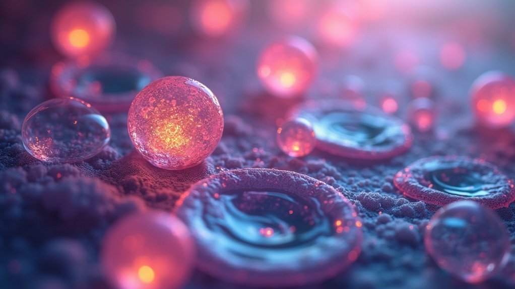
Phase contrast microscopy, pioneered by Frits Zernike in the 1930s, transforms invisible phase shifts into visible contrast differences.
When light passes through live cells, it slows down slightly due to their higher refractive index compared to the surrounding medium. This phase shift is invisible to your eye, but phase contrast technology converts it into amplitude variations you can see.
The microscope achieves this through two critical components: an annular diaphragm that directs the light and a phase plate that creates interference patterns.
This ingenious system lets you observe transparent live cells without staining, preserving their natural state and enabling real-time study of dynamic cellular processes.
Essential Equipment for Live Cell Phase Contrast Imaging

To perform effective live cell phase contrast imaging, you’ll need a properly equipped microscope with specialized objectives and an annular diaphragm that enhances visualization of transparent specimens.
Your setup should include a suitable light source (halogen or LED) and a condenser with annular aperture to achieve the ideal contrast while minimizing heat damage to your cells.
For capturing and documenting your observations, you’ll want to invest in a high-sensitivity digital camera system, such as a CCD camera, which provides superior resolution and reduced noise for clearer images of your living specimens.
Core Microscope Components
Since live cell phase contrast imaging requires specialized equipment to visualize transparent specimens without staining, understanding the essential components is crucial for successful implementation.
You’ll need specialized phase contrast objectives designed to convert phase shifts into amplitude differences, making cellular structures visible in real time.
Your microscope must include a properly equipped condenser with an annular diaphragm to modify the light path and create the necessary phase shifts.
The light source—either halogen or LED—should provide consistent illumination while minimizing photobleaching and preserving sample integrity.
Don’t overlook the importance of proper alignment and adjustment.
You’ll need to carefully align the phase plate and optimize lighting conditions to achieve high-quality imaging results that reveal the subtle details of living cells.
Digital Capture Systems
While observing cells through the eyepiece provides immediate visual feedback, capturing these observations digitally requires specialized equipment that complements your phase contrast setup. Modern digital capture systems typically feature high-sensitivity cameras like CCD or sCMOS, which deliver bright images with minimal exposure time to reduce phototoxicity during live-cell imaging.
| Camera Type | Benefits | Ideal Applications |
|---|---|---|
| CCD | Low noise, high sensitivity | Fixed cell imaging |
| sCMOS | Fast frame rates, wide dynamic range | Rapid cellular processes |
| EMCCD | Extremely low light detection | Fluorescence with phase contrast |
Your system should include software for real-time acquisition and adjustment of parameters during imaging. For extended experiments, integrate environmental controls to maintain ideal temperature and CO2 levels. These digital capture systems transform phase contrast microscopy from observation to quantifiable research, enabling detailed time-lapse studies of cellular dynamics.
Setting Up Your Microscope for Optimal Phase Contrast
Achieving crisp, high-contrast images of living cells requires proper configuration of your phase contrast microscope before you begin imaging.
First, verify your microscope has the essential components: phase contrast objectives and a condenser with an annular aperture that modifies the light path.
For best results, align the annular diaphragm with the objective’s numerical aperture—this matching is vital for enhancing specimen contrast.
Properly calibrate the phase plate to convert phase shifts into visible amplitude differences, allowing you to observe transparent specimens without staining.
Don’t overlook your light source; adjust your halogen or LED illumination for adequate brightness while minimizing photobleaching.
Remember to regularly check the alignment of all optical components to maintain imaging quality and consistency during your live cell observations.
Common Challenges and Troubleshooting Tips

When you’re struggling with image contrast issues, check your phase plate alignment and adjust the condenser to create clearer cellular boundaries without artificial halos.
Focus drift problems often occur during extended imaging sessions, so you’ll need to implement autofocus systems or environmental chambers that minimize thermal fluctuations affecting your sample position.
You can address cell viability concerns by reducing light intensity to prevent photobleaching, maintaining proper temperature and CO₂ levels, and limiting total exposure time to minimize phototoxicity.
Image Contrast Issues
Despite its powerful advantages for observing living cells, phase contrast microscopy presents several image quality challenges that can complicate accurate interpretation.
Unwanted halo effects often appear around cell membranes as light passes through, potentially obscuring critical details. You’ll notice this especially when examining cellular boundaries or thin projections.
When working with phase contrast microscopy, you might encounter inconsistent contrast due to varying refractive indices among different cellular components.
To address this, verify your annular diaphragm is properly sized and positioned—improper settings dramatically reduce image quality. Regular maintenance is equally important; dust on optics can severely degrade contrast.
For thicker specimens, out-of-focus artifacts may blur important structures.
Try adjusting your focal plane carefully or consider Z-stack imaging for clearer visualization of three-dimensional cellular features.
Focus Drift Problems
Clear cellular imaging faces another persistent challenge beyond contrast issues: focus drift.
You’ll notice your once-sharp images gradually becoming blurry during time-lapse experiments as temperature fluctuations affect your sample and microscope components.
To combat focus drift, guarantee stable environmental conditions by minimizing thermal currents and vibrations in your imaging setup.
Regularly calibrate your microscope’s focus mechanism to maintain consistent imaging quality over extended periods. A motorized stage with focus feedback proves invaluable, automatically correcting drift during prolonged sessions.
For best results, implement software with autofocus capabilities that dynamically adjusts focus during time-lapse imaging.
This technology continuously monitors and corrects focal plane changes, delivering crisp images throughout your experiment despite environmental variations that would otherwise compromise your data.
Cell Viability Concerns
Successful live-cell imaging hinges on maintaining cellular health throughout your observation period.
You’ll need to carefully control environmental conditions including temperature, CO2 levels, and humidity to preserve cell viability during extended imaging sessions.
Phototoxicity poses a significant threat to your samples, as excessive light exposure generates damaging free radicals.
Minimize this risk by reducing illumination intensity and exposure time, and consider using low photobleach imaging modes when available.
Your choice of imaging media is equally critical—select formulations specifically designed to support your cell type’s metabolic requirements.
Don’t rely on standard growth media, which may lack the buffering capacity needed for prolonged imaging.
Monitor cell health continuously and be ready to adjust your protocol if you observe morphological changes or reduced motility—early intervention will dramatically improve your experimental outcomes.
Maintaining Cell Viability During Extended Imaging Sessions

When conducting extended phase contrast imaging sessions with live cells, maintaining ideal environmental conditions becomes paramount to the success of your experiment.
You’ll need to monitor temperature, CO2 levels, and humidity consistently to guarantee cell viability throughout your imaging period.
Consider implementing an integrated environmental chamber for real-time control of these conditions within your microscope setup.
To minimize cellular damage, utilize advanced lighting technology with adjustable light intensity and low photobleach modes, which considerably reduces phototoxicity risks during prolonged observation.
Don’t forget to regularly adjust your exposure time and focal plane settings.
These simple modifications allow you to capture high-quality images while minimizing the number of light exposures your samples endure, thereby preserving cell health and extending the viable imaging window for your experiments.
Temperature and Environmental Controls for Live Cell Studies
Building on our discussion of cell viability, let’s now examine the specific environmental parameters that require precise control. When conducting live cell imaging with phase contrast microscopy, you’ll need integrated environmental chambers that maintain ideal conditions directly within your setup.
Keep temperature regulated between 36-37°C, as even minor fluctuations can dramatically alter cell behavior and compromise your results. CO2 levels should be sustained at approximately 5% to preserve physiological pH in your culture media. Proper humidity is equally essential to prevent evaporation during extended imaging sessions.
These environmental chambers enable time-lapse experiments and real-time video capture while ensuring stable conditions. For maximum efficiency, use compatible well-plates to image multiple samples simultaneously under controlled environmental conditions, giving you more data from each experimental run.
Image Acquisition Techniques for Dynamic Cellular Processes

Capturing the intricate dance of living cells requires specialized image acquisition techniques that balance temporal resolution with cellular health.
When you’re setting up phase contrast microscopy for live cells, use dedicated phase contrast objectives paired with the proper annular aperture in your condenser to maximize contrast without compromising resolution.
Your CCD camera selection is vital—high-sensitivity models allow you to reduce light exposure while maintaining image quality during extended time-lapse experiments.
You’ll want to optimize exposure times and acquisition intervals based on your specific cellular processes of interest.
Remember to maintain proper environmental conditions during imaging sessions.
Stable temperature and CO2 levels will guarantee your cells behave naturally, providing authentic insights into cellular dynamics like division, motility, and morphological changes without introducing artifacts from environmental stress.
Data Processing and Analysis of Phase Contrast Images
You’ll need effective noise reduction techniques to clean your phase contrast images before meaningful analysis can begin.
Cell tracking algorithms enable you to monitor dynamic cellular behaviors over time, providing insights into migration patterns and morphological changes.
Quantitative measurement methods transform your visual data into numerical values, allowing for statistical analysis of cell size, shape, and motility across experimental conditions.
Noise Reduction Techniques
Although phase contrast imaging provides excellent visualization of living cells without staining, the technique often suffers from inherent noise that can obscure critical cellular details.
You’ll find that implementing proper noise reduction methods greatly enhances image quality and data reliability.
Start by optimizing your signal-to-noise ratio through careful calibration of exposure time and light intensity before acquisition. For post-processing, apply Gaussian or median filtering to reduce background noise while preserving cellular structures.
When working with time-lapse experiments, temporal averaging across multiple frames effectively minimizes random noise without sacrificing dynamic cellular features.
For more complex challenges, consider using deconvolution algorithms to reverse blurring effects and enhance resolution.
Advanced image processing tools now integrate machine learning capabilities that intelligently distinguish between noise and genuine cellular signals, dramatically improving your analytical outcomes.
Cell Tracking Algorithms
Beyond noise reduction, the real power of phase contrast microscopy emerges when you implement cell tracking algorithms to analyze cellular dynamics over time.
These algorithms transform your images into valuable quantitative data through a sequence of processing steps: background subtraction, edge detection, and cell segmentation.
By isolating individual cells and tracking their positions frame-by-frame, you’ll obtain precise metrics of cell motility, including displacement, velocity, and directional persistence.
Advanced tracking approaches now leverage machine learning techniques to overcome challenges in complex environments, greatly improving accuracy.
Once tracking is complete, you can visualize cell trajectories and generate extensive data on cellular behaviors.
This quantitative foundation enables you to objectively assess how cells interact and respond to various stimuli—turning your phase contrast imaging from merely observational to deeply analytical.
Quantitative Measurement Methods
While visual interpretation provides valuable qualitative insights, transforming phase contrast images into quantitative data reveals the true scientific potential of this technique.
Modern image processing software converts brightness variations into measurable metrics about cellular structures based on refractive index differences.
Your quantitative measurement toolkit includes:
- Cell morphology analysis – Extract precise cell area, volume, and shape parameters to detect subtle structural changes
- Time-lapse imaging sequences – Track cellular movement and responses to stimuli through pixel intensity variations
- Z-stack acquisition – Build three-dimensional reconstructions revealing spatial organization invisible in single-plane views
- Statistical integration – Apply robust statistical methods to your quantified data, enabling objective evaluation of experimental outcomes
Comparison With Other Live Cell Imaging Methods
When researchers evaluate live cell imaging techniques, phase contrast microscopy stands out for its non-invasive approach to visualizing cellular structures.
Unlike fluorescence microscopy, which requires potentially harmful dyes or tags, phase contrast provides clear images without introducing foreign substances that might alter cellular behavior.
Phase contrast delivers unaltered cellular insights by eliminating the need for potentially disruptive fluorescent labels or stains.
You’ll find phase contrast particularly advantageous when studying dynamic cellular processes in real-time.
While widefield fluorescence methods struggle with out-of-focus blur, phase contrast delivers crisp images without background noise.
For thin specimens like individual cells or tissue slices, you’ll achieve higher contrast than with techniques like multiphoton microscopy.
When you need rapid imaging of live specimens, phase contrast excels over traditional brightfield approaches that demand high illumination and longer exposure times—factors that can compromise your sample’s viability during extended observation periods.
Applications in Cell Biology and Developmental Research
Phase contrast microscopy has revolutionized how researchers study living cells by eliminating the need for stains that might alter cellular behavior.
When you’re tracking cellular processes in developmental research, this technique allows you to observe interactions in their natural state.
You’ll find phase contrast microscopy particularly valuable for:
- Monitoring cell motility and morphological changes in real-time without compromising sample integrity
- Observing embryonic development as it unfolds, capturing critical moments in tissue formation
- Studying cellular interactions during differentiation processes with remarkable clarity
- Conducting long-term imaging of live cells and developmental sequences without introducing artifacts
The non-invasive nature of this technique preserves the integrity of your samples, making it ideal for examining thin specimens like cultured cells or small tissue sections during developmental research.
Time-Lapse Strategies for Tracking Cellular Behavior
To truly understand cellular dynamics, time-lapse phase contrast microscopy offers you unprecedented insights into behavioral patterns that single snapshots simply can’t capture.
Witness cells’ living story unfold—revealing truths that static images forever keep hidden.
You’ll observe cells moving through their full cycle without disturbing them with stains or dyes, preserving sample integrity while revealing critical morphological changes.
For peak results, maintain your cells in a controlled environment with stable temperature and CO2 levels throughout your experiment. This approach guarantees cellular behaviors remain natural and physiologically relevant.
Advanced camera technology enables high-speed imaging, allowing you to track rapid movements and interactions in real time.
Don’t just collect visual data—quantitatively analyze brightness changes over time to assess cellular responses, gene expression, and motility patterns.
This analytical approach transforms your observations into meaningful scientific insights about dynamic cellular processes.
Advanced Phase Contrast Techniques for Specialized Studies
While traditional phase contrast methods serve routine cellular imaging well, specialized research demands more sophisticated approaches that push the boundaries of what you can visualize.
Advanced phase contrast microscopy now incorporates specialized objectives and annular apertures that dramatically enhance your ability to observe unstained cellular structures.
For your most demanding studies, consider these transformative techniques:
- Integrate high-sensitivity cameras to capture stunning detail while minimizing phototoxicity
- Employ modified phase plates for thicker samples, eliminating frustrating out-of-focus artifacts
- Implement real-time imaging protocols to witness dramatic cellular interactions as they unfold
- Combine with computational approaches like deconvolution to reveal previously invisible cellular details
These enhancements allow you to extend observation periods while maintaining cell viability—critical when tracking subtle cellular behaviors in response to experimental conditions.
Frequently Asked Questions
What Is the Live Cell Imaging Technique?
You’re observing living cells in real-time with live cell imaging. It lets you monitor dynamic cellular processes without fixation or staining, preserving natural behavior while cells remain alive and functioning.
What Are the Disadvantages of Live Cell Imaging?
When doing live cell imaging, you’ll face limited resolution with thicker specimens, encounter phototoxicity from prolonged light exposure, need specialized equipment, and require technical expertise to maintain imaging conditions during cell dynamics.
What Is the Principle of Phase-Contrast Microscope in Simple Words?
Phase-contrast microscopes turn invisible differences in transparent cells into visible brightness variations. They convert phase shifts from light passing through structures with different refractive indices into contrast you can see without staining specimens.
What Is the Difference Between Live Cell Imaging and Fixed Cell Imaging?
You’ll observe living cells in real-time with live cell imaging, tracking dynamic processes as they happen. Fixed cell imaging shows you preserved, non-viable cells that provide clearer structural details but only static snapshots.
In Summary
You’ll find that phase contrast microscopy revolutionizes your live cell imaging workflow. By mastering the techniques we’ve discussed, you’re now equipped to capture clear, detailed images of living specimens without staining. Don’t hesitate to experiment with the settings—your perfect image is within reach. As you continue your research, you’ll discover phase contrast isn’t just a method; it’s an essential tool for revealing cellular secrets.

Leave a Reply