Phase contrast microscopy lets you observe living cells without staining by converting invisible phase shifts into visible contrast. You’ll need a specialized microscope with proper objectives, an annular diaphragm, and environmental controls to maintain cell viability at 37°C. For best results, guarantee proper alignment of optical components and use high-sensitivity cameras to minimize photodamage. Regular cleaning and optimization help avoid common artifacts like halo effects. The techniques ahead will transform your basic observations into powerful analytical insights.
Numeric List of 12 Second-Level Headings
A thorough understanding of phase contrast imaging requires familiarity with its key components and applications.
When organizing your knowledge about this powerful technique, consider these essential subtopics:
- Principles of Phase Contrast Microscopy
- Setting Up Your Phase Contrast System
- Optimizing Condenser Alignment
- Selecting Appropriate Objectives
- Sample Preparation for Live Cell Imaging
- Maintaining Cell Viability During Observation
- Real-Time Cellular Process Documentation
- Troubleshooting Common Artifacts
- Comparing with Other Microscopy Techniques
- Advanced Applications in Cell Biology
- Limitations with Thicker Specimens
- Digital Image Acquisition and Processing
These headings create a detailed framework for mastering phase contrast microscopy, ensuring you’ll capture clear, artifact-free images of living cells while preserving their natural state throughout your observations.
The Science Behind Phase Contrast Microscopy
Now that we’ve outlined our roadmap for mastering phase contrast techniques, let’s examine the fundamental science that makes this revolutionary imaging method possible.
Developed by Frits Zernike in the 1930s, phase contrast microscopy transforms invisible phase shifts into visible amplitude differences.
When light passes through transparent cellular structures, it experiences subtle phase shifts due to differences in refractive index between cell components. You won’t see these shifts with your eyes, but phase contrast microscopy cleverly converts them into brightness variations.
The system uses an annular diaphragm and phase plate to manipulate light waves, creating contrast without staining.
This makes phase contrast ideal for live cell imaging, allowing you to observe dynamic cellular processes in real time while maintaining sample integrity—something impossible with traditional staining methods.
Essential Equipment for Live Cell Phase Contrast Imaging
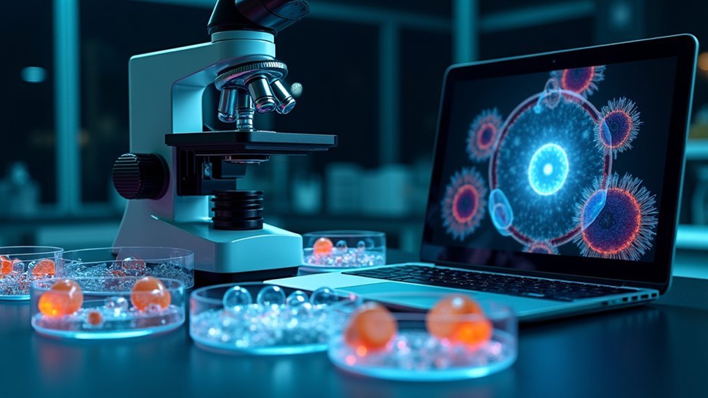
Setting up effective live cell phase contrast imaging requires three core equipment categories for success.
You’ll need a properly configured microscope with specialized phase contrast objectives and an annular diaphragm to visualize transparent cellular structures without staining.
Environmental control systems maintain cell viability during observation, while high-quality image capture technology guarantees you can document and analyze cellular dynamics with precision.
Microscopes and Optical Components
Successful phase contrast imaging of live cells depends fundamentally on specialized equipment that enhances the visibility of otherwise transparent specimens.
You’ll need a dedicated phase contrast microscope equipped with specialized objectives and an annular aperture condenser to create the necessary contrast.
Your optical setup must include a suitable light source—typically halogen or LED—and an annular diaphragm that creates the critical phase shifts in light waves passing through your sample.
Proper alignment of these components isn’t optional; it’s essential for generating clear images without artifacts.
For capturing high-quality images, pair your microscope with high-sensitivity cameras like CCD or EMCCD, especially when working in low-light conditions.
To maintain cell viability during extended imaging sessions, integrate environmental chambers that control temperature, humidity, and CO2 levels without compromising your optical path.
Environmental Control Systems
Three critical environmental factors must be precisely controlled when conducting live cell phase contrast imaging: temperature, CO₂ levels, and humidity.
Environmental control systems guarantee peak cell viability during your long-term observations, allowing you to track cellular dynamics without compromising sample health.
Your imaging setup should include:
- An integrated chamber that fits various sample vessels like well-plates
- Systems for monitoring brightness and movement changes over time
- Controls for oxygen concentration, pH, osmolarity, and viscosity
- Adjustable light intensity with low photobleach modes to reduce phototoxicity
These features maximize efficiency during live-cell imaging by enabling simultaneous observation of multiple locations under different conditions.
You’ll get clearer, more consistent results when your environmental control systems maintain stable conditions throughout your imaging sessions.
Image Capture Technology
Effective live cell phase contrast imaging depends on high-quality image capture technology that balances sensitivity with resolution.
When studying dynamic cellular processes, you’ll need high-sensitivity CCD cameras specifically designed to perform under the low light conditions typical in phase contrast microscopy.
These specialized cameras capture bright, detailed images of transparent specimens without requiring stains that might compromise cell viability. You’ll achieve excellent results by pairing your CCD camera with properly aligned phase contrast objectives and condenser systems.
The camera’s sensitivity allows you to reduce exposure time and light intensity, minimizing photodamage to your live cells.
For best results, verify your image capture setup integrates seamlessly with your environmental control systems, allowing for continuous, stable documentation of cellular activities over extended observation periods.
Optimizing Your Microscope Setup for Clear Contrast
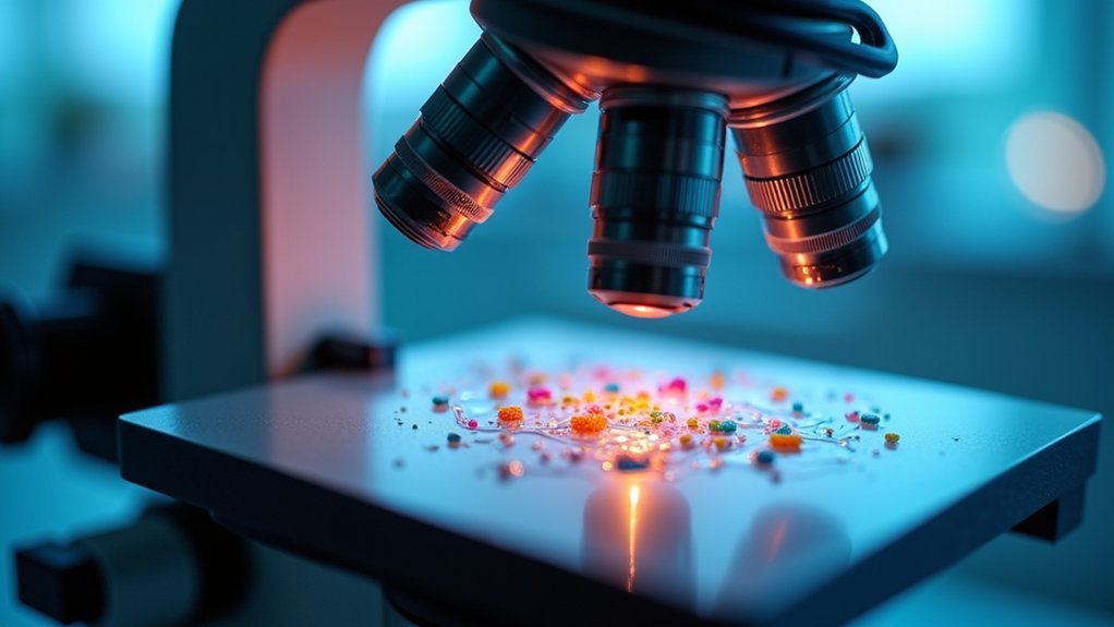
While capturing high-quality phase contrast images of live cells depends on several factors, proper microscope setup forms the foundation of successful imaging. Achieving ideal optical microscopy conditions requires careful alignment and calibration to reveal cellular structures that would otherwise remain invisible.
Precise microscope calibration unveils cellular structures invisible to untrained eyes, making proper setup essential for meaningful observations.
- Verify your microscope has a specialized phase contrast objective and properly aligned annular diaphragm to transform phase shifts into visible contrast.
- Adjust the phase plate settings specifically for your sample’s refractive index, greatly improving imaging of cells without staining.
- Calibrate your focal plane and exposure time regularly to maintain consistency and reduce artifacts.
- Align your light source and condenser precisely, ensuring the light path modification maximizes contrast in transparent specimens.
Environmental Controls for Maintaining Cell Viability
Maintaining precise temperature control is essential for your live cell imaging experiments, as even minor fluctuations can dramatically alter cellular behavior and compromise your results.
You’ll need to establish appropriate oxygen levels and humidity conditions that mimic your cells’ native environment, typically using integrated environmental chambers with CO₂ regulation systems.
These environmental parameters should be carefully calibrated before starting your imaging session and monitored throughout to guarantee consistent cellular viability and authentic physiological responses.
Temperature Regulation Basics
For successful live-cell phase contrast imaging, temperature regulation stands as a cornerstone of environmental control. You’ll need to maintain mammalian cells at approximately 37°C to guarantee ideal cell viability during live-cell imaging sessions. An integrated environmental chamber mounted inside your microscope provides the stable conditions necessary for consistent results.
- Monitor constantly – Even minor temperature fluctuations can considerably alter cellular metabolism and compromise your imaging data.
- Consider heat sources – Choose between heated air systems or infrared radiation based on your specific setup.
- Maintain consistency – Avoid thermal gradients across your sample that could create artifacts.
- Combine with CO₂ control – Temperature regulation works hand-in-hand with CO₂ maintenance to stabilize pH levels in bicarbonate-buffered media.
Oxygen and Humidity Control
Beyond temperature regulation, oxygen and humidity levels represent essential environmental factors that directly impact cell health during live-cell phase contrast imaging.
Maintaining ideal oxygen concentration prevents hypoxia, which can compromise cellular metabolism and yield misleading results in your experiments.
You’ll need robust humidity control to prevent media evaporation, especially during extended imaging sessions. When culture medium evaporates, osmolarity changes can stress your cells and alter their natural behavior.
Consider using integrated environmental chambers that regulate both parameters simultaneously. For most mammalian cell lines, you’ll also want to pair CO₂ with bicarbonate buffers to stabilize pH levels—another critical factor for authentic cellular conditions.
Consistently monitor these environmental parameters throughout your imaging process. This attention to detail greatly improves experimental reliability and maintains the physiological relevance of your observations.
Time-Lapse Techniques for Capturing Cellular Dynamics
Three critical advancements have transformed time-lapse phase contrast imaging into an essential tool for visualizing cellular dynamics.
By combining phase contrast microscopy with environmental controls, you’ll observe live cells over extended periods without staining artifacts. Modern camera technology provides higher frame rates and enhanced sensitivity, allowing you to track rapid cellular movements with unprecedented detail.
Time-lapse phase contrast reveals cellular narratives in their natural state, transforming biological observation into dynamic discovery.
- Set ideal environmental conditions – Maintain temperature and CO2 levels to guarantee cell viability throughout your imaging session
- Adjust acquisition parameters – Balance frame rate and exposure to capture fast processes without photodamage
- Utilize automated tracking software – Quantify morphological changes and migration patterns objectively
- Integrate multi-channel imaging – Combine phase contrast with fluorescence to correlate structure with function
You’ll reveal subtle changes in cellular behavior that would otherwise remain invisible with static imaging techniques.
Common Artifacts and How to Avoid Them
Halo effects around your cells create bright rings that can mask fine structural details, but you’ll minimize these artifacts by properly aligning your phase contrast system and maintaining sample thickness below 30 micrometers.
You can reduce excessive edge contrast issues by fine-tuning your light intensity and using neutral density filters to prevent saturation in bright areas.
Regular cleaning of optical components guarantees the phase ring remains correctly positioned relative to your microscope’s optical axis, greatly improving image clarity and reducing distortion.
Halo Effects Solutions
Bright rings surrounding cellular structures represent one of the most common challenges when working with phase contrast microscopy: the halo effect.
These artifacts occur at specimen boundaries where significant refractive index differences create unwanted phase shifts. You’ll need to address these issues to achieve clearer images for your live cell work.
To minimize halo effects in your phase contrast imaging:
- Align your optical components precisely, ensuring proper positioning of the condenser and phase plate during microscope setup.
- Match the annular diaphragm size to your specific objective magnification.
- Prepare thinner specimens or dilute your samples to reduce phase shifts at cell boundaries.
- Clean all optical surfaces regularly, as dust and oil residue can intensify unwanted halos.
Edge Contrast Issues
While observing live cells through phase contrast microscopy, you’ll inevitably encounter edge contrast issues that can compromise your image interpretation.
These artifacts manifest as halo effects—excessively bright or dark boundaries around cellular structures—resulting from abrupt phase shifts at specimen edges.
To minimize these problematic artifacts, properly align your phase plate and condenser.
Misalignment considerably degrades image quality and amplifies edge distortions.
Optimize your light settings and adjust the annular diaphragm to distribute phase shifts more uniformly across your specimen.
Don’t underestimate the importance of regular calibration and maintenance of your phase contrast microscope.
This preventative approach guarantees consistent image quality during live cell imaging sessions and reduces recurring edge contrast problems that might lead to misinterpretation of cellular morphology.
Image Processing Methods to Enhance Contrast
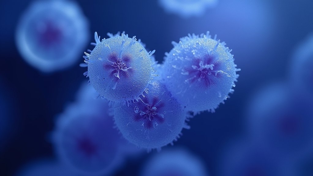
Although phase contrast microscopy inherently provides better visualization of transparent specimens than brightfield microscopy, applying specific image processing techniques can dramatically improve image quality and data interpretation.
You’ll find these methods essential for extracting maximum detail from your samples.
- Deconvolution – Remove out-of-focus light and enhance spatial resolution, making cellular structures appear sharper in your phase contrast images.
- Background subtraction – Eliminate noise and improve visibility of subtle cellular details that might otherwise be missed.
- Histogram equalization – Redistribute intensity values to enhance contrast, highlighting variations between cellular components.
- Adaptive filtering – Target specific features while reducing artifacts, maintaining biological relevance without introducing distortion.
Modern software tools can automate these enhancement processes, ensuring consistent results across multiple images while maintaining both speed and accuracy.
Comparing Phase Contrast With Other Live Cell Techniques
The enhanced images achieved through processing represent just one facet of phase contrast’s capabilities in the cellular imaging landscape.
When you’re deciding between imaging techniques, phase contrast offers distinct advantages for observing live cells without the potential toxicity introduced by fluorescence microscopy, which requires potentially harmful fluorescent tags.
Unlike brightfield microscopy that struggles with transparent specimens, phase contrast converts subtle phase shifts into visible brightness differences, revealing cellular structures in their natural state.
While fluorescence excels at tracking specific molecules, phase contrast enables you to observe dynamic processes over extended periods without compromising cell viability.
For thin samples, you’ll find phase contrast delivers superior contrast compared to differential interference contrast (DIC), though thicker specimens may present challenges due to phase shifts creating unwanted artifacts.
Applications in Cell Culture and Development Studies
Because phase contrast microscopy eliminates the need for staining or fixing specimens, it serves as a cornerstone technology in cell culture monitoring and developmental biology.
You’ll find this technique particularly valuable when you need to observe living cells in their natural state over extended periods.
- Track cellular dynamics – Monitor growth patterns, migration, and cell-cell interactions in real-time without disrupting natural processes
- Observe developmental changes – Witness embryonic development and differentiation as they unfold, capturing critical morphogenetic events
- Study responses to stimuli – Evaluate how live cells react to environmental changes or treatments without invasive interventions
- Examine transparent specimens – Visualize details in plant tissues and microbial cells that would otherwise be invisible under conventional microscopy
Advanced Quantification of Cellular Behavior
Building on simple observational capabilities, modern phase contrast microscopy now offers sophisticated quantitative analysis of living cells. You’ll gain precise measurements of RGB brightness changes that reveal gene expression variations during your experiments.
When tracking cellular movement, you can analyze brightness, hue, and spatial appearance to understand dynamic interactions. Automated coordinate recording provides extensive data on cell travel range and speed, enabling detailed motility studies.
For best results, utilize Z-stack data to select the best focal planes, reducing out-of-focus artifacts that might compromise your measurements.
Time-series data outputs let you evaluate how treatments affect cellular behavior over time.
Advanced quantification transforms phase contrast imaging from merely observational to deeply analytical, giving you quantitative insights into complex cellular processes previously impossible to measure.
Troubleshooting Poor Image Quality in Phase Contrast
Identifying the root causes of poor phase contrast images allows you to quickly resolve visual problems during live cell observations.
When your images lack clarity, check these critical elements that directly impact image quality:
- Alignment verification – Confirm proper alignment between the phase ring and annular diaphragm to prevent contrast reduction.
- Condenser optimization – Position the annular aperture to match your specific phase contrast objective.
- Light intensity adjustment – Find the sweet spot where illumination reveals detail without creating glare.
- Optical component maintenance – Clean lenses and filters regularly to prevent dust and smudges from degrading visualization.
Remember that selecting appropriate objectives for your specimen thickness is equally important, as using incompatible objectives can introduce artifacts and obscure cellular structures.
Frequently Asked Questions
What Is the Live Cell Imaging Technique?
Live cell imaging is a technique that lets you observe living cells in real-time without staining them. You’ll see dynamic cellular processes as they happen, though you must minimize light exposure to prevent cell damage.
What Are the Disadvantages of Live Cell Imaging?
You’ll face phototoxicity and heat damage to cells from intense illumination, limited resolution in phase contrast, compromised image quality from reduced light exposure, and challenges in maintaining consistent temperature and pH during experiments.
What Is the Principle of Phase-Contrast Microscope in Simple Words?
Phase-contrast microscopes convert invisible phase differences into visible brightness variations. You’ll see transparent cells without staining because light passing through different cellular densities creates contrast, making structures appear bright or dark against the background.
What Is the Difference Between Live Cell Imaging and Fixed Cell Imaging?
In live cell imaging, you’re observing cells in their natural state in real-time, capturing dynamic processes. With fixed cell imaging, you’re viewing chemically preserved cells as static snapshots, sacrificing natural behavior for easier staining.
In Summary
You’ve now mastered the fundamentals of live cell phase contrast imaging. With proper equipment, optimization, and environmental controls, you’ll capture clear, dynamic cellular behaviors without staining or labeling. Don’t hesitate to troubleshoot image issues as they arise. Whether you’re studying cell culture, development, or quantifying cellular behaviors, phase contrast provides an invaluable window into the living cellular world that other techniques simply can’t match.


Leave a Reply