For fluorescent specimen images, you’ll achieve best results by: 1) Optimizing fixation and permeabilization protocols specific to your target location, 2) Selecting compatible fluorophores with high quantum efficiency and using appropriate blocking agents, and 3) Fine-tuning microscope settings including exposure time, gain, and pixel size to maximize signal-to-noise ratio. Adding anti-fade reagents prevents photobleaching during extended imaging sessions. These techniques together guarantee crisp, quantifiable images with minimal background interference.
Optimizing Fixation and Permeabilization Protocols
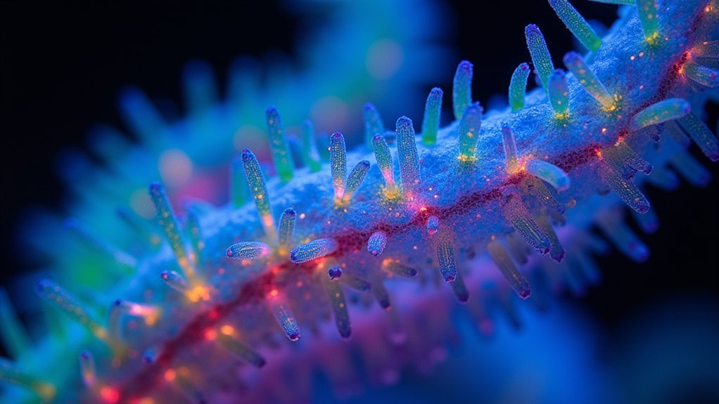
When capturing fluorescent specimen images, your fixation and permeabilization methods can make or break your results. Choose aldehyde-based fixatives like formaldehyde to preserve cellular structures, or alcohol fixatives like methanol to expose hidden epitopes.
Be careful—over-fixation causes false negatives while under-fixation produces blurred images.
For intracellular targets after aldehyde fixation, you’ll need permeabilization. Triton® X-100 in PBS improves antibody accessibility to internal structures.
Don’t skip blocking—use normal serum or BSA to minimize non-specific binding and enhance your signal-to-noise ratio.
Always confirm your target’s cellular location before selecting protocols. The right combination of fixation, permeabilization, and blocking agents creates the foundation for fluorescent microscopy techniques that deliver the ideal image with minimal background signal and maximum specificity.
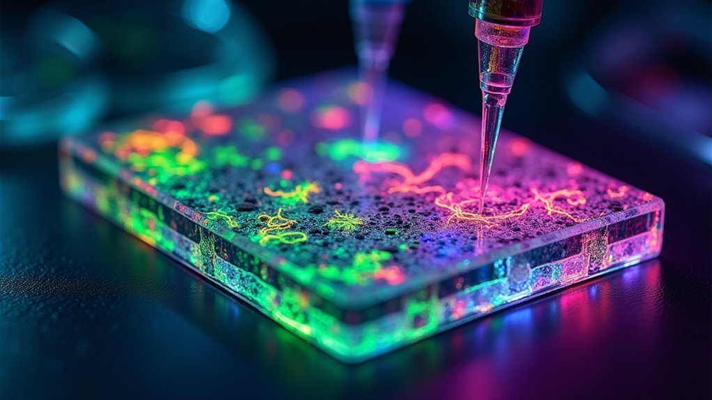
Selecting Appropriate Fluorophores and Blocking Agents
Because your fluorophore selection directly impacts image quality, you’ll need to choose molecules with ideal quantum efficiency and appropriate excitation/emission profiles for your microscopy system.
Verify that your fluorophores exhibit minimal background fluorescence and remain compatible with your imaging setup to maximize signal-to-noise ratio.
When using primary antibodies in fluorescent microscopy, perform antibody titration to determine best concentrations. Excessive antibody can increase non-specific staining and degrade image quality.
For secondary antibodies, employ blocking agents like species-matched normal serum or BSA to prevent non-specific binding. This enhances the specificity of your fluorescent signals.
Don’t forget to incorporate anti-fade reagents during sample preparation. These compounds preserve fluorophores from photobleaching, extending their lifespan and allowing for prolonged observation periods with consistent signal intensity.
Mastering Microscope Settings for Maximum Signal-to-Noise Ratio
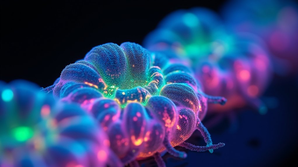
Ideal microscope settings serve as the foundation for high-quality fluorescent imaging, directly impacting your signal-to-noise ratio (SNR) and overall image interpretation.
Maintain consistent excitation intensity and exposure time across all samples to guarantee reliable quantitative comparisons in your images.
While adjusting gain settings can amplify your fluorescence signal, remember that it equally boosts background noise—balance is key.
Consider increasing pixel size or binning to enhance light capture and SNR, though this trades off some spatial resolution.
Apply anti-fade reagents to preserve signal strength throughout imaging sessions, minimizing the background noise that occurs when signals fade.
Regularly check your image histogram to prevent signal saturation, which compromises detail and reduces SNR.
These optimizations will help you capture crisp, quantifiable fluorescent images with excellent signal-to-noise ratios.
Frequently Asked Questions
What Are the Methods of Fluorescence Imaging?
You can use Fluorescent Widefield, Point Scanning Confocal, Two-Photon, Light Sheet, or TIRF microscopy for fluorescence imaging. Each method offers unique advantages for visualizing fluorescent specimens with different requirements.
What Is the Best Camera for Fluorescence Microscopy?
For fluorescence microscopy, you’ll want a camera with high quantum efficiency (>90%), sCMOS technology for low noise, large pixel sizes, and sensitivity that matches your fluorophores’ emission spectrum.
How to Take Immunofluorescence Images?
To take immunofluorescence images, you’ll need to properly fix and permeabilize cells, block non-specific binding, apply optimized antibody dilutions, and capture images quickly using appropriate exposure settings to prevent photobleaching.
What Kind of Microscope Is Used for Fluorescence Imaging?
You’ll need a fluorescence microscope for fluorescence imaging. Common types include epi-fluorescence, confocal, two-photon, and light sheet microscopes, each offering different advantages for resolution, depth penetration, and live specimen imaging.
In Summary
You’ve now mastered the trifecta of fluorescent imaging: optimized fixation protocols, strategic fluorophore selection, and fine-tuned microscope settings. By implementing these three techniques in your workflow, you’ll consistently capture clearer, more vibrant specimens with minimal background interference. Don’t forget that practice makes perfect—each imaging session builds your expertise and intuition for producing publication-quality fluorescent images that truly showcase your research specimens.


Leave a Reply