
The best fixative depends on your specimen type. For most soft tissues, 10% Neutral Buffered Formalin works excellently. Use TRIPLEfix for fatty tissues like breast samples, while B-5 Substitute (mercury-free) is ideal for lymphoid specimens. Electron microscopy requires 2-4% glutaraldehyde, and cytological specimens benefit from ethanol-based fixatives. Your fixation time matters too—generally 12-24 hours guarantees proper preservation. The right fixative-tissue pairing dramatically impacts your analysis quality and results.
What Are The Best Fixatives For Biological Specimens?
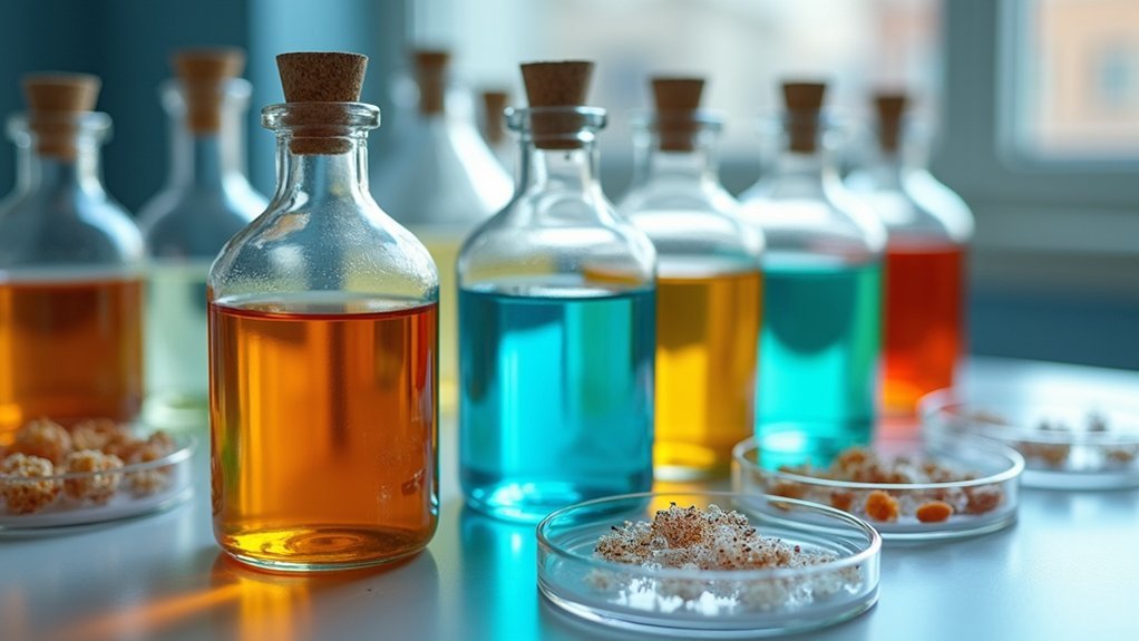
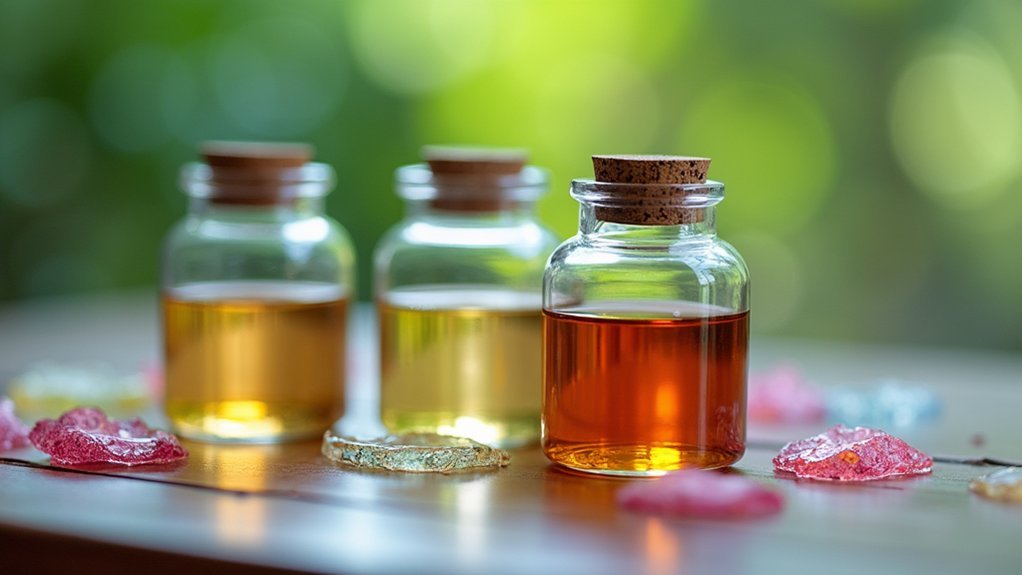
When preserving biological specimens, choosing the right fixative is essential to maintain cellular structure and guarantee accurate analysis.
10% neutral buffered formalin (NBF) stands as the gold standard for soft tissues, offering excellent preservation properties and compatibility with most histological stains.
For hard tissue specimens, you can use either 10% NBF or unbuffered formalin, though the buffered version prevents unwanted pigment formation in blood-rich samples.
If you’re working with fatty tissue specimens like breast samples, consider TRIPLEfix, a buffered alcohol mixture that better stabilizes cellular components.
Lymphoid and hematopoietic tissues benefit from mercury-free fixatives such as B-5 Substitute, which preserves detailed cellular structures without hazardous chemicals.
For cytological specimens, ethanol serves as the primary fixative, often enhanced with Saccomanno Fluid for superior preservation and staining results.
Understanding Fixation Fundamentals in Specimen Preparation
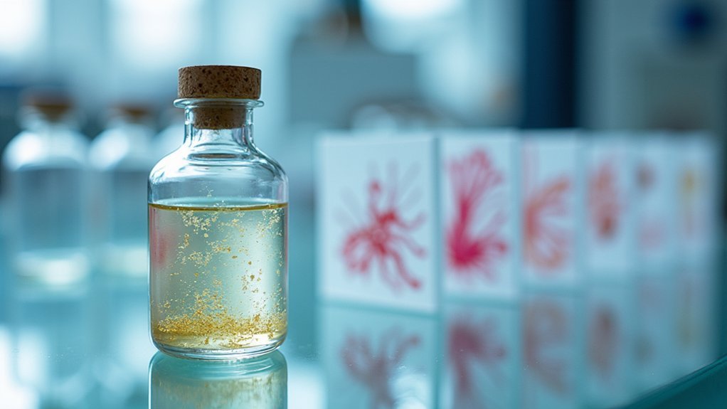
Because preservation quality directly affects analytical outcomes, mastering fixation fundamentals is essential for any laboratory professional. Proper fixation prevents autolysis and maintains cellular detail through protein cross-linking, ensuring specimens retain their scientific value.
When preparing biological samples, remember these critical points:
- Select appropriate fixatives based on tissue type—10% neutral buffered formalin works well for most soft tissues, while specialized fixatives serve specific purposes.
- Consider fixation duration carefully—most tissues require 12-24 hours for ideal results.
- Use alcohol-based fixatives for cytological smears to preserve cellular structures effectively.
- Choose mercury-free alternatives like B-5 Substitute for lymphoid tissues to maintain cellular detail without hazardous chemicals.
Understanding these fundamentals helps you produce consistently high-quality specimens for accurate diagnostic and research applications.
Formaldehyde-Based Fixatives: The Gold Standard for Routine Histology
For over a century, formaldehyde-based fixatives have dominated routine histology due to their unmatched ability to preserve tissue architecture. The most widely used preparation, 10% neutral buffered formalin (NBF), contains 4% formaldehyde in phosphate buffer at pH 6.8, preventing problematic formalin pigment formation while delivering exceptional morphological preservation.
Optimal fixation time with formaldehyde typically ranges from 12-24 hours, ensuring proper penetration and cellular stabilization.
Histological excellence demands patience—formaldehyde requires 12-24 hours to fully penetrate and stabilize cellular architecture.
When you need faster results, take into account alcoholic formalin, which combines 40% formaldehyde with 95% ethanol for rapid protein preservation. Similarly, formol acetic alcohol accelerates fixation time for urgent specimens.
While these alternative formaldehyde preparations work quickly, they may produce formalin pigment—a tradeoff to weigh when selecting the appropriate fixative for your specific histological needs.
Glutaraldehyde Fixation for Ultrastructural Preservation
While formaldehyde excels at routine histology, glutaraldehyde stands as the premier fixative for electron microscopy and ultrastructural studies. This powerful fixative creates extensive protein cross-links that perfectly preserve cellular architecture at the nanoscale level.
For ideal ultrastructural preservation, you’ll need to:
- Prepare a 2-4% glutaraldehyde solution in PBS
- Fix your specimens for 1-2 hours at room temperature
- Rinse thoroughly with buffer to remove excess fixative
- Follow with post-fixation treatments like osmium tetroxide for enhanced contrast
Remember that glutaraldehyde penetrates tissues more slowly than formaldehyde due to its larger size, so you may need longer fixation times for thicker specimens.
The reward is worth the wait—glutaraldehyde delivers exceptional preservation of fine cellular details, membranes, and organelles critical for electron microscopy analysis.
Alcoholic Fixatives for Rapid Processing and Cytological Specimens
When time is of the essence, alcoholic fixatives emerge as the go-to solution for cytological specimens and rapid processing workflows. Absolute ethanol and methanol efficiently denature proteins and preserve cellular morphology in just 5-10 minutes of fixation time.
For cytologic smears, you’ll find ethanol particularly effective, especially when combined with ready-to-use solutions like Saccomanno Fluid for enhanced preservation.
These alcoholic fixatives excel at preserving nucleic acids and cytoplasmic contents, making your specimens ideal for subsequent staining and molecular analysis.
Be cautious about fixation duration—avoid prolonged exposure to alcoholic fixatives as over-fixation can compromise tissue integrity and staining quality.
With proper timing, you’ll achieve excellent preservation of cellular details while maintaining the specimen’s molecular composition for thorough diagnostic evaluation.
Mercury-Based Fixatives: B-5 and Zenker’s for Hematopoietic Tissues
Mercury-based fixatives like B-5 and Zenker’s contain mercuric chloride compounds that provide exceptional nuclear detail and cellular preservation in hematopoietic tissues.
You’ll achieve superior staining results with these fixatives when specimens are fixed for 4-8 hours and subsequently stored in 70% ethanol.
When working with these powerful fixatives, you must follow strict safety protocols due to the toxic nature of mercury compounds, including proper waste disposal and personal protective equipment use.
Composition and Mechanism
Two powerful mercury-based fixatives dominate the field of hematopoietic tissue preservation: B-5 and Zenker’s solutions.
These formulations provide excellent nuclear detail through their mercuric chloride base, which crosslinks proteins more effectively than formalin for extended periods.
B-5 contains 12g mercuric chloride and 2.5g sodium acetate in 200ml distilled water, while Zenker’s combines 50g mercuric chloride with 25g potassium dichromate and 50ml glacial acetic acid.
The mechanism of fixation of hematopoietic tissues involves:
- Protein denaturation via mercury ion binding to sulfhydryl groups
- Cross-linking of adjacent protein molecules
- Stabilization of nuclear chromatin patterns
- Preservation of cytoplasmic granules in blood cells
Both fixatives require post-fixation mercury removal and storage in 70% ethanol, with ideal fixation times ranging from 4-24 hours depending on tissue type.
Fixation Quality Advantages
Despite their handling challenges, B-5 and Zenker’s fixatives remain the gold standard for hematopoietic tissue preservation due to their superior quality outcomes.
When you’re working with lymphoid tissues, these mercury-based solutions deliver excellent nuclear detail that’s unmatched by mercury-free alternatives.
The mercuric chloride in B5 and Zenker’s enhances chromatin patterns and nuclear boundaries, making cellular assessment remarkably precise.
You’ll notice the difference particularly in bone marrow biopsies and lymph nodes, where diagnostic accuracy depends on clear visualization of cellular morphology.
These chemical fixation methods require specific timing (4-8 hours) to achieve ideal results without overfixation.
After processing, you’ll need to store specimens in 70% ethanol rather than formalin to maintain the exceptional tissue architecture and prevent degradation that could compromise your histological analysis.
Safety Precautions Needed
While the preservation quality of B-5 and Zenker’s fixatives is exceptional, working with these solutions demands rigorous safety protocols due to their mercury content.
You’ll need to handle these fixatives with extreme caution due to the toxicity of mercuric chloride (12g in B-5 and 50g in Zenker’s).
Always follow these essential safety precautions:
- Wear appropriate protective equipment including gloves and eye protection when handling.
- Work in a properly functioning fume hood to prevent inhalation of toxic vapors.
- Store processed tissues in 70% ethanol to prevent mercury pigment formation.
- Collect all waste in designated hazardous waste containers—never pour down the sink.
Adhere to your institution’s safety guidelines when using these fixatives to protect yourself and the environment from mercury contamination.
Acidic Fixatives: Bouin’s and Carnoy’s Solutions for Special Applications
Acidic fixatives represent specialized tools in the histologist’s arsenal, with Bouin’s and Carnoy’s solutions standing out for their unique preservation capabilities. When you’re working with specimens requiring specific detail retention, these fixatives offer distinct advantages.
| Feature | Bouin’s Solution | Carnoy’s Solution |
|---|---|---|
| Composition | Picric acid, formaldehyde, acetic acid | Ethanol, chloroform, acetic acid |
| Best Application | Glycogen preservation, cytoplasmic detail | Nucleic acids, cytological specimens |
| Fixation Time | 4-24 hours | 1-4 hours |
| Advantages | Clear cytoplasmic detail | Rapid penetration and action |
| Cautions | Hazardous picric acid; thorough washing required | Can cause excessive shrinkage |
You’ll need thorough washing protocols after using either fixative to prevent staining artifacts. Remember that both can cause tissue hardening, so carefully control fixation duration to maintain specimen integrity.
Fixation Protocols for Hard Tissues: Bone and Cartilage
Hard tissues present unique challenges that require specialized fixation approaches. When preserving bone and cartilage specimens, you’ll achieve ideal results with 10% neutral buffered formalin (NBF) rather than unbuffered alternatives, which can form undesirable brownish-black pigments in blood-rich tissues.
For effective hard tissue fixation:
- Maintain specimen thickness at 2-3mm to guarantee proper fixative penetration.
- Allow a fixation time of 12-24 hours for complete preservation.
- Use buffered solutions to prevent formic acid formation and pigment formation.
- Implement thorough washing post-fixation to maintain tissue integrity.
The buffering component in NBF prevents acidification that would occur with unbuffered formalin, protecting your specimens from artifacts and preserving cellular structures for accurate histological examination and long-term storage.
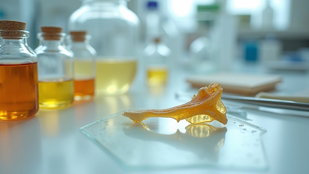
Optimal Fixatives for Fatty and Adipose Tissues
Fatty and adipose tissues require specific fixation methods that differ from those used for hard tissues. When preserving these specimens, 10% neutral buffered formalin (NBF) serves as an effective primary choice, preserving cellular morphology while ensuring good contrast during histological staining procedures.
For best results, you’ll need to monitor fixation time carefully—typically several hours to overnight—to achieve proper preservation without compromising tissue integrity. Inadequate timing often leads to artifacts that distort microscopic examination.
As an alternative, consider TRIPLEfix, a specialized mixture of buffered alcohols designed specifically for fatty specimens. This fixative stabilizes cellular components and enhances clarity in adipose tissues.
Whichever method you choose, proper fixation techniques remain essential for maintaining the life-like appearance of fat cells during subsequent analysis.
Specialized Fixatives for Lymphoid and Immune System Specimens
Lymphoid tissues demand selective fixation methods that preserve delicate cellular structures while enabling detailed immunological analysis. When working with these tissues, you’ll want to choose B-5 Substitute as your primary fixative. This mercury-free alternative delivers excellent morphological preservation without the hazards of mercuric chloride.
For ideal results with lymphoid tissues:
- Limit fixation time to 4-8 hours to prevent over-fixation while ensuring complete preservation.
- Transfer specimens to 70% ethanol for storage after initial fixation.
- Prepare tissues for immunohistochemistry by using B-5 Substitute to maintain antigenic sites.
- Remember that B-5 Substitute allows for superior visualization of cellular architecture in lymphocytes and related immune cells.
You’ll achieve better special staining outcomes and more reliable immunohistochemical results compared to standard formalin fixation.
Microwave-Assisted Fixation Techniques for Time-Sensitive Preparations
When rapid results are essential, microwave-assisted fixation revolutionizes your specimen preparation workflow by reducing hours-long processes to mere minutes. This technique harnesses microwave energy to enhance fixative penetration while maintaining uniform heating throughout your samples.
For ideal results, your fixative must be compatible with microwave technology—10% neutral buffered formalin works excellently in this application. The process typically requires only 5-30 minutes, depending on your tissue type and microwave settings, while preserving cellular morphology and antigenicity.
You’ll appreciate the consistent fixation outcomes that microwave assistance provides, particularly when preparing specimens for downstream immunohistochemistry (IHC) or molecular analysis.
The technique effectively preserves nucleic acids and proteins, ensuring your time-sensitive preparations maintain high quality while markedly reducing processing time.
Proprietary and Commercial Fixative Formulations: Benefits and Limitations
Although traditional fixatives like formalin remain laboratory staples, proprietary commercial formulations have emerged to address specific preservation challenges while reducing health hazards.
When selecting proprietary fixatives for your specimens, you’ll need to weigh several factors:
- Reduced toxicity – Many commercial formulations substitute harmful ingredients with zinc or barium salts, offering safer alternatives to standard commercial formaldehyde mixtures.
- Enhanced antigen preservation – Proprietary solutions often boast superior performance for immunohistochemistry (IHC) applications by maintaining epitope integrity.
- Faster processing – Some commercial products integrate microwave-assisted fixation technology, reducing preparation time while improving penetration.
- Undisclosed compositions – You’ll need to validate these products for your specific applications since manufacturers rarely publish complete ingredient lists.
Remember that each proprietary formulation requires thorough evaluation before implementing it in your workflow.
Environmental and Safety Considerations in Fixative Selection
You’ll need to implement strict hazard mitigation strategies when working with traditional fixatives such as formaldehyde, including proper ventilation systems and consistent use of appropriate personal protective equipment.
Your laboratory must comply with regulatory requirements for the handling, storage, and disposal of hazardous fixatives to protect staff and the environment.
Consider switching to less toxic alternative fixatives that maintain specimen quality while reducing health risks and simplifying compliance with environmental regulations.
Hazard Mitigation Strategies
Selecting the right fixatives requires careful consideration of their environmental and safety impacts.
You’ll need to implement strategic approaches to minimize risks associated with these chemicals in your laboratory setting.
- Choose mercury-free alternatives like B-5 Substitute instead of traditional fixatives containing mercuric chloride, greatly reducing toxic exposure risks while maintaining specimen quality.
- Always work in well-ventilated areas and wear appropriate PPE (gloves, masks) when handling fixatives, especially those containing formaldehyde, to prevent inhalation and skin contact.
- Establish proper disposal protocols for your fixative waste, as improper disposal can lead to environmental contamination of soil and water systems.
- Store fixatives properly using secondary containment measures and clear labeling to prevent accidental spills and promote quick identification of hazardous materials.
Regulatory Compliance Requirements
When working with biological fixatives, you’re subject to stringent regulatory frameworks established by agencies like OSHA and EPA. These regulations govern how you handle, store, and dispose of hazardous materials like formaldehyde and mercuric compounds commonly used in specimen preservation.
Your laboratory must develop thorough safety protocols that include proper PPE usage, adequate ventilation systems, and detailed MSDS documentation for each fixative. Risk assessments are essential for regulatory compliance and protecting staff from carcinogenic exposure.
Consider selecting proprietary fixatives with lower formaldehyde concentrations or alternative compounds that reduce health risks while maintaining preservation quality.
Remember that all fixative waste requires specialized disposal methods to prevent environmental contamination. Failure to comply with these regulations can result in significant penalties and, more importantly, compromise the health of your personnel and the environment.
Frequently Asked Questions
Which Is the Most Ideal Fixative?
For routine histopathology, you’ll find 10% neutral buffered formalin (NBF) is the most ideal fixative. It provides excellent preservation and contrast for your tissue specimens when using H&E staining techniques.
What Is the Fixative for Pathological Specimens?
For pathological specimens, you’ll primarily use 10% neutral buffered formalin (NBF). It’s excellent for preserving tissue morphology. For lymphoid tissues, consider B-5 Substitute, and for glycogen preservation, you might need Bouin’s Solution instead.
What Are the Most Commonly Used Fixatives in Histology?
You’ll find that 10% neutral buffered formalin is the most commonly used fixative in histology. Mercuric fixatives like B-5, alcoholic formalin, Bouin’s solution, and proprietary alternatives are also frequently used for specific tissue types.
What Is the Most Popular Fixative?
The most popular fixative you’ll encounter is 10% neutral buffered formalin (NBF). It’s widely used in histopathology because it excellently preserves tissues and works well with various staining techniques you might need to perform.
In Summary
You’ll find no perfect universal fixative for all specimens. Choose your fixative based on your specific research needs, biological material, and planned analytical techniques. Balance preservation quality against safety concerns and accessibility. Whether you’re using classic formaldehyde-based solutions or exploring newer alternatives, proper fixation protocol adherence remains critical for reliable results. Remember, it’s your specific research question that should ultimately determine your fixative selection.
Leave a Reply