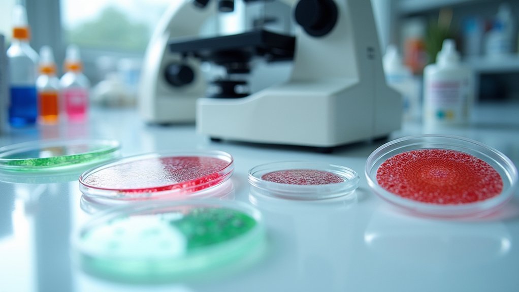
For perfect double-staining slides, select primary antibodies from different species to prevent cross-reactivity. Optimize antibody concentrations (1:100-1:1000) for ideal signal-to-noise ratios. Use 2-4% paraformaldehyde fixation for 10-20 minutes to preserve antigen integrity. Apply blocking serum matching your secondary antibody species to minimize background. Fine-tune incubation periods (1-2 hours at room temperature or overnight at 4°C for primary antibodies). These technical adjustments will transform your immunohistochemistry results from adequate to exceptional.
5 Pro Tips for Perfect Double-Staining Slides
When preparing double-stained slides, selecting primary antibodies from different species is absolutely crucial to prevent cross-reactivity.
You’ll need to optimize each antibody’s concentration, typically starting with dilutions between 1:100 and 1:1000, to achieve the best signal-to-noise ratio.
Always use blocking serum from a species different than your primary antibodies to reduce non-specific binding.
Selecting blocking serum from unrelated species prevents unwanted antibody interactions, dramatically improving staining specificity.
Your washing protocol between incubation steps is critical—thorough rinses with PBS-Tween 20 will minimize background staining.
Consider the order of antibody application carefully, especially when working with similar target antigens.
Choose chromogens with distinctly different colors to guarantee clear visualization of each target without interference.
This optimization process may require several trials, but the results—beautifully distinct double staining with minimal background—are worth the effort.
Selecting Compatible Primary Antibodies for Optimal Visualization
Successful double staining begins with choosing compatible primary antibodies that work harmoniously together.
Always select antibodies from different species to prevent cross-reactivity when applying species-specific secondary antibodies.
You’ll need to verify each antibody’s specificity and sensitivity, as these factors directly impact visual contrast in your final images.
Use antibodies that have been validated for simultaneous application to guarantee compatibility.
Don’t overlook the importance of ideal concentrations—follow recommended dilution ranges for balanced staining intensity across both targets.
Before committing to your full experiment, run preliminary tests to determine the best application order and blocking conditions.
This preparatory work helps minimize non-specific binding and maximizes signal detection.
Remember that even small adjustments to your protocol can dramatically improve visualization results and research outcomes.
Mastering Fixation Techniques for Preserved Antigenicity
Proper fixation forms the foundation of successful double staining, directly influencing antigen preservation and ultimate staining quality. For ideal results, use 2%–4% paraformaldehyde for 10–20 minutes at room temperature to maintain membrane-bound and cytoskeletal antigens.
While organic solvents like methanol or acetone can be utilized, they require longer exposure and may compromise certain antigenic sites.
Standardization of fixation times across experiments is critical to guarantee consistent antigenicity and reliable double-staining outcomes.
Don’t use protein-based adhesives when mounting slides as they can block charged surfaces and impair antibody binding.
You’ll achieve superior results by regularly monitoring fixation conditions, particularly pH and temperature, as less-than-ideal parameters can greatly reduce staining intensity and specificity in your double-staining protocols.
Strategic Blocking Methods to Minimize Background Interference
Effective blocking represents the backbone of double-staining protocols, directly influencing signal-to-noise ratios and ultimately determining staining clarity.
You’ll achieve best results by using blocking serum that matches your secondary antibody’s species—for example, goat serum when using mouse primary antibodies reduces non-specific binding considerably.
Incubate your sections with 1-5% BSA, milk powder, or serum for 30-60 minutes at room temperature to create a protective layer that minimizes background staining.
Verify your blocking solution contains proteins from different species than your primary antibody to prevent cross-reactivity and enhance specificity.
Different fixation methods demand different blocking strategies—aldehyde-fixed tissues require different conditions than organic solvent-fixed samples.
Don’t hesitate to refine blocking concentrations based on your specific experimental conditions to minimize interference and achieve clean double-staining with maximum specificity.
Fine-Tuning Incubation Parameters for Balanced Staining Intensity
When both antibody signals compete for visibility in double-staining protocols, carefully balanced incubation parameters become essential to your experimental success.
Test primary antibody incubation times between 1-2 hours at room temperature or overnight at 4°C to achieve ideal staining intensity without background noise.
Your secondary antibody incubation should range from 30-60 minutes, with dilutions calculated precisely based on primary antibody concentration for peak signal amplification.
Use a primary antibody dilution range of 1:100-1:1000, adjusting for each antibody’s sensitivity and your target antigen’s abundance.
Include 1%-5% blocking serum concentration in your buffers to minimize non-specific binding.
Consistently monitor temperature and timing throughout all pretreatment steps, as subtle variations can dramatically impact your double staining protocol results.
Frequently Asked Questions
How to Perform Double Staining?
To perform double staining, you’ll need to apply two primary antibodies from different species, follow each with species-specific secondary antibodies conjugated to different fluorophores, then wash thoroughly between steps to prevent cross-reactivity.
How to Optimize Immunostaining?
To optimize immunostaining, you’ll need to use appropriate antibody dilutions, implement thorough washing steps, select species-specific blocking serum, and guarantee minimal cross-reactivity between your primary and secondary antibodies for better signal-to-noise ratios.
How Do You Prepare a Slide for Staining?
You’ll need to snap freeze tissues, cut 4-8µm cryostat sections, mount on superfrost slides, warm at room temperature for 30 minutes, fix in ice-cold acetone, and rinse with PBS-Tween 20 before staining.
How to Do Double Immunostaining?
For double immunostaining, you’ll need to use primary antibodies from different species, block thoroughly, optimize antibody concentrations, permeabilize appropriately, and use secondary antibodies with distinct fluorophores. Wash extensively between steps to minimize background staining.
In Summary
You’ve now got the essential tools for successful double-staining. Remember, it’s not just about selecting compatible antibodies but optimizing each step from fixation to final visualization. When you’re troubleshooting, focus on one variable at a time. Don’t rush the process—precision pays off in crisp, interpretable results. With practice and these techniques, you’ll consistently produce publication-quality immunofluorescence slides that clearly distinguish multiple targets.


Leave a Reply