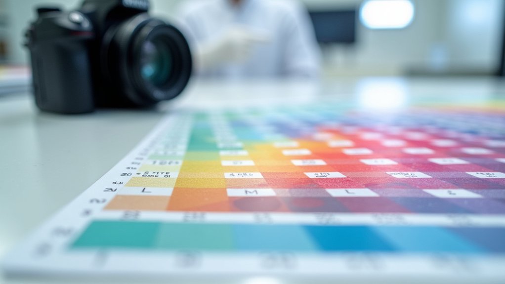
Color calibration in digital pathology is critical for accurate diagnoses. You’ll need to follow FDA guidelines using bio-polymer slides that mimic tissue properties and implement ICC profiles specific to your scanner model. Perform regular calibration checks according to manufacturer schedules, document all validation results, and compare scanned images against established color profiles. Proper calibration reduces inter-scanner variability, enhances AI algorithm performance, and guarantees consistent tissue representation. Discover how these techniques can appreciably improve your diagnostic confidence and standardization.
Color Calibration Guide for Digital Pathology Imaging

The microscope’s lens reveals a world where color accuracy can make the difference between correct and missed diagnoses.
When working with whole slide imaging systems, you’ll need to implement proper color calibration to guarantee consistent representation of stained tissue across different laboratories.
Your imaging system’s color fidelity directly impacts diagnostic confidence. To achieve standardization, utilize device-specific ICC profiles instead of generic RGB standards. These specialized profiles greatly enhance color accuracy in digital pathology applications.
Consider using calibration tools like the Sierra device with bio-polymers that mimic actual tissue characteristics.
Establish a regular calibration schedule using dedicated calibration slides and software to maintain consistency between scanners. This disciplined approach guarantees your digital pathology workflow delivers reliable, standardized results that pathologists can trust regardless of where slides are examined.
Understanding Color Variation Between Scanner Models
While examining digitized tissue under virtual microscopy, you’ll notice that images from different scanner models often display subtle but diagnostically significant color variations. This stems from fundamental differences in hardware components and how each scanner processes light information.
Understanding these variations is critical for maintaining diagnostic accuracy across multiple scanner sources:
- Camera sensors in different scanner models capture distinct parts of the light spectrum, creating inherent color variation in tissue representation.
- Even scanners from the same vendor may produce inconsistent color outputs due to manufacturing differences.
- Advanced color management techniques in newer models like the Aperio GT 450 aim to better match traditional microscope views.
When working across multiple scanner platforms, you’ll need to account for these differences to guarantee consistent interpretation of histopathological features.
The Science Behind Tissue Stain Color Representation

When pathologists examine digitized tissue specimens, they’re interpreting a complex interplay of light interactions with histological stains.
These stains bind to specific cellular components, creating distinctive color patterns that reveal critical diagnostic information.
Your understanding of how these colors are digitally captured is essential for accurate interpretation.
Each tissue sample’s true appearance can be compromised by variations in scanner hardware, potentially leading to diagnostic inconsistencies.
Proper color calibration guarantees that what you see digitally mirrors what you’d observe under a microscope.
FDA Recommendations for WSI Color Calibration
The FDA requires your WSI systems to undergo rigorous color calibration using target slides containing measurable color patches that mimic tissue stain spectral characteristics.
You’ll need to build validation datasets that document truth values, intended colors, and actual output colors to comply with international calibration standards.
Your ongoing collaboration with vendors is essential to maintain color fidelity across different scanning devices, ensuring diagnostic accuracy in your digital pathology workflow.
FDA Recommendations for WSI Color Calibration
To guarantee diagnostic accuracy in digital pathology, FDA recommendations for whole slide imaging (WSI) color calibration focus on rigorous testing procedures.
You’ll need to incorporate target slides with measurable color patches that reflect the spectral characteristics of stained tissue when validating pathology scanners.
For compliant WSI systems, you should:
- Create validation datasets that include truth, intended color, and output color measurements
- Implement ongoing validation processes to confirm consistent color fidelity in digital pathology images
- Collaborate with vendors to address color variation challenges across different scanner systems
The FDA emphasizes adherence to internationally accepted calibration standards, which helps build pathologist confidence in diagnostic accuracy regardless of which laboratory produced the images.
Measurable Targets Required
Essential to FDA compliance, measurable target slides serve as the cornerstone of effective color calibration in digital pathology. You’ll need to implement these targets to replicate the spectral characteristics of stained tissue, ensuring your WSI systems deliver consistent diagnostic accuracy across different laboratories.
| Target Type | Purpose | Validation Frequency | Impact on Diagnosis |
|---|---|---|---|
| Color Patches | Truth establishment | Monthly | High fidelity reproduction |
| Spectral Mimics | Tissue simulation | Quarterly | Reduced interpretation errors |
| Calibration Slides | System alignment | After maintenance | Consistent reporting |
| Reference Standards | Cross-lab consistency | Annually | Enhanced collaborative diagnosis |
Spectral Similarity Standards
While developing your WSI color calibration protocol, you’ll need to comply with FDA recommendations that specify target slides must exhibit spectral characteristics nearly identical to actual stained tissue.
This similarity guarantees diagnostic accuracy across different imaging systems and laboratories.
To meet these standards effectively:
- Create calibration datasets that include truth values, intended colors, and actual output colors to establish proper color management.
- Implement continuous validation processes to maintain color fidelity as technology evolves.
- Use measurable color patches that accurately mimic histological stains’ spectral properties.
Implementing ICC Color Profiles in Digital Pathology
ICC color profiles serve as the foundation for accurate color representation in your digital pathology workflow, transforming standard RGB output into device-specific calibrated images that closely match original tissue appearances.
You’ll need to understand both scanner-specific and display-specific profiles to implement an extensive color management system that meets CIE and ISO standards.
When properly applied, these profiles will greatly reduce inter-scanner variability and create the consistent imaging environment necessary for reliable AI-based diagnostic applications.
ICC Profile Fundamentals
Color accuracy forms the foundation of reliable digital pathology imaging, making International Color Consortium (ICC) profiles indispensable tools for standardization.
When you implement ICC profiles in your whole slide imaging workflow, you’re ensuring that tissue colors match what pathologists see under the microscope, markedly enhancing image fidelity and diagnostic reliability.
ICC profiles deliver three key advantages:
- Superior precision compared to standard RGB values, resulting in more accurate color representation across various viewing devices
- Standardized color management that minimizes discrepancies between different scanners and displays
- Enhanced validation of AI algorithms used in automated diagnostics, ensuring color consistency
Color calibration using ICC profiles adheres to rigorous CIE and ISO standards, providing the consistency needed for confident clinical interpretations in digital pathology.
Calibration Best Practices
To successfully implement ICC color profiles in your digital pathology workflow, you’ll need to follow several established best practices that extend beyond basic profile creation.
Start by implementing device-specific ICC standards for each scanner in your laboratory to guarantee consistent representation of stained tissues across platforms.
Schedule regular calibration sessions with qualified calibration specialists who understand ICC, CIE, and ISO standards. This systematic approach maintains color accuracy throughout your digital pathology ecosystem, reducing variations caused by different scanner hardware.
Document all calibration procedures for ongoing validation and quality assurance. This documentation supports regulatory compliance while building diagnostic confidence among pathologists reviewing your digital slides.
Remember that properly calibrated systems with ICC profiles produce markedly closer matches to original tissue colors than standard RGB values, potentially improving diagnostic outcomes.
Physical Calibration Methods Using Bio-polymer Slides

While traditional color calibration methods have limitations in digital pathology, bio-polymer slides represent a significant advancement in the field. The Sierra Colour Calibration Device employs a specialized bio-polymer that mimics tissue spectral properties when stained, ensuring accurate color fidelity in whole slide imaging systems.
You’ll benefit from these bio-polymer calibration methods in three key ways:
- Consistent color representation – Bio-polymer slides provide ground-truth reference data unaffected by illumination variations.
- Enhanced diagnostic accuracy – Your scanned images will closely match the true colors seen under a microscope.
- Standardized results – Your laboratory’s color management will align with others, enabling reliable collaboration.
Regular implementation of these calibration slides during routine scanner checks maintains peak color accuracy essential for reliable pathological assessment.
Impact of Color Standardization on AI Algorithm Performance
Implementing proper color standardization can dramatically boost your AI diagnostic accuracy, as evidenced by improved κ values from 0.439 to 0.619 after calibration.
You’ll notice significant performance enhancements when your AI algorithms process standardized whole slide images rather than uncalibrated scans from different sources.
This consistency across multiple clinical settings guarantees your AI tools deliver more reliable diagnostics, ultimately supporting better patient outcomes through standardized imaging conditions.
Calibration Boosts Diagnostics
The impact of color standardization on AI algorithm performance represents a significant breakthrough in digital pathology. By implementing physical color calibration across your WSI systems, you’ll dramatically improve diagnostic reliability and concordance between AI algorithms and pathologists’ assessments.
The evidence is compelling:
- Calibration increased κ values from 0.439 to 0.619 at Stavanger University Hospital, demonstrating substantial improvement in diagnostic accuracy.
- Karolinska University Hospital saw even more dramatic results, with κ values jumping from 0.354 to 0.738 after calibration.
- These improvements guarantee consistent image quality regardless of which scanning system you’re using.
Beyond enhancing AI performance, calibration enables broader clinical application of these technologies, ultimately improving patient outcomes through more reliable diagnostics across diverse clinical environments.
AI Performance Improvements
Across multiple clinical trials, color standardization has emerged as a pivotal factor in maximizing AI algorithm performance for digital pathology applications.
Studies reveal striking improvements in AI systems for prostate cancer Gleason grading, with concordance values (κ) jumping from 0.439 to 0.619 after implementing color calibration protocols.
You’ll find that physical color calibration methods markedly enhance AI diagnostic reliability across diverse clinical settings.
This standardization addresses the inherent variations between different whole slide imaging systems that previously limited AI effectiveness. By establishing consistent color representation, you’re enabling AI algorithms to perform with greater precision regardless of where images originate.
The impact on diagnostic accuracy can’t be overstated—properly calibrated imaging directly translates to more reliable AI-based diagnostics and, ultimately, improved patient outcomes through better clinical decision-making.
Steps for Routine Color Validation in Laboratory Settings

When establishing reliable digital pathology workflows, routine color validation becomes essential for maintaining diagnostic accuracy and consistency.
You’ll need to implement standardized procedures using calibration slides that mimic stained tissue to guarantee consistent color accuracy across your scanners.
To maintain proper color validation in your lab:
- Regularly assess and adjust scanner settings through image viewing software like Aperio ImageScope to guarantee uniform color representation.
- Schedule calibration checks according to manufacturer guidelines, integrating these into your routine maintenance protocols.
- Document validation results systematically to track changes over time, helping maintain FDA compliance.
This systematic approach allows you to identify discrepancies by comparing scanned images against established ICC color profiles, ultimately improving diagnostic reliability in your digital pathology practice.
Addressing Color Discrepancies Across Multiple Imaging Systems
Following proper validation protocols, you’ll likely encounter color variations when working with multiple imaging systems in your lab. These color discrepancies stem from hardware differences between whole slide imaging scanners, each capturing and rendering colors uniquely.
To overcome this challenge, implement device-specific ICC profiles instead of standard RGB formats. Regular color calibration using specialized tools like the Sierra Colour Calibration Device can notably improve consistency across your systems.
| Scanner Type | Common Color Issue | Recommended Solution |
|---|---|---|
| Brightfield | Yellow tint bias | Custom ICC profile |
| Fluorescence | Green channel shift | Spectral calibration |
| Multi-spectral | Cross-talk artifacts | Reference slide validation |
| High-throughput | Batch inconsistency | Daily calibration routine |
| Low-cost | Poor H&E differentiation | Bio-polymer calibration slide |
Remember that human color perception differs from scanner interpretations, making standardized calibration essential for reliable diagnostic outcomes.
Color Management Best Practices for Diagnostic Accuracy

Reliable diagnostic assessments in digital pathology depend on consistent and accurate color representation throughout your imaging workflow.
To maintain color fidelity across your WSI systems, implement device-specific ICC profiles rather than relying on standard RGB settings. These profiles greatly enhance scan accuracy and improve diagnostic reliability.
Device-specific ICC profiles are essential for true color fidelity in digital pathology, ensuring diagnostic confidence beyond standard RGB limitations.
Incorporate these calibration essentials into your laboratory protocols:
- Utilize bio-polymer calibration devices (like the Sierra Colour Calibration Device) that mimic stained tissue for ideal color matching.
- Establish a regular schedule for scanner calibration and validation, aligning with FDA recommendations and international standards.
- Train your team to recognize scanner-specific color variations and adjust interpretation accordingly when working across multiple systems.
Consistent application of these practices guarantees your digital pathology workflow delivers the color accuracy needed for confident diagnoses.
Frequently Asked Questions
What Is Dicom Standard for Digital Pathology?
DICOM standard for digital pathology is your framework for storing, transmitting, and sharing medical imaging data. It guarantees interoperability between systems while managing essential metadata and integrating with EHRs for seamless clinical workflows.
How Does Color Calibration Work?
Color calibration works by using specialized target slides with measurable color patches. You’ll create ICC profiles for your scanner that match stained tissue colors, ensuring consistency across different imaging systems during regular maintenance checks.
What Is a Digital Pathology Scanner?
A digital pathology scanner is a device you’d use to capture high-resolution digital images of tissue slides. It transforms physical specimens into whole slide images for pathologists to analyze and diagnose digitally.
In Summary
By standardizing color calibration in your digital pathology workflow, you’ll guarantee consistent image quality across different scanners. You’re not just improving visual comparability—you’re enhancing diagnostic accuracy and AI algorithm performance. Don’t overlook regular validation protocols. When you implement ICC profiles and follow FDA recommendations, you’re safeguarding patient care through reliable color representation that pathologists can trust regardless of which imaging system captured the slide.


Leave a Reply